
Main menu

  • Home
  • Content
    • Current
    • Ahead of print
    • Past Issues
    • JNM Supplement
    • SNMMI Annual Meeting Abstracts
    • Continuing Education
    • JNM Podcasts
  • Subscriptions
    • Subscribers
    • Institutional and Non-member
    • Rates
    • Journal Claims
    • Corporate & Special Sales
  • Authors
    • Submit to JNM
    • Information for Authors
    • Assignment of Copyright
    • AQARA requirements
  • Info
    • Reviewers
    • Permissions
    • Advertisers
  • About
    • About Us
    • Editorial Board
    • Contact Information
  • More
    • Alerts
    • Feedback
    • Help
    • SNMMI Journals
  • SNMMI
    • JNM
    • JNMT
    • SNMMI Journals
    • SNMMI

User menu

  • Subscribe
  • My alerts
  • Log in
  • My Cart

Search

  • Advanced search
Journal of Nuclear Medicine
  • SNMMI
    • JNM
    • JNMT
    • SNMMI Journals
    • SNMMI
  • Subscribe
  • My alerts
  • Log in
  • My Cart
Journal of Nuclear Medicine

Advanced Search

  • Home
  • Content
    • Current
    • Ahead of print
    • Past Issues
    • JNM Supplement
    • SNMMI Annual Meeting Abstracts
    • Continuing Education
    • JNM Podcasts
  • Subscriptions
    • Subscribers
    • Institutional and Non-member
    • Rates
    • Journal Claims
    • Corporate & Special Sales
  • Authors
    • Submit to JNM
    • Information for Authors
    • Assignment of Copyright
    • AQARA requirements
  • Info
    • Reviewers
    • Permissions
    • Advertisers
  • About
    • About Us
    • Editorial Board
    • Contact Information
  • More
    • Alerts
    • Feedback
    • Help
    • SNMMI Journals
  • View or Listen to JNM Podcast
  • Visit JNM on Facebook
  • Join JNM on LinkedIn
  • Follow JNM on Twitter
  • Subscribe to our RSS feeds
Meeting ReportInstrumentation & Data Analysis Track

Attenuation Correction of PET/MR Using Deep Neural Network Based on Dixon and ZTE MR Images

Kuang Gong, Jaewon Yang, Kyungsang Kim, Georges El Fakhri, Youngho Seo and Quanzheng Li
Journal of Nuclear Medicine May 2018, 59 (supplement 1) 650;
Kuang Gong
1Massachusetts General Hospital Boston MA United States
3Massachusetts General Hospital Boston MA United States
  • Find this author on Google Scholar
  • Find this author on PubMed
  • Search for this author on this site
Jaewon Yang
5UCSF Radiology and Biomedical Imaging San Francisco CA United States
  • Find this author on Google Scholar
  • Find this author on PubMed
  • Search for this author on this site
Kyungsang Kim
2Massachusetts General Hospital Allston MA United States
  • Find this author on Google Scholar
  • Find this author on PubMed
  • Search for this author on this site
Georges El Fakhri
4Massachusetts General Hospital, Harvard Medical Sc Boston MA United States
  • Find this author on Google Scholar
  • Find this author on PubMed
  • Search for this author on this site
Youngho Seo
6University of California, San Francisco San Francisco CA United States
  • Find this author on Google Scholar
  • Find this author on PubMed
  • Search for this author on this site
Quanzheng Li
1Massachusetts General Hospital Boston MA United States
3Massachusetts General Hospital Boston MA United States
  • Find this author on Google Scholar
  • Find this author on PubMed
  • Search for this author on this site
  • Article
  • Figures & Data
  • Info & Metrics
Loading

Abstract

650

Objectives: To obtain meaningful quantitative results from PET images, attenuation correction is necessary during image reconstruction. For PET/MR hybrid systems, attenuation correction is challenging as MR images do not reflect attenuation coefficients directly. To address this issue, we present a deep neural network to derive the continuous attenuation coefficients for brain PET/MR imaging using both Dixon and zero echo time (ZTE) MR images. To make fully use of multiple MR images, a new network structure was proposed to extract the features from Dixon and ZTE images independently at early layers and combine them together at later layers.

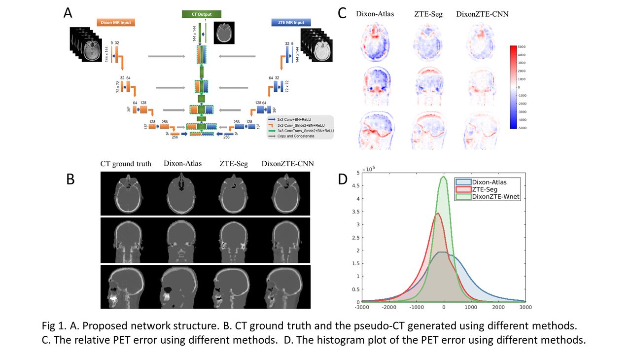
Methods: Fourteen patients (age 60.3y±10.2, F=6, M=8) who had whole-body PET/CT scan, followed by additional PET/MRI scanning without second tracer administration, were included in this study. Both Dixon and ZTE images were acquired during the PET/MRI scanning with average scan duration of 259.3 ± 94.2 s. The average administered FDG dose was 275.9 ± 60.6 MBq. When preparing the network training pairs, we first registered CT images and ZTE images to the Dixon MR images through rigid transformation using the ANTs software. Then random rotation and permutation was performed on the training pairs to avoid over-fitting. Later quantitative analysis was based on Seven-Fold cross validation. The proposed network structure is shown in Fig. 1(A), which extracts features from different inputs independently and combines them together in later stage. To make full use of the axial information, nine neighboring axial slices were stacked, occupying nine input channels, to reduce the axial aliasing artifacts. The network structures were implemented in TensorFlow 1.1.0. Batch size was set to 30 and 1000 epochs were run. This method is denoted as DixonZTE-CNN. The atlas method based on Dixon MR image (denoted as Dixon-Atlas) and the ZTE segmentation method based on ZTE MR image (denoted as ZTE-Seg) were adopted as baseline methods. Regional as well as global relative PET errors were used to compare the performance of different methods.

Results: The proposed DixonZTE-CNN can generate pseudo-CT images with more structure details (as presented in Fig 1). Quantitative analysis (as presented in Table 1) shows that DixonZTE-CNN achieved the lowest whole brain error (2.31% ± 0.60%) compared with Dixon-Atlas (5.50% ± 1.46%) and ZTE-Seg (3.76% ± 1.43%)). Regional analysis in cerebellum, brain lobes and inner regions also demonstrates the improvements by using DixonZTE-CNN. Conclusion: We have proposed a deep neural network to generate continuous attenuation map for brain PET/MR imaging based on both Dixon and ZTE images. Analysis using real data sets shows that the neural network can produce smaller quantification errors in PET image compared to other standard methods, and thus provide a promising new approach for the attenuation correction of PET/MR

Figure
  • Download figure
  • Open in new tab
  • Download powerpoint
View this table:
  • View inline
  • View popup

Relative PET error for different regions

Previous
Back to top

In this issue

Journal of Nuclear Medicine
Vol. 59, Issue supplement 1
May 1, 2018
  • Table of Contents
  • Index by author
Article Alerts
Sign In to Email Alerts with your Email Address
Email Article

Thank you for your interest in spreading the word on Journal of Nuclear Medicine.

NOTE: We only request your email address so that the person you are recommending the page to knows that you wanted them to see it, and that it is not junk mail. We do not capture any email address.

Enter multiple addresses on separate lines or separate them with commas.
Attenuation Correction of PET/MR Using Deep Neural Network Based on Dixon and ZTE MR Images
(Your Name) has sent you a message from Journal of Nuclear Medicine
(Your Name) thought you would like to see the Journal of Nuclear Medicine web site.
Citation Tools
Attenuation Correction of PET/MR Using Deep Neural Network Based on Dixon and ZTE MR Images
Kuang Gong, Jaewon Yang, Kyungsang Kim, Georges El Fakhri, Youngho Seo, Quanzheng Li
Journal of Nuclear Medicine May 2018, 59 (supplement 1) 650;

Citation Manager Formats

  • BibTeX
  • Bookends
  • EasyBib
  • EndNote (tagged)
  • EndNote 8 (xml)
  • Medlars
  • Mendeley
  • Papers
  • RefWorks Tagged
  • Ref Manager
  • RIS
  • Zotero
Share
Attenuation Correction of PET/MR Using Deep Neural Network Based on Dixon and ZTE MR Images
Kuang Gong, Jaewon Yang, Kyungsang Kim, Georges El Fakhri, Youngho Seo, Quanzheng Li
Journal of Nuclear Medicine May 2018, 59 (supplement 1) 650;
Twitter logo Facebook logo LinkedIn logo Mendeley logo
  • Tweet Widget
  • Facebook Like
  • Google Plus One
Bookmark this article

Jump to section

  • Article
  • Figures & Data
  • Info & Metrics

Related Articles

  • No related articles found.
  • Google Scholar

Cited By...

  • No citing articles found.
  • Google Scholar

More in this TOC Section

Instrumentation & Data Analysis Track

  • Deep Learning Based Kidney Segmentation for Glomerular Filtration Rate Measurement Using Quantitative SPECT/CT
  • Preclinical validation of a single-scan rest/stress imaging technique for 13NH3 cardiac perfusion studies
  • Comparison of 22 partial volume correction methods for amyloid PET imaging with 11C-PiB
Show more Instrumentation & Data Analysis Track

PET/MR Quantitation (Image Generation)

  • Impact of attenuation map generation schemes in lesion quantification in prostate cancer patients with PET/MRI
  • PET-MR Attenuation Correction using 3D Multi-Echo Dixon-Based UTE Sequence
Show more PET/MR Quantitation (Image Generation)

Similar Articles

SNMMI

© 2025 SNMMI

Powered by HighWire