
Main menu

  • Home
  • Content
    • Current
    • Ahead of print
    • Past Issues
    • JNM Supplement
    • SNMMI Annual Meeting Abstracts
    • Continuing Education
    • JNM Podcasts
  • Subscriptions
    • Subscribers
    • Institutional and Non-member
    • Rates
    • Journal Claims
    • Corporate & Special Sales
  • Authors
    • Submit to JNM
    • Information for Authors
    • Assignment of Copyright
    • AQARA requirements
  • Info
    • Reviewers
    • Permissions
    • Advertisers
  • About
    • About Us
    • Editorial Board
    • Contact Information
  • More
    • Alerts
    • Feedback
    • Help
    • SNMMI Journals
  • SNMMI
    • JNM
    • JNMT
    • SNMMI Journals
    • SNMMI

User menu

  • Subscribe
  • My alerts
  • Log in
  • My Cart

Search

  • Advanced search
Journal of Nuclear Medicine
  • SNMMI
    • JNM
    • JNMT
    • SNMMI Journals
    • SNMMI
  • Subscribe
  • My alerts
  • Log in
  • My Cart
Journal of Nuclear Medicine

Advanced Search

  • Home
  • Content
    • Current
    • Ahead of print
    • Past Issues
    • JNM Supplement
    • SNMMI Annual Meeting Abstracts
    • Continuing Education
    • JNM Podcasts
  • Subscriptions
    • Subscribers
    • Institutional and Non-member
    • Rates
    • Journal Claims
    • Corporate & Special Sales
  • Authors
    • Submit to JNM
    • Information for Authors
    • Assignment of Copyright
    • AQARA requirements
  • Info
    • Reviewers
    • Permissions
    • Advertisers
  • About
    • About Us
    • Editorial Board
    • Contact Information
  • More
    • Alerts
    • Feedback
    • Help
    • SNMMI Journals
  • View or Listen to JNM Podcast
  • Visit JNM on Facebook
  • Join JNM on LinkedIn
  • Follow JNM on Twitter
  • Subscribe to our RSS feeds
Research ArticleFocus on Molecular Imaging

Recent Advances in 18F Radiochemistry: A Focus on B-18F, Si-18F, Al-18F, and C-18F Radiofluorination via Spirocyclic Iodonium Ylides

Vadim Bernard-Gauthier, Mathieu L. Lepage, Bjoern Waengler, Justin J. Bailey, Steven H. Liang, David M. Perrin, Neil Vasdev and Ralf Schirrmacher
Journal of Nuclear Medicine April 2018, 59 (4) 568-572; DOI: https://doi.org/10.2967/jnumed.117.197095
Vadim Bernard-Gauthier
1Division of Nuclear Medicine and Molecular Imaging, Massachusetts General Hospital and Department of Radiology, Harvard Medical School, Boston, Massachusetts
  • Find this author on Google Scholar
  • Find this author on PubMed
  • Search for this author on this site
Mathieu L. Lepage
2Chemistry Department, University of British Columbia, Vancouver, British Columbia, Canada
  • Find this author on Google Scholar
  • Find this author on PubMed
  • Search for this author on this site
Bjoern Waengler
3Molecular Imaging and Radiochemistry, Department of Clinical Radiology and Nuclear Medicine, Medical Faculty Mannheim, Heidelberg University, Mannheim, Germany; and
  • Find this author on Google Scholar
  • Find this author on PubMed
  • Search for this author on this site
Justin J. Bailey
4Department of Oncology, Division of Oncological Imaging, University of Alberta, Edmonton, Alberta, Canada
  • Find this author on Google Scholar
  • Find this author on PubMed
  • Search for this author on this site
Steven H. Liang
1Division of Nuclear Medicine and Molecular Imaging, Massachusetts General Hospital and Department of Radiology, Harvard Medical School, Boston, Massachusetts
  • Find this author on Google Scholar
  • Find this author on PubMed
  • Search for this author on this site
David M. Perrin
2Chemistry Department, University of British Columbia, Vancouver, British Columbia, Canada
  • Find this author on Google Scholar
  • Find this author on PubMed
  • Search for this author on this site
Neil Vasdev
1Division of Nuclear Medicine and Molecular Imaging, Massachusetts General Hospital and Department of Radiology, Harvard Medical School, Boston, Massachusetts
  • Find this author on Google Scholar
  • Find this author on PubMed
  • Search for this author on this site
Ralf Schirrmacher
4Department of Oncology, Division of Oncological Imaging, University of Alberta, Edmonton, Alberta, Canada
  • Find this author on Google Scholar
  • Find this author on PubMed
  • Search for this author on this site
  • Article
  • Figures & Data
  • Info & Metrics
  • PDF
Loading

Abstract

Straightforward radiosynthesis protocols for 18F-labeled radiopharmaceuticals are an indispensable but often overlooked prerequisite to successfully perform molecular imaging studies in vivo by PET. In recent years, thanks to the expansion of the 18F chemical toolbox, structurally diverse and novel clinically relevant radiopharmaceuticals have been synthesized with both high efficiency and ready implementation. This article provides an overview of recent 18F-labeling methodologies, specifically for B-18F, Si-18F, Al-18F, and iodine (III)-mediated radiofluorination via the spirocyclic iodonium ylide technology.

  • radiochemistry
  • fluorine-18
  • radiofluorination
  • spirocyclic iodonium

For nearly 60 y, radiochemists have devised numerous methodologies to introduce 18F into organic compounds to support the development and clinical translation of PET radiopharmaceuticals (1). Radiochemists have long sought simplified radiochemical methods to preferably introduce the 18F radionuclide in a kit-like manner, similar to the production of 99mTc-based imaging agents for SPECT. Here, we review 4 promising new radiochemistries based on B-18F, Si-18F, Al-18F formation and spirocyclic iodonium ylide (SCIDY) use coming close to that premise.

BORON–18F RADIOCHEMISTRY

In seeking new methods, several teams have eschewed C–F bond formation in favor of other heteroatom–fluorine bond-forming strategies. Ting et al. hypothesized that an organotrifluoroborate, readily prepared in water under mildly acidic conditions (pH 2–3), would afford late-stage, one-step labeling in a manner similar to radiometal chelation (2). Advantages of RBF3s include hydrophilicity (as anionic salts, they would enhance in vivo clearance for increased-contrast images compared with more lipophilic organofluorides that retard clearance), bioorthogonality (the strong B–F bond, being bioorthogonal, would be metabolically stable), nontoxicity (little in the vast literature would suggest that these are acutely toxic (3)), and the potential for tripling the molar activity (Am) of the fluoride used (because the pool of fluoride ions would condense on one third as many molecules to give the trifluoroborate) (Fig. 1A) (4). In addition, since peptides are deprotected in acid (often, concentrated hydrofluoric acid) and are high-performance liquid chromatography–purified in acidic medium, organotrifluoroborate synthesis would pose few problems to the structural integrity of most peptides being labeled.

FIGURE 1.
  • Download figure
  • Open in new tab
  • Download powerpoint
FIGURE 1.

(A) RBF3 radiofluorination. (B) Structure of 18F-AMBF3-rhodamine-bisRGD. DMF = dimethylformamide.

A concerted effort to apply both synthetic methods and physical organic chemistry led Perrin’s group to address the molecular basis for ensuring the kinetic stability of RBF3 in water, ultimately leading to several synthons that could be fluorinated relatively rapidly and in reasonable radiochemical yields (RCYs) while providing sufficient stability in vitro and ultimately in vivo (5,6). Early radiofluorination methods toward this goal provided preliminary signs of success with the 18F labeling and PET imaging of biotin, marimastat (7), arginylglycylaspartic acid (RGD) (8), bombesin (9), and bisRGD (10). Although going beyond simple proof of concept to show in vivo stability and target specificity with blocking controls, in certain cases bone uptake was seen, and in others, tumor uptake values were low. Furthermore, synthesis was still not readily user-friendly, often requiring the addition of carrier 19F-fluoride, and tumor uptake values were too low (<2% of the injected dose per gram). These observations were attributed to the extremely polar nature of the organotrifluoroborate and to the metabolic instability of the linker arm in the case of a bombesin analog.

With promising work from the Schirrmacher lab on isotope exchange for 18F labeling of silylfluorides—work that was recapitulated on boron first by Gabbai (11)—Perrin et al. applied this technique to existing organotrifluoroborates while also establishing a range of other useful organotrifluoroborate prosthetic compositions. Although there is no thermodynamic driving force for isotope exchange, this technique provides exceptional simplicity, because the precursor is identical to the product. This advantage has been particularly useful in the context of organotrifluoroborates in terms of precursor production, isolation, shelf stability, and characterization because organotrifluoroborates are stable, easily isolated, and characterized by 19F NMR spectroscopy. Use of isotope exchange on several new prosthetics, which were grafted onto new ligands, enabled successful labeling of several molecularly complex peptides, including Tyr3-octreotate (TATE), bradykinin, bombesin, and a fluorescent dimeric RGD (12). In the case of TATE, high tumor uptake was observed (≤16% of injected dose per gram), with very high contrast ratios (13). Excitingly, this method also enabled the radiolabeling of a fluorescent-dimeric-RGD (Fig. 1B) (14). Tumor uptake increased 10-fold over that found in a previous study with a dimeric-RGD-organotrifluoroborate construct.

SILICON–18F RADIOCHEMISTRY

In the early days of silicon-fluoride-acceptor (SiFA) radiochemistry, the 18F labeling of the SiFA building block (Fig. 2A) worked surprisingly well and proved that revisiting isotopic exchange as a strategy to obtain radiotracers of high Am was a good choice despite the historical prevalent bias against isotopic exchange (isotopic exchange usually does not yield 18F-radiotracers of high Am) (15). Isotopic exchange of the nonradioactive 19F with anionic 18F− in various solvents ranging from acetonitrile to water gave not only access to simple labeled SiFA model compounds but also to a SiFA-modified TATE, a peptide well known to diagnose somatostatin receptor–positive tumors. This first SiFA-Tyr3-TATE was 18F-labeled in high RCYs and with surprisingly high Am for an isotopic exchange reaction (16). However, when injected into an AR42J tumor–bearing rat, a major drawback of the SiFA building block became apparent, namely its inherently high lipophilicity originating from its unique chemical structure, leading to an almost exclusive accumulation in the liver. The 2 tertiary butyl-groups, shielding the 18F–silicon bond from being exposed to water and becoming hydrolyzed in vivo, unfortunately are a prerequisite sine qua non to impart chemical stability to the 18F-SiFA structure. The high lipophilicity has serious implications for in vivo imaging simply because lipophilic compounds are siphoned out efficiently by the liver and accumulate there, extracting the radiotracer from the blood and making it unavailable for target engagement. At this point, the SiFA methodology was just an example of a new niche radiochemistry unambiguously failing in vivo as a result of SiFA’s chemical nature adversely influencing its biodistribution. An obvious solution to this problem was either changing the SiFA structure toward lower lipophilicity while still ensuring hydrolytic stability or adding hydrophilic modules counteracting SiFA’s lipophilicity. Wängler et al. chose an acetylated amino-sugar (AcNH-β-Glc) attached to an asparagine (Asn) side chain to add a hydrophilic moiety to the SiFA-TATE (17). The 18F-SiFA-Asn(AcNH-β-Glc)-PEG-Tyr3-TATE accumulated far more favorably in AR42J tumors in a mouse xenograft model. Eight percent of the injected dose per gram was taken up by the tumor, albeit there was a still-high liver uptake. Apparently, the reduction of lipophilicity was a step in the right direction although not yet drastic enough. This desired step was achieved by introducing SiFAlin, a positively charged SiFA building block, 2 highly hydrophilic Asn side chains, and the aforementioned AcNH-β-Glc (18). The LogD of this conjugate dropped to −1.21, being in the same range as 68Ga-DOTATATE. In a comparative small-animal PET/CT study of 68Ga-DOTATATE and 18F-SiFAlin-Asn(AcNH-Glc)-Asp2-PEG1-TATE (Fig. 2B) in an AR42J tumor xenograft model, the SUV of both compounds was almost identical, at 5.50 versus 7.80, respectively (19). This conjugate is currently being evaluated in a toxicity assessment, and pending positive results, a first-in-human PET study is planned in a healthy control group and in patients bearing a somatostatin receptor–positive tumor. A SiFA compound for prostate-specific membrane antigen imaging in LNCaP tumor–bearing mice has recently been reported in a symposium abstract (20). Besides the SiFA approach, Honer et al. have introduced an impressive preclinical line-up of 18F-silicon compounds (21).

FIGURE 2.
  • Download figure
  • Open in new tab
  • Download powerpoint
FIGURE 2.

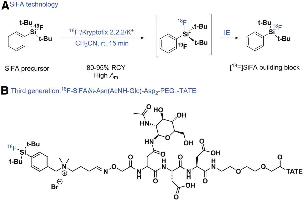
(A) SiFA labeling concept based on isotopic exchange. (B) Third generation of SiFA-derivatized TATE for diagnosis of somatostatin-expressing tumors. rt = room temperature.

ALUMINUM–18F RADIOCHEMISTRY

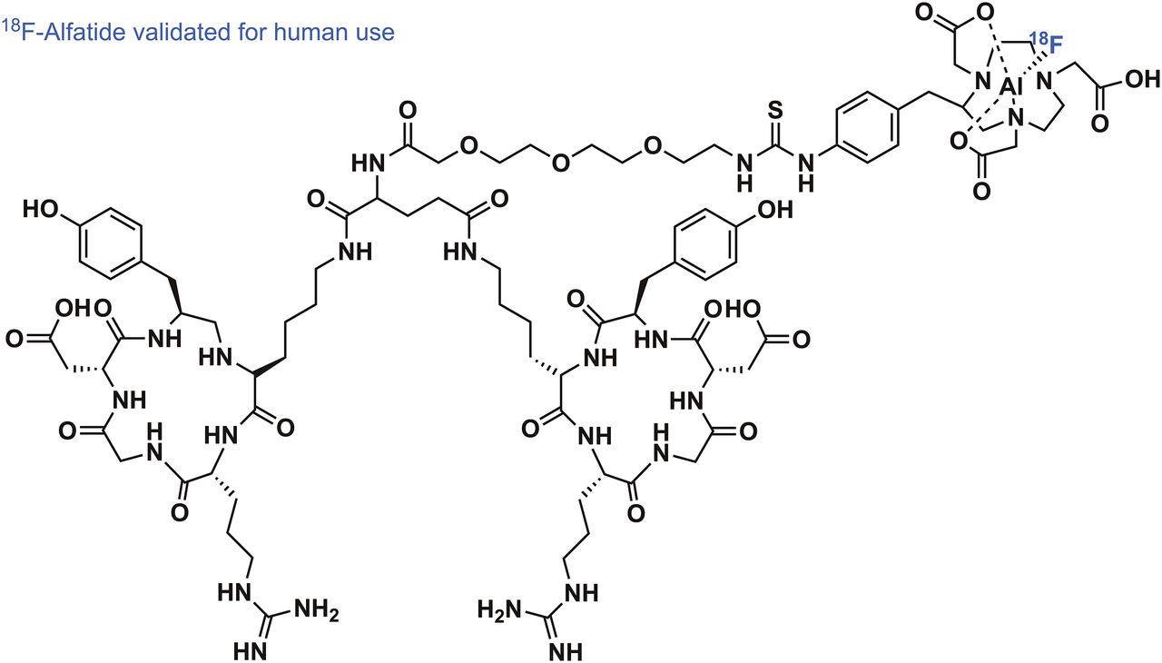
The Al–18F radiochemistry, first introduced by McBride et al. in 2009 (22), admittedly came a long way in a short time—from the disclosure of the basic radiochemical concept to the first reported applications in human PET imaging (23). The labeling mechanic of the Al–18F radiochemistry is rooted in the preformation of aluminum mono-18F-fluoride cation (18F-AlF2+) under aqueous conditions. The subsequent chelation of 18F-AlF2+ by well-known chelators such as pentetic acid, 1,4,7-triazacyclononane-N,N′,N″-triacetic acid (NOTA), and 1,4,7-triazacyclononane-N,N′-diacetic acid (NODA), all familiar from radiometal complexation chemistry, has invigorated strong clinical translational efforts to simply mimic 68Ga or 64Cu labeling of established radiometal-based PET tracers with 18F. The driving force for the efficient formation of 18F-AlF2+ is the exceptionally high binding energy between aluminum and fluorine of more than 670 kJ/mol. This radiochemistry works fully under aqueous conditions and has been thoroughly reviewed alongside its preclinical applications (24). Pentadentate chelators, which donate 3 nitrogen and 2 oxygen atoms to the binding of 18F-AlF2+, have been used first for PET tracer development of peptide and protein nature. These first-generation chelators for 18F− complexation, however, required high reaction temperatures of more than 100°C, potentially detrimental to the integrity of some compounds of higher molecular weight. Although high temperatures can be a general drawback in radiolabeling, impressive first-in-human PET imaging of integrin ανβ3–expressing tumors with RGD peptides derivatized with NOTA validated the use of 18F-AlF2+ in clinical PET imaging. The RGD derivative 18F-AlF-NOTA-PRGD2 (18F-alfatide) could be labeled in high RCYs of more than 40% (decay-corrected) and with a radiochemical purity of more than 95% within 20 min (Fig. 3) (23). Integrin-expressing lung tumors in several patients could be clearly delineated using 18F-alfatide, with a tumor-to-muscle and tumor-to-blood ratio of 2.9 and 5.87, respectively. After these encouraging results, a pilot study by Gao et al., aimed at imaging integrin ανβ3 in suspected lung cancer patients, further used 18F-alfatide successfully in a clinical setting (25). Seventeen patients with lung cancer were accurately identified among true-negative (hamartoma) as well as false-positive (chronic inflammation and inflammatory pseudotumor) cases. The question of whether 18F-alfatide PET/CT imaging can predict the short-term outcome of concurrent chemoradiotherapy in patients with advanced non–small cell lung cancer was investigated by Luan et al. (26). Responders and nonresponders to chemotherapy could be distinguished with high sensitivity, specificity, and accuracy, confirming that 18F-alfatide could be a useful tool in therapy response prediction. The electronically uncharged nature of 18F-AlF-NOTA complexes prompted the investigation of 18F-alfatide II, a derivative of 18F-alfatide, extended by one more polyethylene glycol unit, for imaging brain metastasis (27). In 9 patients with a total of 20 metastatic brain tumors, it was demonstrated that 18F-alfatide II exhibited a tumor-to-background ratio superior to that of 18F-FDG. Even though there is no direct clinical advantage to the use of 18F-alfatide derivatives in comparison to an already-established RGD tracer such as 18F-galacto-RGD, the ease of synthesis and reliable labeling methodology make 18F-alfatide derivatives a serious competitor. This view was further substantiated recently by Zhang et al., who evaluated the use of 18F-alfatide PET/CT to predict sensitivity to chemotherapy in patients with recently diagnosed glioblastoma (28). Twenty-five patients diagnosed with glioblastoma multiform underwent surgical resection. 18F-alfatide PET/CT successfully delineated the residual lesions of the glioblastoma. Baseline scans, as well as follow-up scans after 3 wk, were available and confirmed the predictable power of 18F-alfatide PET/CT for response evaluation after chemotherapy. Among others (29), recent advancements in chelator development resulted in the introduction of racemic H3RESCA, a new chelator complexing 18F-AlF2+ at room temperature. Besides extensive preclinical evaluation, a first-in-human study using a HER2-targeting H3RESCA-derivatized Affibody (PEP04314; Affibody AB) has been reported (30).

FIGURE 3.
  • Download figure
  • Open in new tab
  • Download powerpoint
FIGURE 3.

Structure of 18F-alfatide, clinically used radiotracer for imaging of integrin-expressing tumors.

IODINE(III)-MEDIATED RADIOFLUORINATION: SCIDY TECHNOLOGY

Since 2014, there were several reports of a 1-step, regioselective method for 18F incorporation via hypervalent iodine(III)-mediated precursors to afford 18F-arenes from 18F-fluoride in high RCYs and high Am (31–33). The novel technology relies on SCIDY technology to produce labeling precursors that regioselectively introduce 18F-fluoride ion into nonactivated aromatic rings with wide substrate scope under metal-free conditions (Fig. 4A). The technique involves stable, easily purified SCIDY precursors, is readily implemented with standard workup procedures, is easily adopted in any laboratory, and has since been applied for producing 18F-radiopharmaceuticals for PET imaging studies in humans. Notably, 18F-FPEB (3-fluoro-5-[(pyridin-3-yl)ethynyl]benzonitrile), an important radiopharmaceutical for imaging metabotropic glutamate receptor 5 in human that is challenged by low RCYs, was validated using the SCIDY-radiofluorination approach. A dramatically increased RCY for 18F-FPEB—from 1%–2% to 20% (a 10-fold increase)—was realized, and the product, which had an Am of 666 GBq/μmol, represents the first radiopharmaceutical prepared by such technologies (Fig. 4B) (34).

FIGURE 4.
  • Download figure
  • Open in new tab
  • Download powerpoint
FIGURE 4.

SCIDY-based radiofluorination and its translation into clinically relevant PET tracers. (A) Concept of 18F-radiofluorination. (B) 18F-FPEB for clinical PET imaging. (C) 18F-lorlatinib synthesized using SCIDY.

Two promising spirocyclic auxiliaries, namely, cyclopentyl (33) and adamantyl (35), demonstrated excellent incorporation yields on challenging electron-neutral or -donating arenes. SCIDY methodology proved to be efficient for radiolabeling a diverse range of nonactivated functionalized arenes and heteroarenes, with remarkably broad functional group compatibility. Several 18F-PET tracers and building blocks were successfully synthesized from 18F-fluoride for the first time, or the corresponding radiosyntheses were significantly simplified and improved via SCIDY methods. For example, the first synthesis of 5-18F-fluorouracil from 18F-fluoride was achieved in 11% RCY (33), and there are new 18F building blocks such as fluoroaromatic azides (36,37) that have wide applications in click-radiosynthesis. The conceptual advantages of excellent regioselectivity, and the viability of incorporating 18F into a wide array of nonactivated (hetero)arenes, make this methodology suitable for routine radiopharmaceutical production. Furthermore, a number of PET radiopharmaceuticals for routine clinical research, including 18F-lorlatinib (38), 18F-FDPA (39), 18F-6-fluoro-l-m-tyrosine, and 18F-mFBG (35), which are challenging to prepare using conventional labeling methods, have been isolated in high RCYs by this methodology (Fig. 4C). The promise of these newly developed reactants for radiofluorination is demonstrated for the wide range of nitrogen- and oxygen-containing heterocycles and drug fragments that have been labeled with 18F-fluoride, including basic amine functional groups, which have been a longstanding challenge in 18F-radiochemistry.

CONCLUSION

Amid the inherent technical and chemical challenges associated with efficiently delivering innovative 18F-radiopharmaceuticals, the methodologies described here represent major advances that will likely have an impact on translational imaging research as further steps toward the clinic are taken in coming years.

DISCLOSURE

No potential conflict of interest relevant to this article was reported.

Footnotes

  • Published online Dec. 28, 2017.

  • © 2018 by the Society of Nuclear Medicine and Molecular Imaging.

REFERENCES

  1. 1.↵
    1. Ametamey SM,
    2. Honer M,
    3. Schubiger PA
    . Molecular imaging with PET. Chem Rev. 2008;108:1501–1516.
    OpenUrlCrossRefPubMed
  2. 2.↵
    1. Ting R,
    2. Adam MJ,
    3. Ruth TJ,
    4. Perrin DM
    . Arylfluoroborates and alkylfluorosilicates as potential PET imaging agents: high-yielding aqueous biomolecular 18F-labeling. J Am Chem Soc. 2005;127:13094–13095.
    OpenUrlCrossRefPubMed
  3. 3.↵
    1. Molander GA,
    2. Ellis N
    . Organotrifluoroborates: protected boronic acids that expand the versatility of the Suzuki coupling reaction. Acc Chem Res. 2007;40:275–286.
    OpenUrlCrossRefPubMed
  4. 4.↵
    1. Liu Z,
    2. Li Y,
    3. Lozada J,
    4. et al
    . Stoichiometric leverage: rapid 18F-aryltrifluoroborate radiosynthesis at high specific activity for click conjugation. Angew Chem Int Ed English. 2013;52:2303–2307.
    OpenUrlCrossRef
  5. 5.↵
    1. Ting R,
    2. Harwig C,
    3. Lo J,
    4. et al
    . Substituent effects on aryltrifluoroborate solvolysis in water: implications for Suzuki-Miyaura coupling and the design of stable 18F-labeled aryltrifluoroborates for use in PET imaging. J Org Chem. 2008;73:4662–4670.
    OpenUrlCrossRefPubMed
  6. 6.↵
    1. Liu Z,
    2. Chao D,
    3. Li Y,
    4. Ting R,
    5. Oh J,
    6. Perrin DM
    . From minutes to years: predicting organotrifluoroborate solvolysis rates. Chemistry. 2015;21:3924–3928.
    OpenUrl
  7. 7.↵
    1. auf dem Keller U,
    2. Bellac CL,
    3. Li Y,
    4. et al
    . Novel matrix metalloproteinase inhibitor F-18 marimastat-aryltrifluoroborate as a probe for in vivo positron emission tomography imaging in cancer. Cancer Res. 2010;70:7562–7569.
    OpenUrlAbstract/FREE Full Text
  8. 8.↵
    1. Li Y,
    2. Guo J,
    3. Tang S,
    4. Lang L,
    5. Chen X,
    6. Perrin DM
    . One-step and one-pot-two-step radiosynthesis of cyclo-RGD-18F-aryltrifluoroborate conjugates for functional imaging. Am J Nucl Med Mol Imaging. 2013;3:44–56.
    OpenUrl
  9. 9.↵
    1. Li Y,
    2. Liu Z,
    3. Pourghiasian M,
    4. et al
    . Alkyne-18F-ArBF3 for one-pot click 18F-labeling of bombesin for in vivo PET imaging of tumors expressing the GRP-receptor. Am J Nucl Med Mol Imaging. 2013;3:57–70.
    OpenUrl
  10. 10.↵
    1. Li Y,
    2. Liu ZB,
    3. Lozada J,
    4. et al
    . Single step F-18-labeling of dimeric cycloRGD for functional PET imaging of tumors in mice. Nucl Med Biol. 2013;40:959–966.
    OpenUrl
  11. 11.↵
    1. Li ZB,
    2. Chansaenpak K,
    3. Liu SL,
    4. Wade CR,
    5. Conti PS,
    6. Gabbai FP
    . Harvesting F-18-fluoride ions in water via direct F-18-F-19 isotopic exchange: radiofluorination of zwitterionic aryltrifluoroborates and in vivo stability studies. MedChemComm. 2012;3:1305–1308.
    OpenUrlCrossRef
  12. 12.↵
    1. Liu ZB,
    2. Pourghiasian M,
    3. Radtke MA,
    4. et al
    . An organotrifluoroborate for broadly applicable one-step F-18-labeling. Angew Chem Int Ed English. 2014;53:11876–11880.
    OpenUrlCrossRef
  13. 13.↵
    1. Liu Z,
    2. Pourghiasian M,
    3. Benard F,
    4. Pan J,
    5. Lin K-S,
    6. Perrin DM
    . Preclinical evaluation of a high-affinity 18F-trifluoroborate octreotate derivative for somatostatin receptor imaging. J Nucl Med. 2014;55:1499–1505.
    OpenUrlAbstract/FREE Full Text
  14. 14.↵
    1. Liu Z,
    2. Radtke MA,
    3. Wong MQ,
    4. Lin K-S,
    5. Yapp DT,
    6. Perrin DM
    . Dual mode fluorescent 18F-PET tracers: efficient modular synthesis of rhodamine-cRGD 2-18F-organotrifluoroborate, rapid, and high yielding one-step 18F-labeling at high specific activity, and correlated in vivo PET imaging and ex vivo fluorescence. Bioconjugate Chem. 2014;25:1951–1962.
    OpenUrlCrossRefPubMed
  15. 15.↵
    1. Wängler C,
    2. Kostikov A,
    3. Zhu J,
    4. Chin J,
    5. Wängler B,
    6. Schirrmacher R
    . Silicon-[18F]fluorine radiochemistry: basics, applications and challenges. Appl Sci (Basel). 2012;2:277–302.
    OpenUrl
  16. 16.↵
    1. Schirrmacher R,
    2. Bradtmöller G,
    3. Schirrmacher E,
    4. et al
    . 18F-labeling of peptides by means of an organosilicon-based fluoride acceptor. Angew Chem Int Ed Eng. 2006;45:6047–6050.
    OpenUrlCrossRefPubMed
  17. 17.↵
    1. Wängler C,
    2. Waser B,
    3. Alke A,
    4. et al
    . One-step 18F-labeling of carbohydrate-conjugated octreotate-derivatives containing a silicon-fluoride-acceptor (SiFA): in vitro and in vivo evaluation as tumor imaging agents for positron emission tomography (PET). Bioconjug Chem. 2010;21:2289–2296.
    OpenUrlCrossRefPubMed
  18. 18.↵
    1. Wängler C,
    2. Niedermoser S,
    3. Chin J,
    4. et al
    . One-step 18F-labeling of peptides for positron emission tomography imaging using the SiFA methodology. Nat Protoc. 2012;7:1946–1955.
    OpenUrlCrossRefPubMed
  19. 19.↵
    1. Niedermoser S,
    2. Chin J,
    3. Wängler C,
    4. et al
    . In vivo evaluation of 18F-SiFAlin–modified TATE: a potential challenge for 68Ga-DOTATATE, the clinical gold standard for somatostatin receptor imaging with PET. J Nucl Med. 2015;56:1100–1105.
    OpenUrlAbstract/FREE Full Text
  20. 20.↵
    1. Bailey JJ,
    2. Wuest M,
    3. Bouvet C,
    4. et al
    . Silicon/fluorine-18/PSMA: a winning team for PET imaging of prostate cancer [abstract]. J Label Compd Radiopharm. 2017;60(suppl 1):S353.
    OpenUrl
  21. 21.↵
    1. Honer M,
    2. Mu L,
    3. Stellfeld T,
    4. et al
    . 18F-labeled bombesin analog for specific and effective targeting of prostate tumors expressing gastrin-releasing peptide receptors. J Nucl Med. 2011;52:270–278.
    OpenUrlAbstract/FREE Full Text
  22. 22.↵
    1. McBride WJ,
    2. Sharkey RM,
    3. Karacay H,
    4. et al
    . A novel method of 18F radiolabeling for PET. J Nucl Med. 2009;50:991–998.
    OpenUrlAbstract/FREE Full Text
  23. 23.↵
    1. Wan W,
    2. Guo N,
    3. Pan D,
    4. et al
    . First experience of 18F-alfatide in lung cancer patients using a new lyophilized kit for rapid radiofluorination. J Nucl Med. 2013;54:691–698.
    OpenUrlAbstract/FREE Full Text
  24. 24.↵
    1. McBride WJ,
    2. Sharkey RM,
    3. Goldenberg DM
    . Radiofluorination using aluminum-fluoride (Al18F). EJNMMI Res. 2013;3:36.
    OpenUrlCrossRefPubMed
  25. 25.↵
    1. Gao S,
    2. Wu H,
    3. Li W,
    4. et al
    . A pilot study imaging integrin αvβ3 with RGD PET/CT in suspected lung cancer patients. Eur J Nucl Med Mol Imaging. 2015;42:2029–2037.
    OpenUrlCrossRefPubMed
  26. 26.↵
    1. Luan X,
    2. Huang Y,
    3. Gao S,
    4. et al
    . 18F-alfatide PET/CT may predict short-term outcome of concurrent chemoradiotherapy in patients with advanced non-small cell lung cancer. Eur J Nucl Med Mol Imaging. 2016;43:2336–2342.
    OpenUrl
  27. 27.↵
    1. Yu C,
    2. Pan D,
    3. Mi B,
    4. et al
    . 18F-Alfatide II PET/CT in healthy human volunteers and patients with brain metastases. Eur J Nucl Med Mol Imaging. 2015;42:2021–2028.
    OpenUrl
  28. 28.↵
    1. Zhang H,
    2. Liu N,
    3. Gao S,
    4. et al
    . Can an 18F-ALF-NOTA-PRGD2 PET/CT scan predict treatment sensitivity to concurrent chemoradiotherapy in patients with newly diagnosed glioblastoma? J Nucl Med. 2016;57:524–529.
    OpenUrlAbstract/FREE Full Text
  29. 29.↵
    1. Cleeren F,
    2. Lecina J,
    3. Billaud EMF,
    4. Ahamed M,
    5. Verbruggen A,
    6. Bormans GM
    . New chelators for low temperature Al18F-labeling of biomolecules. Bioconjug Chem. 2016;27:790–798.
    OpenUrl
  30. 30.↵
    1. Cleeren F,
    2. Lecina J,
    3. Ahamed M,
    4. et al
    . Al18F-labeling of heat-sensitive biomolecules for positron emission tomography imaging. Theranostics. 2017;7:2924–2939.
    OpenUrl
  31. 31.↵
    1. Cardinale J,
    2. Ermert J,
    3. Humpert S,
    4. Coenen HH
    . Iodonium ylides for one-step, no-carrier-added radiofluorination of electron rich arenes, exemplified with 4-(([18F]fluorophenoxy)-phenylmethyl)piperidine NET and SERT ligands. RSC Advances. 2014;4:17293–17299.
    OpenUrl
  32. 32.
    1. Zlatopolskiy BD,
    2. Zischler J,
    3. Krapf P,
    4. et al
    . Copper-mediated aromatic radiofluorination revisited: efficient production of PET tracers on a preparative scale. Chemistry. 2015;21:5972–5979.
    OpenUrl
  33. 33.↵
    1. Rotstein BH,
    2. Stephenson NA,
    3. Vasdev N,
    4. Liang SH
    . Spirocyclic hypervalent iodine(III)-mediated radiofluorination of non-activated and hindered aromatics. Nat Commun. 2014;5:4365.
    OpenUrlPubMed
  34. 34.↵
    1. Stephenson NA,
    2. Holland JP,
    3. Kassenbrock A,
    4. et al
    . Iodonium ylide-mediated radiofluorination of 18F-FPEB and validation for human use. J Nucl Med. 2015;56:489–492.
    OpenUrlAbstract/FREE Full Text
  35. 35.↵
    1. Rotstein BH,
    2. Wang L,
    3. Liu RY,
    4. et al
    . Mechanistic studies and radiofluorination of structurally diverse pharmaceuticals with spirocyclic iodonium(III) ylides. Chem Sci. 2016;7:4407–4417.
    OpenUrl
  36. 36.↵
    1. Wang L,
    2. Jacobson O,
    3. Avdic D,
    4. et al
    . Ortho-stabilized 18F-azido click agents and their application in PET imaging with single-stranded DNA aptamers. Angew Chem Int Ed Engl. 2015;54:12777–12781.
    OpenUrl
  37. 37.↵
    1. Jacobson O,
    2. Weiss ID,
    3. Wang L,
    4. et al
    . 18F-labeled single-stranded DNA aptamer for PET imaging of protein tyrosine kinase-7 expression. J Nucl Med. 2015;56:1780–1785.
    OpenUrlAbstract/FREE Full Text
  38. 38.↵
    1. Collier TL,
    2. Normandin MD,
    3. Stephenson NA,
    4. et al
    . Synthesis and preliminary PET imaging of 11C and 18F isotopologues of the ROS1/ALK inhibitor lorlatinib. Nat Commun. 2017;8:15761.
    OpenUrl
  39. 39.↵
    1. Wang L,
    2. Cheng R,
    3. Fujinaga M,
    4. et al
    . A facile radiolabeling of [18F]FDPA via spirocyclic iodonium ylides: preliminary PET imaging studies in preclinical models of neuroinflammation. J Med Chem. 2017;60:5222–5227.
    OpenUrl
  • Received for publication October 11, 2017.
  • Accepted for publication December 24, 2017.
PreviousNext
Back to top

In this issue

Journal of Nuclear Medicine: 59 (4)
Journal of Nuclear Medicine
Vol. 59, Issue 4
April 1, 2018
  • Table of Contents
  • Table of Contents (PDF)
  • About the Cover
  • Index by author
Print
Download PDF
Article Alerts
Sign In to Email Alerts with your Email Address
Email Article

Thank you for your interest in spreading the word on Journal of Nuclear Medicine.

NOTE: We only request your email address so that the person you are recommending the page to knows that you wanted them to see it, and that it is not junk mail. We do not capture any email address.

Enter multiple addresses on separate lines or separate them with commas.
Recent Advances in 18F Radiochemistry: A Focus on B-18F, Si-18F, Al-18F, and C-18F Radiofluorination via Spirocyclic Iodonium Ylides
(Your Name) has sent you a message from Journal of Nuclear Medicine
(Your Name) thought you would like to see the Journal of Nuclear Medicine web site.
Citation Tools
Recent Advances in 18F Radiochemistry: A Focus on B-18F, Si-18F, Al-18F, and C-18F Radiofluorination via Spirocyclic Iodonium Ylides
Vadim Bernard-Gauthier, Mathieu L. Lepage, Bjoern Waengler, Justin J. Bailey, Steven H. Liang, David M. Perrin, Neil Vasdev, Ralf Schirrmacher
Journal of Nuclear Medicine Apr 2018, 59 (4) 568-572; DOI: 10.2967/jnumed.117.197095

Citation Manager Formats

  • BibTeX
  • Bookends
  • EasyBib
  • EndNote (tagged)
  • EndNote 8 (xml)
  • Medlars
  • Mendeley
  • Papers
  • RefWorks Tagged
  • Ref Manager
  • RIS
  • Zotero
Share
Recent Advances in 18F Radiochemistry: A Focus on B-18F, Si-18F, Al-18F, and C-18F Radiofluorination via Spirocyclic Iodonium Ylides
Vadim Bernard-Gauthier, Mathieu L. Lepage, Bjoern Waengler, Justin J. Bailey, Steven H. Liang, David M. Perrin, Neil Vasdev, Ralf Schirrmacher
Journal of Nuclear Medicine Apr 2018, 59 (4) 568-572; DOI: 10.2967/jnumed.117.197095
Twitter logo Facebook logo LinkedIn logo Mendeley logo
  • Tweet Widget
  • Facebook Like
  • Google Plus One
Bookmark this article

Jump to section

  • Article
    • Abstract
    • BORON–18F RADIOCHEMISTRY
    • SILICON–18F RADIOCHEMISTRY
    • ALUMINUM–18F RADIOCHEMISTRY
    • IODINE(III)-MEDIATED RADIOFLUORINATION: SCIDY TECHNOLOGY
    • CONCLUSION
    • DISCLOSURE
    • Footnotes
    • REFERENCES
  • Figures & Data
  • Info & Metrics
  • PDF

Related Articles

  • This Month in JNM
  • PubMed
  • Google Scholar

Cited By...

  • No citing articles found.
  • Google Scholar

More in this TOC Section

  • From Genome to Phenome: Opportunities and Challenges of Molecular Imaging
  • Imaging the Activity of Efflux Transporters at the Blood–Brain Barrier in Neurologic Diseases: Radiotracer Selection Criteria
  • Molecular Imaging of Pulmonary Fibrosis
Show more Focus on Molecular Imaging

Similar Articles

Keywords

  • radiochemistry
  • fluorine-18
  • radiofluorination
  • spirocyclic iodonium
SNMMI

© 2025 SNMMI

Powered by HighWire