


Article Figures & Data
Figures
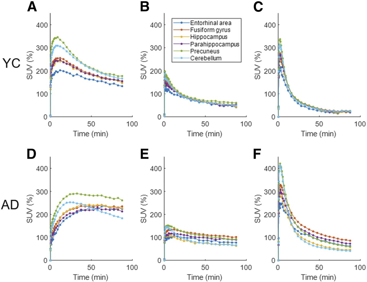
- FIGURE 1.
Line plots of time–activity curves in SUV of selected brain regions of 11C-RO-963 (A and D), 11C-RO-643 (B and E), and 18F-RO-948 (C and F) for YCs (panels A–C) and AD subjects (panels D–F).
- FIGURE 2.
Sagittal SUVR images of candidate radioligands, applied to same AD subjects and YCs. Cb = cerebellar cortex.
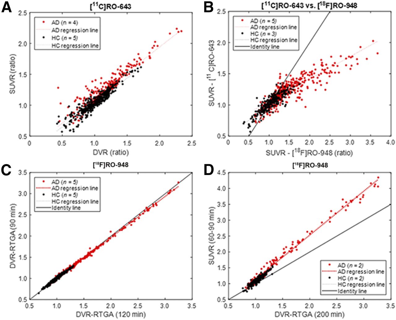
- FIGURE 3.
Scatterplots of SUVR vs. DVR for 11C-RO-643 (A) and SUVR data for 11CRO-643 vs. 18F-RO-948 for subjects who had both scans (B). Scatterplots of DVR (reference tissue graphical analysis [RTGA]) for 90- vs. 120-min circulation times for data analysis (C) and SUVR vs. DVR for 18F-RO-948 (D).
- FIGURE 4.
(A) Surface projection maps of mean 18F-RO-948 SUVR images of left and right hemispheres of AD subjects (from top to bottom: lateral, medial, and ventral views). (B) Box plot of SUVR data comparing AD subjects and OCs in 8 regions in which AD subjects showed highest mean SUVRs, as well as 3 Braak anterior regions (hippocampus, entorhinal area, and parahippocampus). Dots represent points lying outside ±2.7 SDs, assuming normal distributions. ER = entorhinal area; FO = frontal operculum; Fs = fusiform gyrus; Hp = hippocampus; IC = isthmus/cingulate; iPa = inferior parietal lobe; iTp = inferior temporal lobe; lOc = lateral occipital lobe; mFr = middle frontal lobe; mTp = middle temporal cortex; PH = parahippocampus; pCg = posterior cingulate gyrus; Pr = precuneus; sPa = superior parietal lobe; rFr = rostral frontal lobe; SM = supramarginal gyrus.
- FIGURE 5.
(A–D) Two roughly symmetrical SPM clusters (AD > OC) overlaid on 3 orthogonal views of standard MRI. (E) Line plots of volumes (across 1-cm-thick coronal slices) of over-the-threshold voxels of SPM analysis, hippopotamus, and amygdala along anterior–posterior axis. The 2 blue vertical lines indicate extent of anterior slice of Braak anterior block (19). (F–K) Clusters of high 18F-RO-948 tau-positive frequencies (>7 of 11 AD subjects) that spatially agreed with the 2 SPM clusters (F–H). Additional high-frequency clusters were noted in precuneus and posterior cingulate gyrus (I), and in superior and inferior parietal lobes, and supramarginal gyrus (J and K).
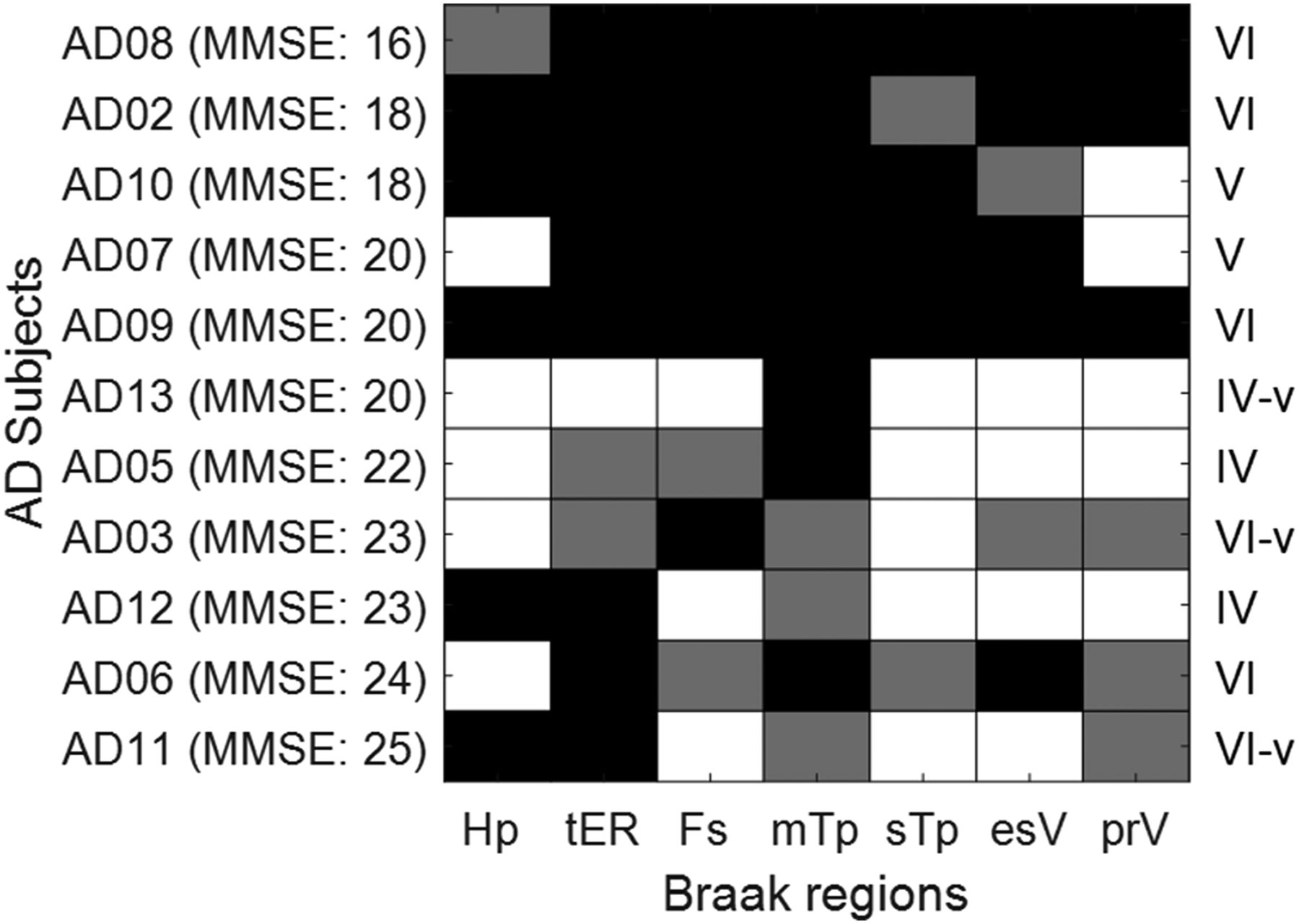
- FIGURE 6.
Checkerboard plot showing 18F-RO-948-positivity results (black cells: bilaterally positive; gray cells: unilaterally positive) of anterior and posterior Braak regions (19), and estimated Braak stages (right column) (20). esV =extrastriatal visual cortex; Fs = fusiform gyrus; Hp = hippocampus; mTp = middle temporal cortex; prV = primary visual cortex; sTp = superior temporal cortex; tER = transentorhinal cortex.
- FIGURE 7.
SUVR-cerebellar cortex images of 18F-RO-948 for all 11 AD subjects, arranged in descending order of global mean SUVR, alongside corresponding Aβ image (11C-Pittsburgh compound B SUVR, except for subjects AD02, AD05, and AD13, who had 18F-AV45 scans).


Tables
Control AD Part Radioligand Subject Plasma data (+) Age ± SD (y) Subject Plasma data (+) Age ± SD (y) 1 11C-RO-963 2 (1F) 2 30 ± 31 2 (1F) 1 67 ± 69 11C-RO-643 2 2 11C-RO-643 2 (0F) 2 25 ± 26 5 (2F) 2 75.0 ± 7.8 18F-RO-948 2 4 11C-RO-963 3 (1F) 3 32 ± 38 0 18F-RO-948 2 2A 18F-RO-948 5 (0F) 4 62 ± 8 5 (1F) 3 64.0 ± 6 Test–retest 4 3 2B 18F-RO-948 6 (3F) NA 58 ± 9 Dosimetry Parameter Side Hp ER PH mFr Pr lOc Fs SM mTp iTp iPa Separation* Left Present Absent Present Absent Absent Absent Present Absent Present Absent Absent Right Absent Present Present Absent Absent Absent Absent Absent Absent Absent Absent Statistical differences† Left Absent Present Present Present Absent Absent Present Absent Present Present Absent Right Present Present Present Absent Absent Absent Present Absent Present Present Present ↵* AD > maximal OC.
↵† AD > OC; Mann–Whitney test.
Hp = hippocampus; ER = entorhinal area; PH = parahippocampus; mFr = middle frontal lobe; Pr = precuneus; lOc = lateral occipital lobe; Fs = fusiform gyrus; SM = supramarginal gyrus; mTp = middle temporal cortex; iTp = inferior temporal lobe; iPa = inferior parietal lobe.
Supplemental Data
Files in this Data Supplement:


Jump to section
Related Articles
Cited By...
- Imaging {alpha}-synuclein pathologies in animal models and patients with Parkinsons and related diseases
- The Use of Tau PET to Stage Alzheimer Disease According to the Braak Staging Framework
- The Use of Tau PET to Stage Alzheimer Disease According to the Braak Staging Framework
- TauIQ: A Canonical Image Based Algorithm to Quantify Tau PET Scans