
Main menu

  • Home
  • Content
    • Current
    • Ahead of print
    • Past Issues
    • JNM Supplement
    • SNMMI Annual Meeting Abstracts
    • Continuing Education
    • JNM Podcasts
  • Subscriptions
    • Subscribers
    • Institutional and Non-member
    • Rates
    • Journal Claims
    • Corporate & Special Sales
  • Authors
    • Submit to JNM
    • Information for Authors
    • Assignment of Copyright
    • AQARA requirements
  • Info
    • Reviewers
    • Permissions
    • Advertisers
  • About
    • About Us
    • Editorial Board
    • Contact Information
  • More
    • Alerts
    • Feedback
    • Help
    • SNMMI Journals
  • SNMMI
    • JNM
    • JNMT
    • SNMMI Journals
    • SNMMI

User menu

  • Subscribe
  • My alerts
  • Log in
  • My Cart

Search

  • Advanced search
Journal of Nuclear Medicine
  • SNMMI
    • JNM
    • JNMT
    • SNMMI Journals
    • SNMMI
  • Subscribe
  • My alerts
  • Log in
  • My Cart
Journal of Nuclear Medicine

Advanced Search

  • Home
  • Content
    • Current
    • Ahead of print
    • Past Issues
    • JNM Supplement
    • SNMMI Annual Meeting Abstracts
    • Continuing Education
    • JNM Podcasts
  • Subscriptions
    • Subscribers
    • Institutional and Non-member
    • Rates
    • Journal Claims
    • Corporate & Special Sales
  • Authors
    • Submit to JNM
    • Information for Authors
    • Assignment of Copyright
    • AQARA requirements
  • Info
    • Reviewers
    • Permissions
    • Advertisers
  • About
    • About Us
    • Editorial Board
    • Contact Information
  • More
    • Alerts
    • Feedback
    • Help
    • SNMMI Journals
  • View or Listen to JNM Podcast
  • Visit JNM on Facebook
  • Join JNM on LinkedIn
  • Follow JNM on Twitter
  • Subscribe to our RSS feeds
Research ArticlePhysics and Instrumentation

Improving the Accuracy of Simultaneously Reconstructed Activity and Attenuation Maps Using Deep Learning

Donghwi Hwang, Kyeong Yun Kim, Seung Kwan Kang, Seongho Seo, Jin Chul Paeng, Dong Soo Lee and Jae Sung Lee
Journal of Nuclear Medicine October 2018, 59 (10) 1624-1629; DOI: https://doi.org/10.2967/jnumed.117.202317
Donghwi Hwang
1Department of Biomedical Sciences, Seoul National University, Seoul, Korea
2Department of Nuclear Medicine, Seoul National University, Seoul, Korea
  • Find this author on Google Scholar
  • Find this author on PubMed
  • Search for this author on this site
Kyeong Yun Kim
1Department of Biomedical Sciences, Seoul National University, Seoul, Korea
2Department of Nuclear Medicine, Seoul National University, Seoul, Korea
  • Find this author on Google Scholar
  • Find this author on PubMed
  • Search for this author on this site
Seung Kwan Kang
1Department of Biomedical Sciences, Seoul National University, Seoul, Korea
2Department of Nuclear Medicine, Seoul National University, Seoul, Korea
  • Find this author on Google Scholar
  • Find this author on PubMed
  • Search for this author on this site
Seongho Seo
3Department of Neuroscience, College of Medicine, Gachon University, Incheon, Korea
  • Find this author on Google Scholar
  • Find this author on PubMed
  • Search for this author on this site
Jin Chul Paeng
2Department of Nuclear Medicine, Seoul National University, Seoul, Korea
4Institute of Radiation Medicine, Medical Research Center, Seoul National University, Seoul, Korea; and
  • Find this author on Google Scholar
  • Find this author on PubMed
  • Search for this author on this site
Dong Soo Lee
2Department of Nuclear Medicine, Seoul National University, Seoul, Korea
4Institute of Radiation Medicine, Medical Research Center, Seoul National University, Seoul, Korea; and
5Department of Molecular Medicine and Biopharmaceutical Sciences, Graduate School of Convergence Science and Technology, Seoul National University, Suwon, Korea
  • Find this author on Google Scholar
  • Find this author on PubMed
  • Search for this author on this site
Jae Sung Lee
1Department of Biomedical Sciences, Seoul National University, Seoul, Korea
2Department of Nuclear Medicine, Seoul National University, Seoul, Korea
4Institute of Radiation Medicine, Medical Research Center, Seoul National University, Seoul, Korea; and
  • Find this author on Google Scholar
  • Find this author on PubMed
  • Search for this author on this site
  • Article
  • Figures & Data
  • Info & Metrics
  • PDF
Loading

Article Figures & Data

Figures

  • Tables
  • FIGURE 1.
    • Download figure
    • Open in new tab
    • Download powerpoint
    FIGURE 1.

    CNN architectures used to learn μ-CT from λ-MLAA and μ-MLAA. (A) CAE. (B) Unet. (C) Hybrid network of CAE and Unet. Green and red vertical strips at far left indicate inputs to CNN, and red stripes at right indicate output. Each box represents multichannel feature map. Number of feature maps and dimension of each feature map are denoted on interior and bottom of box. Data flow is left to right through contracting path to capture context and symmetric expanding path to recover image. Arrows stand for copying feature maps, and sky-blue boxes are copied feature map.
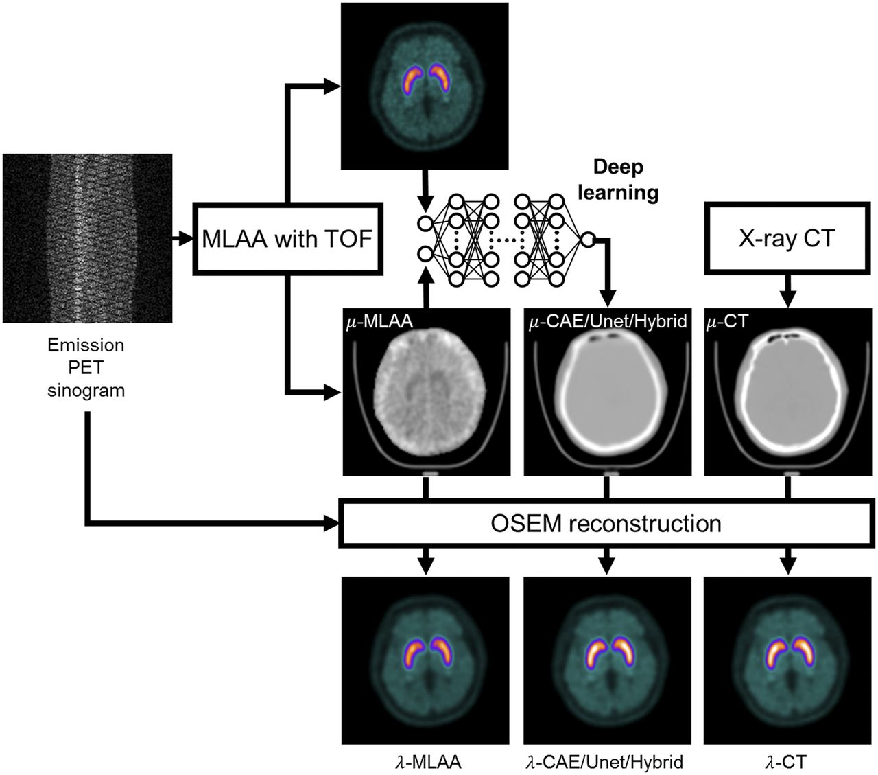
  • FIGURE 2.
    • Download figure
    • Open in new tab
    • Download powerpoint
    FIGURE 2.

    Flow chart of image analysis. For comparison, emission PET sinogram was reconstructed using μ-maps obtained using MLAA before (μ-MLAA) and after (μ-CAE, μ-Unet, and μ-Hybrid) applying deep CNNs and ground truth μ-CT. TOF = time of flight.
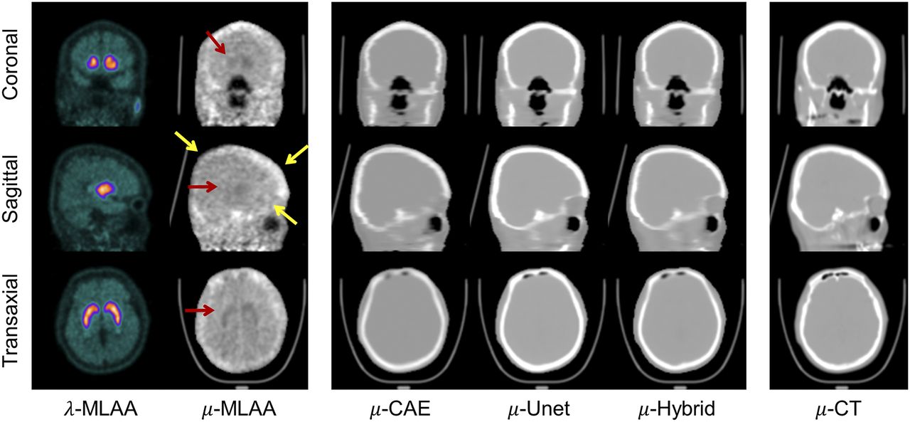
  • FIGURE 3.
    • Download figure
    • Open in new tab
    • Download powerpoint
    FIGURE 3.

    Comparison of CNN outputs (μ-CAE, μ-Unet, and μ-Hybrid) to μ-MLAA and μ-CT. Red and yellow arrows indicate, respectively, crosstalk artifacts and bone estimation error shown in μ-MLAA.
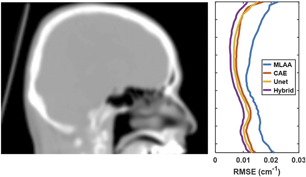
  • FIGURE 4.
    • Download figure
    • Open in new tab
    • Download powerpoint
    FIGURE 4.

    Root-mean square errors (RMSE) relative to μ-CT plotted across slice axial location (average of 40 test sets).
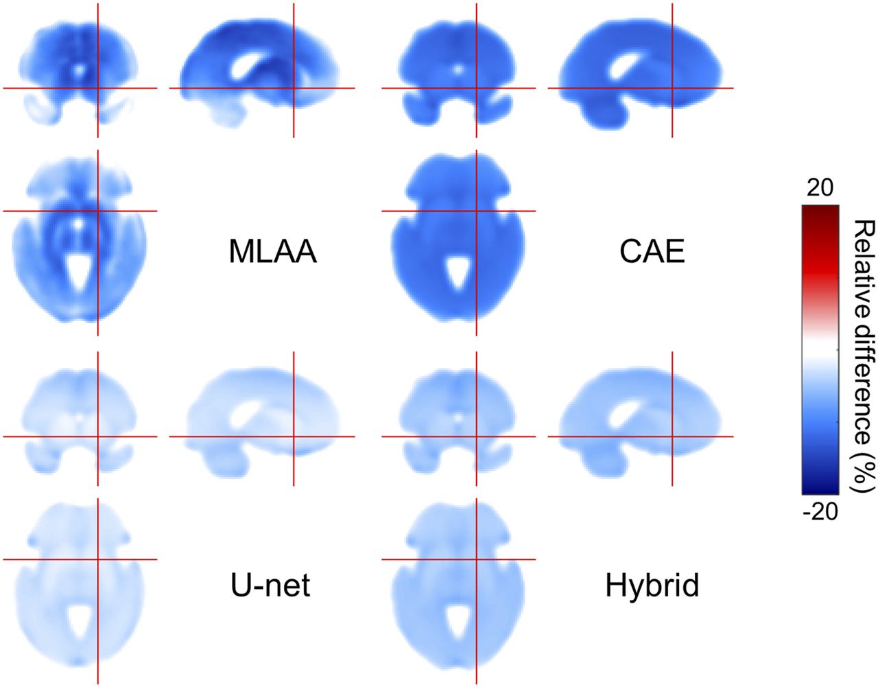
  • FIGURE 5.
    • Download figure
    • Open in new tab
    • Download powerpoint
    FIGURE 5.

    Percentage error map of spatially normalized activity distribution (average of 40 test sets).
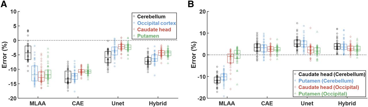
  • FIGURE 6.
    • Download figure
    • Open in new tab
    • Download powerpoint
    FIGURE 6.

    Percentage error in activity (A) and binding ratio (B) estimation relative to ground truth (OSEM with μ-CT). Each horizontal bar and vertical box indicates median and SD, respectively. In B, specific and nonspecific regions for binding ratio calculation are indicated as “specific (nonspecific).”

Tables

  • Figures
    • View popup
    TABLE 1

    Dice Similarity Coefficients with μ-CT for Whole Head and Cranial Bone Region

    Whole headCranial region
    MethodBoneAirBoneAir
    MLAA0.374 ± 0.0580.317 ± 0.0700.399 ± 0.0630.426 ± 0.062
    CNN (CAE)0.717 ± 0.0470.513 ± 0.0570.747 ± 0.0470.523 ± 0.063
    CNN (Unet)0.787 ± 0.0420.575 ± 0.0470.801 ± 0.0430.580 ± 0.053
    CNN (Hybrid)0.794 ± 0.0370.718 ± 0.0480.810 ± 0.0380.738 ± 0.044
    • Data are mean ± SD. Results of analysis of variation and post hoc tests are shown in Supplemental Figure 3.

PreviousNext
Back to top

In this issue

Journal of Nuclear Medicine: 59 (10)
Journal of Nuclear Medicine
Vol. 59, Issue 10
October 1, 2018
  • Table of Contents
  • Table of Contents (PDF)
  • About the Cover
  • Index by author
Print
Download PDF
Article Alerts
Sign In to Email Alerts with your Email Address
Email Article

Thank you for your interest in spreading the word on Journal of Nuclear Medicine.

NOTE: We only request your email address so that the person you are recommending the page to knows that you wanted them to see it, and that it is not junk mail. We do not capture any email address.

Enter multiple addresses on separate lines or separate them with commas.
Improving the Accuracy of Simultaneously Reconstructed Activity and Attenuation Maps Using Deep Learning
(Your Name) has sent you a message from Journal of Nuclear Medicine
(Your Name) thought you would like to see the Journal of Nuclear Medicine web site.
Citation Tools
Improving the Accuracy of Simultaneously Reconstructed Activity and Attenuation Maps Using Deep Learning
Donghwi Hwang, Kyeong Yun Kim, Seung Kwan Kang, Seongho Seo, Jin Chul Paeng, Dong Soo Lee, Jae Sung Lee
Journal of Nuclear Medicine Oct 2018, 59 (10) 1624-1629; DOI: 10.2967/jnumed.117.202317

Citation Manager Formats

  • BibTeX
  • Bookends
  • EasyBib
  • EndNote (tagged)
  • EndNote 8 (xml)
  • Medlars
  • Mendeley
  • Papers
  • RefWorks Tagged
  • Ref Manager
  • RIS
  • Zotero
Share
Improving the Accuracy of Simultaneously Reconstructed Activity and Attenuation Maps Using Deep Learning
Donghwi Hwang, Kyeong Yun Kim, Seung Kwan Kang, Seongho Seo, Jin Chul Paeng, Dong Soo Lee, Jae Sung Lee
Journal of Nuclear Medicine Oct 2018, 59 (10) 1624-1629; DOI: 10.2967/jnumed.117.202317
Twitter logo Facebook logo LinkedIn logo Mendeley logo
  • Tweet Widget
  • Facebook Like
  • Google Plus One
Bookmark this article

Jump to section

  • Article
    • Abstract
    • MATERIALS AND METHODS
    • RESULTS
    • DISCUSSION
    • CONCLUSION
    • DISCLOSURE
    • Footnotes
    • REFERENCES
  • Figures & Data
  • Info & Metrics
  • PDF

Related Articles

  • This Month in JNM
  • PubMed
  • Google Scholar

Cited By...

  • Improving 18F-FDG PET Quantification Through a Spatial Normalization Method
  • Nuclear Medicine and Artificial Intelligence: Best Practices for Algorithm Development
  • PET/MRI, Part 2: Technologic Principles
  • Machine Learning in Nuclear Medicine: Part 2--Neural Networks and Clinical Aspects
  • Denoising of Scintillation Camera Images Using a Deep Convolutional Neural Network: A Monte Carlo Simulation Approach
  • Intelligent Imaging: Artificial Intelligence Augmented Nuclear Medicine
  • Generation of PET Attenuation Map for Whole-Body Time-of-Flight 18F-FDG PET/MRI Using a Deep Neural Network Trained with Simultaneously Reconstructed Activity and Attenuation Maps
  • Google Scholar

More in this TOC Section

  • Performance Evaluation of the uMI Panorama PET/CT System in Accordance with the National Electrical Manufacturers Association NU 2-2018 Standard
  • A Multicenter Study on Observed Discrepancies Between Vendor-Stated and PET-Measured 90Y Activities for Both Glass and Resin Microsphere Devices
  • Ultra-Fast List-Mode Reconstruction of Short PET Frames and Example Applications
Show more Physics and Instrumentation

Similar Articles

Keywords

  • deep learning
  • simultaneous reconstruction
  • crosstalk
  • denoising
  • quantification
SNMMI

© 2025 SNMMI

Powered by HighWire