


Article Figures & Data
Figures
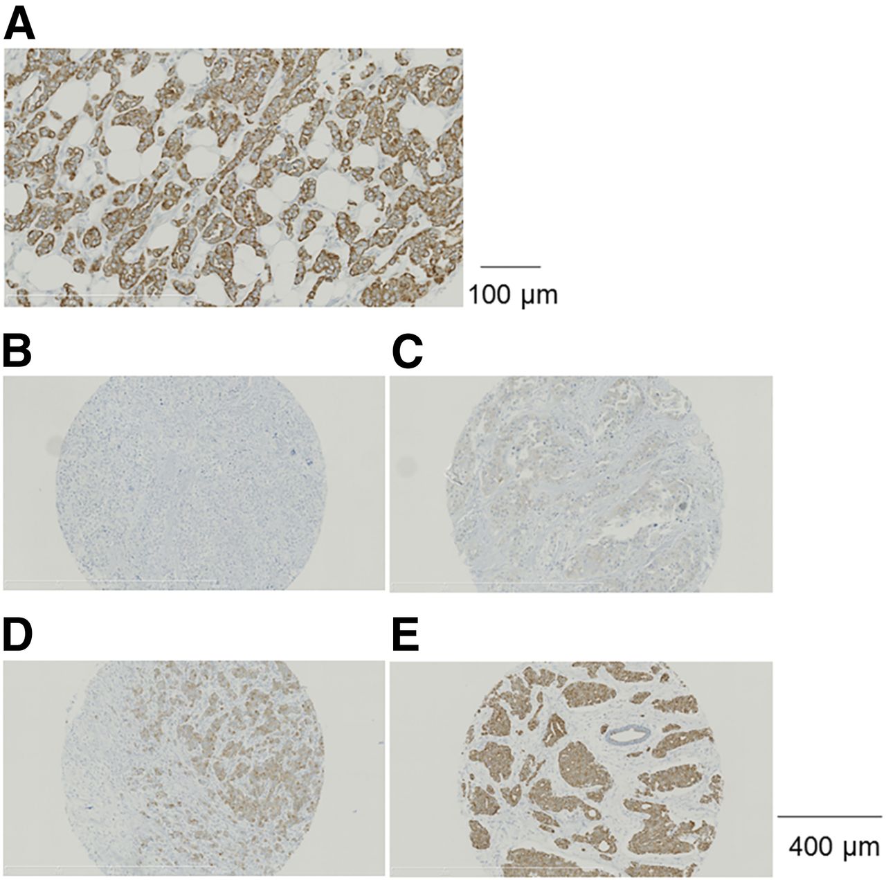
- FIGURE 1.
(A) Membrane and cytoplasmic localization of GRPR immunostaining in a breast cancer specimen (magnification, ×20). (B–E) Representative samples of breast cancer specimens showing the 4 categories of GRPR immunostaining results: no GRPR expression (IRS score 0 or 1) (B), weak GRPR expression (IRS score 2 or 3) (C), moderate GRPR expression (IRS score 4–8) (D), and strong GRPR expression (IRS score 9–12) (E) (magnification, ×10).
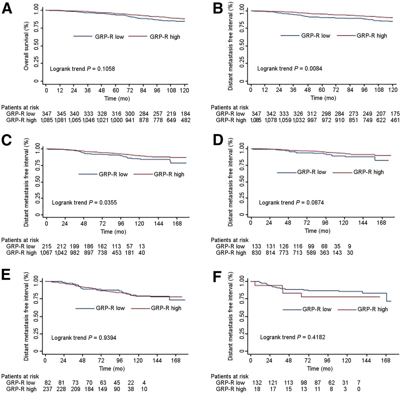
- FIGURE 2.
(A) Association of GRPR expression in primary tumors with OS in 1,432 patients. (B) DMFI in 1,432 patients according to GRPR expression in primary breast tumor. (C) DMFI in ER-positive tumors as a function of GRPR expression. (D) DMFI in luminal-A tumors as a function of GRPR expression. (E) DMFI in luminal-B tumors as a function of GRPR expression. (F) Distant MFI in ER-negative tumors as a function of GRPR expression.
Tables
- TABLE 1
Distribution of Low and High GRPR Expression Within Different Categories of Breast Cancers
GRPR expression Characteristic Category No. of patients Low % High % P Age (y) ≤40 y 85 45.9 54.1 <0.00001 >40 y 1,347 22.9 77.1 Histology* IDC 1,221 25.4 74.6 0.09 ILC 161 19.3 80.7 Pathologic size (mm) <20 961 20.9 79.1 0.00003 ≥20 471 31.0 69.0 SBR grade* I 358 10.6 89.4 <0.00001† II 661 16.9 83.1 III 397 48.6 51.4 Ki-67 <20% 1,115 17.0 83.0 <0.00001 ≥20% 317 49.8 50.2 Lymph node status* N0 848 23.3 76.7 0.03 N+ 472 28.8 71.2 ER 0% 150 88.0 12.0 <0.00001 ≥1% 1,282 16.8 83.2 PR 0% 266 60.9 39.1 <0.00001 ≥1% 1,166 15.9 84.1 AR <10% 184 63.0 37.0 <0.00001 ≥10% 1,248 18.5 81.5 HER2 overexpression No 1,286 22.8 77.2 0.0001 Yes 146 37.0 63.0 ↵* Patients with other histologic subtypes (n = 50) are excluded from statistical analysis. Number of patients with missing information regarding items: SBR grade, n = 16; lymph node status (Nx), n = 112. Statistical analysis based on χ2 test.
↵† P value was calculated for SBR grade I + II vs. III.
IDC = invasive ductal carcinoma; ILC = invasive lobular carcinoma.
Univariate analysis Multivariate analysis Characteristic Category No. of patients P Odds ratio P Odds ratio Age (y) <40 85 <0.0001 1 Nonsignificant (not retained) 41–50 287 <0.0001 2.897 (1.742–4.816) >50 1,060 <0.0001 2.851 (1.818–4.472) Histology IDC 1,221 0.0389 1 Not retained ILC 161 0.0910 1.427 (0.945–2.155) Other 43 0.0479 2.586 (1.009–6.628) Pathologic size (mm) ≥20 471 <0.0001 1 Not retained <20 961 1.699 [1.324 ; 2.180] SBR grade Grade III 397 <0.0001 1 0.0005 1 Grade II 661 <0.0001 4.637 (3.495–6.154) 0.0023 1.831 (1.242–2.699) Grade I 358 <0.0001 7.965 (5.395–11.761) 0.0002 2.617 (1.591–4.303) Ki-67 ≥20% 317 <0.0001 1 0.0196 1 <20% 1,115 4.869 (3.716–6.378) 1.584 (1.076–2.330) Lymph node status N+ 472 0.0006 1 0.0350 1 N0 848 0.0289 1.329 (1.030–1.715) 0.3117 1.173 (0.861–1.597) Missing 112 0.0003 3.082 (1.672–5.681) 0.0103 2.636 (1.257–5.526) ER 0% 150 <0.0001 1 <0.0001 1 ≥1% 1,282 36.394 (21.772–60.837) 11.998 (5.980–24.075) PR 0% 266 <0.0001 1 0.0429 1 ≥1% 1,166 8.259 (6.167–11.061) 1.652 (1.016–2.686) AR <10% 184 <0.0001 1 0.0027 1 ≥10% 1,248 7.509 (5.389–10.462) 2.011 (1.275–3.173) HER2 overexpression Yes 146 0.0002 1 0.0038 1 No 1,286 1.989 (1.387–2.852) 0.461 (0.273–0.779) IDC = invasive ductal carcinoma; ILC = invasive lobular carcinoma.
Statistical analysis based on Cox model; P < 0.05 was considered significant. Data in parentheses are 95% CIs.
- TABLE 3
Distribution of Low and High GRPR Expression Within Different Categories of ER-Positive Tumors
GRPR expression Characteristic Category No. of patients Low % High % P Age (y) ≤40 65 29.2 70.8 0.006 >40 1,217 16.1 83.9 Histology* IDC 1,080 17.1 82.9 0.70 ILC 158 18.4 81.6 Pathologic size (mm) <20 883 14.9 85.1 0.009 ≥20 399 20.8 79.2 SBR grade* I 356 10.1 89.9 <0.00001† II 643 14.9 85.1 III 267 29.6 70.4 Ki-67 <20% 1,072 13.3 86.7 <0.00001 ≥20% 210 30.0 70.0 Lymph node status* N0 762 16.0 84.0 0.052 N+ 414 20.5 79.5 Estrogen receptor 1%–9% 18 44.4 55.6 0.005‡ ≥10% 1,264 16.4 83.6 PR 0% 116 25.9 74.1 0.006 ≥1% 1,166 15.9 84.1 AR <10% 91 33.0 67.0 0.00002 ≥10% 1,191 15.5 84.5 HER2 overexpression No 1,183 16.7 83.3 0.91 Yes 99 17.2 82.8 ↵* Patients with other histologic subtypes (n = 44) are excluded from statistical analysis. Number of patients with missing information regarding items: SBR grade, n = 16; lymph node status (Nx), n = 106.
↵† P value was calculated for SBR grade I + II vs. III.
↵‡ Statistical analysis based on χ2 or Fischer test.
IDC = invasive ductal carcinoma; ILC = invasive lobular carcinoma.
- TABLE 4
Distribution of Low and High GRPR Expression Within Different Molecular Subtype of Breast Cancers
GRPR expression Subtype No. of patients Low % High % P Luminal A 963 13.8 86.2 <0.00001 Luminal B HER2− 220 29.5 70.5 Luminal B HER2+ 99 17.2 82.8 HER2-enriched 47 78.7 21.3 Triple-negative 103 92.2 7.8
Supplemental Data
Files in this Data Supplement:


Jump to section
Related Articles
Cited By...
- A Vision for Gastrin-Releasing Peptide Receptor Targeting for Imaging and Therapy: Perspective from Academia and Industry
- Diagnostic Potential of 68Ga-NeoB PET/CT with Estrogen Receptor- and Progesterone Receptor-Positive Breast Cancer Undergoing Staging or Restaging for Metastatic Disease
- Gastrin-Releasing Peptide Receptor Imaging and Therapy in the Era of Personalized Medicine
- Preclinical Investigation of [212Pb]Pb-DOTAM-GRPR1 for Peptide Receptor Radionuclide Therapy in a Prostate Tumor Model
- Comparison of 68Ga-PSMA-617 PET/CT and 68Ga-RM2 PET/CT in Patients with Localized Prostate Cancer Who Are Candidates for Radical Prostatectomy: A Prospective, Single-Arm, Single-Center, Phase II Study
- Substitution of L-Tryptophan by {alpha}-Methyl-L-Tryptophan in 177Lu-RM2 Results in 177Lu-AMTG, a High-Affinity Gastrin-Releasing Peptide Receptor Ligand with Improved In Vivo Stability
- A Radiotracer for Molecular Imaging and Therapy of Gastrin-Releasing Peptide Receptor-Positive Prostate Cancer
- Imaging the Distribution of Gastrin-Releasing Peptide Receptors in Cancer
- Somatostatin Antagonists for Radioligand Therapy of Nonendocrine Tumors