


Article Figures & Data
Figures
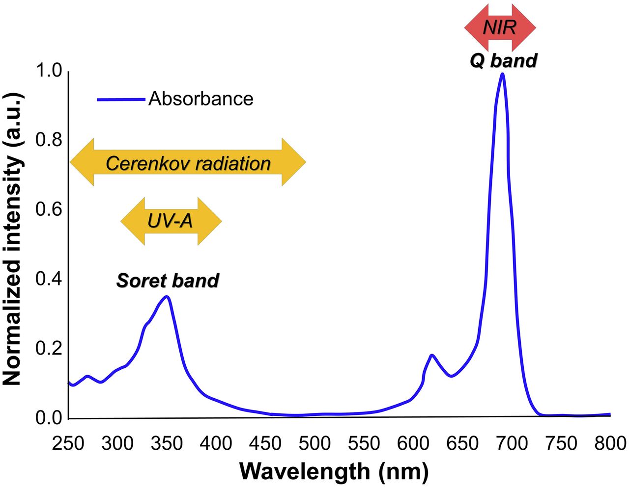
- FIGURE 1.
Absorption spectrum of IRDye 700DX.
- FIGURE 2.
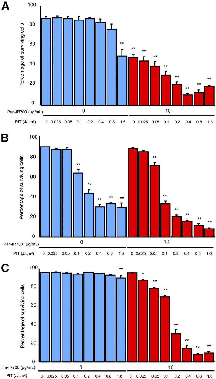
Evaluation of in vitro UV-A PIT (*P < 0.05, **P < 0.01, vs. untreated control). (A) Percentage of surviving cells decreased in light dose-dependent manner although 50% of A431-luc cells died when pan-IR700 alone without UV-A light was administered. (B) Propidium iodide staining showed membrane damage in MDAMB468-luc cells induced by UV-A PIT although significant cytotoxicity associated with UV-A light alone was observed. (C) With tra-IR700, percentage of surviving 3T3/HER2-luc cells decreased in light dose-dependent manner.
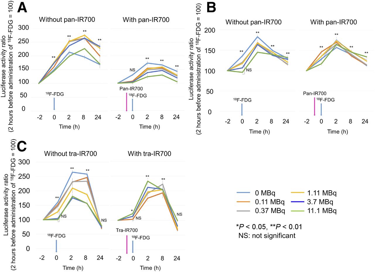
- FIGURE 3.
Evaluation of in vitro CR-PIT using 18F-FDG. (A) When A431-luc cells were used, suppression of luciferase activity in cells was seen in CR-PIT and the degree of suppression increased in a dose-dependent manner. (B) When MDAMB468-luc cells were used, CR-PIT did not significantly change the level of luciferase activity compared with 18F-FDG alone. (C) When 3T3/HER2-luc cells were used, CR-PIT also did not significantly change the level of luciferase activity compared with 18F-FDG alone.
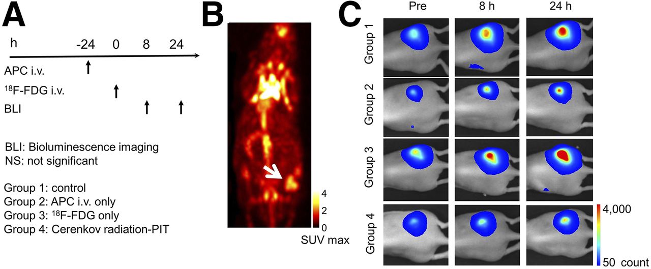
- FIGURE 4.
In vivo effect of CR-PIT on A431-luc tumor. (A) CR-PIT regimen is shown. (B) 18F-FDG PET imaging of tumor-bearing mice. Tumor showed high accumulation of 18F-FDG (arrow). (C) Bioluminescence imaging of tumor-bearing mice.
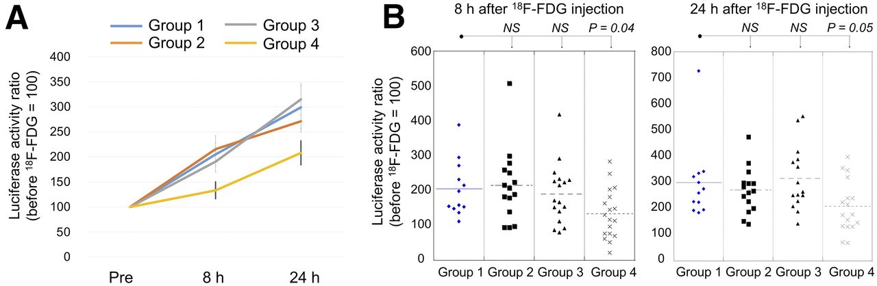
- FIGURE 5.
In vivo effect of CR-PIT on A431-luc tumor. (A) Luciferase activity ratio was suppressed in CR-PIT group compared with control group. (B) Significant suppression of luciferase activity ratio was seen in CR-PIT group compared with control group. NS = not significant.
- FIGURE 6.
Resected tumors stained with hematoxylin and eosin. Cellular necrosis and microhemorrhage were seen within a background of live but damaged tumor cells after CR-PIT whereas little damage was observed in tumors receiving 18F-FDG administration alone. No obvious damage was observed in the tumor receiving only APC but no 18F-FDG administration.


Additional Files
Supplemental Data
Files in this Data Supplement:


Jump to section
Related Articles
Cited By...
- No citing articles found.