


Article Figures & Data
Figures
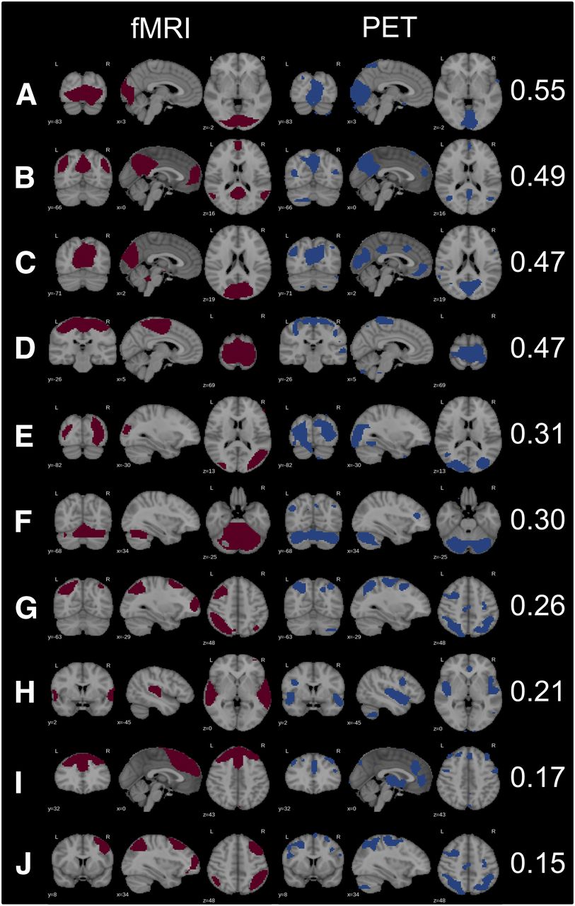
- FIGURE 1.
Matched RSNs in fMRI and PET data with corresponding Dice coefficients: primary visual posterior (A), DMN (B), primary visual (posterior) (C), sensorimotor (D), secondary visual (E), cerebellar (F), central executive right (G), auditory (H), executive control (I), central executive left (J). Because right and left CENs were captured as 1 independent component in PET data, G and J for PET are the same.
- FIGURE 2.
Unmatched (unique to either fMRI or PET data) RSNs and noise components: salience (A), spatial attention (B), primary visual (anterior) (C), anterior insular (D), temporopolar (E), lateral motor (F), (anterior) default mode (G), basal ganglia (H), nucleus accumbens (I), secondary visual (J), mesial parietal/prefrontal (K).


Additional Files
Supplemental Data
Files in this Data Supplement:


Jump to section
Related Articles
Cited By...
- Multimodal Neuroimaging of the Effect of Serotonergic Psychedelics on the Brain
- The long distance relationship of regional amyloid burden and tau pathology spread
- Early-Phase 18F-Florbetapir and 18F-Flutemetamol Images as Proxies of Brain Metabolism in a Memory Clinic Setting
- A new framework for metabolic connectivity mapping using bolus [18F]FDG PET and kinetic modelling
- Mechanisms of Network Changes in Cognitive Impairment in Multiple Sclerosis
- Metabolic and haemodynamic resting-state connectivity of the human brain: a high-temporal resolution simultaneous BOLD-fMRI and FDG-fPET multimodality study
- Integrity of Neurocognitive Networks in Dementing Disorders as Measured with Simultaneous PET/Functional MRI
- Analysis of continuous infusion functional PET (fPET) in the human brain
- Simultaneous task-based BOLD-fMRI and [18-F] FDG functional PET for measurement of neuronal metabolism in the human visual cortex
- Neurometabolic Resting-State Networks Derived from Seed-Based Functional Connectivity Analysis
- Reply: Neurometabolic Resting-State Networks Derived from Seed-Based Functional Connectivity Analysis