
Main menu

  • Home
  • Content
    • Current
    • Ahead of print
    • Past Issues
    • JNM Supplement
    • SNMMI Annual Meeting Abstracts
    • Continuing Education
    • JNM Podcasts
  • Subscriptions
    • Subscribers
    • Institutional and Non-member
    • Rates
    • Journal Claims
    • Corporate & Special Sales
  • Authors
    • Submit to JNM
    • Information for Authors
    • Assignment of Copyright
    • AQARA requirements
  • Info
    • Reviewers
    • Permissions
    • Advertisers
  • About
    • About Us
    • Editorial Board
    • Contact Information
  • More
    • Alerts
    • Feedback
    • Help
    • SNMMI Journals
  • SNMMI
    • JNM
    • JNMT
    • SNMMI Journals
    • SNMMI

User menu

  • Subscribe
  • My alerts
  • Log in
  • My Cart

Search

  • Advanced search
Journal of Nuclear Medicine
  • SNMMI
    • JNM
    • JNMT
    • SNMMI Journals
    • SNMMI
  • Subscribe
  • My alerts
  • Log in
  • My Cart
Journal of Nuclear Medicine

Advanced Search

  • Home
  • Content
    • Current
    • Ahead of print
    • Past Issues
    • JNM Supplement
    • SNMMI Annual Meeting Abstracts
    • Continuing Education
    • JNM Podcasts
  • Subscriptions
    • Subscribers
    • Institutional and Non-member
    • Rates
    • Journal Claims
    • Corporate & Special Sales
  • Authors
    • Submit to JNM
    • Information for Authors
    • Assignment of Copyright
    • AQARA requirements
  • Info
    • Reviewers
    • Permissions
    • Advertisers
  • About
    • About Us
    • Editorial Board
    • Contact Information
  • More
    • Alerts
    • Feedback
    • Help
    • SNMMI Journals
  • View or Listen to JNM Podcast
  • Visit JNM on Facebook
  • Join JNM on LinkedIn
  • Follow JNM on Twitter
  • Subscribe to our RSS feeds
LetterLetters to the Editor

Neurometabolic Resting-State Networks Derived from Seed-Based Functional Connectivity Analysis

Nicola Trotta, Kristof Baete, Koen Van Laere, Serge Goldman, Xavier De Tiège and Vincent Wens
Journal of Nuclear Medicine October 2018, 59 (10) 1642-1643; DOI: https://doi.org/10.2967/jnumed.118.212878
Nicola Trotta
*CUB—Hôpital Erasme 808 Route de Lennik Brussels, Belgium 1070 Email:
  • Find this author on Google Scholar
  • Find this author on PubMed
  • Search for this author on this site
  • For correspondence: vwens@ulb.ac.be
Kristof Baete
*CUB—Hôpital Erasme 808 Route de Lennik Brussels, Belgium 1070 Email:
  • Find this author on Google Scholar
  • Find this author on PubMed
  • Search for this author on this site
  • For correspondence: vwens@ulb.ac.be
Koen Van Laere
*CUB—Hôpital Erasme 808 Route de Lennik Brussels, Belgium 1070 Email:
  • Find this author on Google Scholar
  • Find this author on PubMed
  • Search for this author on this site
  • For correspondence: vwens@ulb.ac.be
Serge Goldman
*CUB—Hôpital Erasme 808 Route de Lennik Brussels, Belgium 1070 Email:
  • Find this author on Google Scholar
  • Find this author on PubMed
  • Search for this author on this site
  • For correspondence: vwens@ulb.ac.be
Xavier De Tiège
*CUB—Hôpital Erasme 808 Route de Lennik Brussels, Belgium 1070 Email:
  • Find this author on Google Scholar
  • Find this author on PubMed
  • Search for this author on this site
  • For correspondence: vwens@ulb.ac.be
Vincent Wens
*CUB—Hôpital Erasme 808 Route de Lennik Brussels, Belgium 1070 Email:
  • Find this author on Google Scholar
  • Find this author on PubMed
  • Search for this author on this site
  • For correspondence: vwens@ulb.ac.be
  • Article
  • Figures & Data
  • Info & Metrics
  • PDF
Loading

TO THE EDITOR: We read with great interest the paper by Savio et al. (1) on the imaging of resting-state networks (RSNs) from simultaneous 18F-FDG PET/functional MRI (fMRI) data. The authors applied an independent component analysis (ICA) commonly used in fMRI and reported fair cross-modality agreement for several RSNs. In view of the distinct nature of the neurovascular and neurometabolic couplings, this tends to confirm the neural basis of RSNs, in line with recent magnetoencephalographic studies. Interestingly, some networks were only identified in one modality or the other, for example, the 18F-FDG PET ICA reported by Savio et al. (1) failed at detecting the salience/insular or temporopolar RSNs disclosed in the corresponding fMRI ICA. This leaves the question of their neural underpinning pending.

Methodologically, this study focused on the ICA technique. A complementary approach is seed-based functional connectivity (sbFC) whereby a seed location is selected a priori as part of the sought network, and correlation maps are estimated between the seed and all other voxel activities. Compared with ICA, sbFC is straightforward to interpret and avoids the issues of selecting the number of components (which affects ICA decompositions) and visually discriminating between physiologic and noise components. Furthermore, sbFC can be subjected to the rigorous statistical framework of random field theory (RFT) (2), henceforth eliminating the usage of somewhat arbitrary thresholds. Supplementing ICA with sbFC thus appears necessary for robust inferences about RSNs.

We hereby report that sbFC analysis of 18F-FDG PET data do allow statistical mapping of most RSNs, including those unidentified from the 18F-FDG PET ICA (1).

Specifically, we considered a resting-state (eyes closed) 18F-FDG PET dataset of 50 healthy adults (27 women; age range, 18–43 y) whose acquisition and preprocessing procedures have been detailed in a previous publication (3). We used SPM8 (http://www.fil.ion.ucl.ac.uk/spm/, Wellcome Trust Centre for Neuroimaging) to design voxelwise general linear models of the 18F-FDG PET images with metabolism at the seed location as covariate of interest. One-sided t tests were then applied to identify significantly positive sbFC, both at P < 0.05 with the whole-brain familywise error rate controlled by RFT and at P < 0.001 uncorrected.

Figure 1 illustrates the statistically masked sbFC t-maps obtained from selected seeds. Several RSNs emerged at RFT significance, from low-level (e.g., sensorimotor, visual) to cognitive (e.g., language, executive) and subcortical (e.g., cerebellar, basal ganglia) networks. Other RSNs were identified only partially but recovered at P < 0.001 uncorrected (e.g., auditory, default-mode, and frontoparietal networks). In line with the discussion in Savio et al. (1), this may be due to the nondynamic nature of our 18F-FDG PET data. Indeed, compared with fMRI, the loss of temporal samples strongly limits the sensitivity of correlation estimates. Importantly, the salience/insular and temporopolar RSNs missing in the 18F-FDG PET ICA (1) were identified as well. The reason may simply be that our dataset contains approximately 2.3 times more subjects, leading to approximately 33% less correlation noise. Indeed, repeating our sbFC analyses on half our population failed at revealing these 2 RSNs. Besides, this increase in sampling size would enable computing extra 18F-FDG PET ICA components possibly disclosing these RSNs.

FIGURE 1.
  • Download figure
  • Open in new tab
  • Download powerpoint
FIGURE 1.

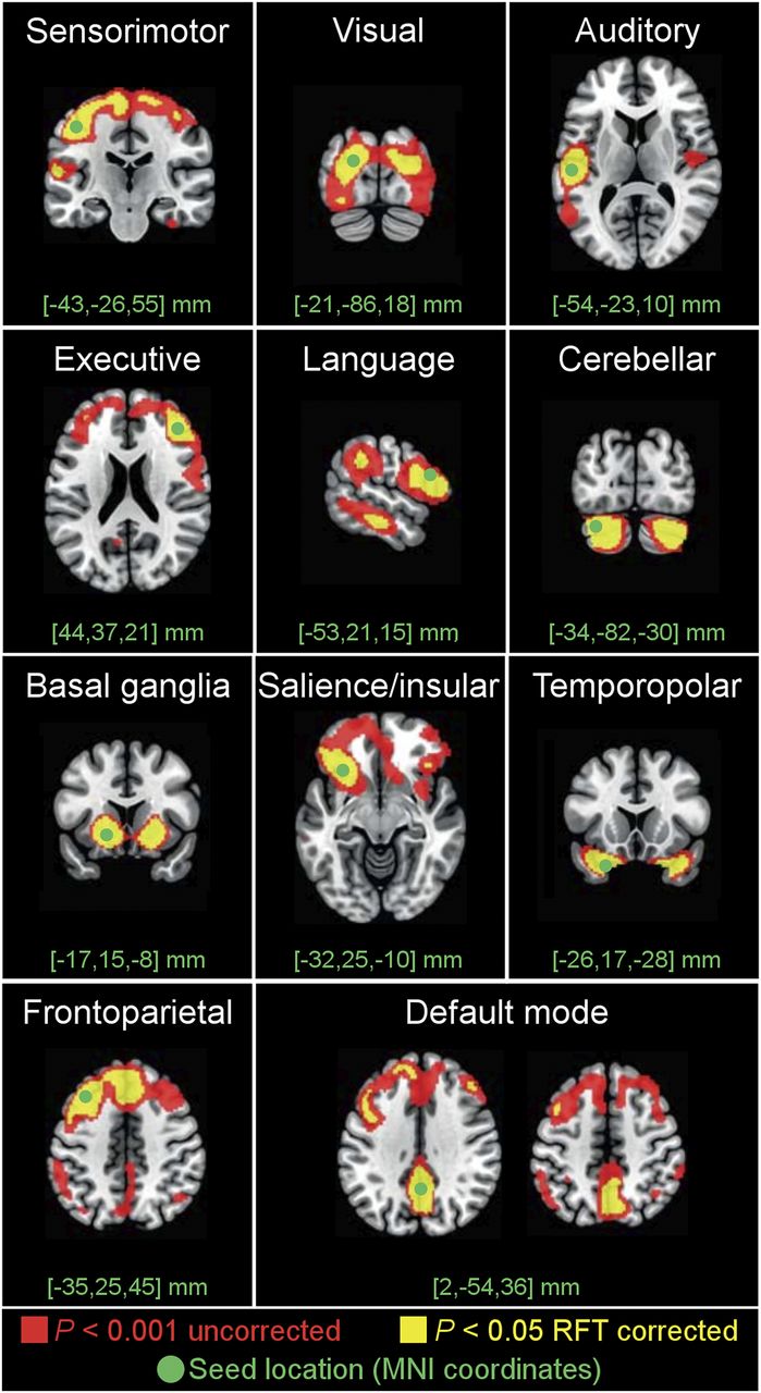
Statistically masked sbFC t-maps obtained from selected seeds.

Together with previous seminal studies that had some shortcomings limiting the interpretations of their results (4,5), Savio et al.’s study (1) and our 18F-FDG PET data obtained by statistical sbFC mapping bring novel evidence that the field of RSNs—up to now exclusive to fMRI and to a lesser extent extracranial electrophysiology—can be expanded to the realm of neurometabolism and thus pervades all functional neuroimaging. In particular, the combination of sbFC with ICA applied to resting-state 18F-FDG PET data opens novel scientific and clinical lines of research on the neurometabolic processes associated with functional integration and its pathologic disruptions by brain disorders.

Footnotes

  • Published online Apr. 26, 2018.

  • © 2018 by the Society of Nuclear Medicine and Molecular Imaging.

REFERENCES

  1. 1.↵
    1. Savio A,
    2. Fünger S,
    3. Tahmasian M,
    4. et al
    . Resting state networks as simultaneously measured with fMRI and PET. J Nucl Med. 2017;58:1314–1317.
    OpenUrlAbstract/FREE Full Text
  2. 2.↵
    1. Worsley KJ,
    2. Marrett S,
    3. Neelin P,
    4. Vandal AC,
    5. Friston KJ,
    6. Evans AC
    . A unified statistical approach for determining significant signals in images of cerebral activation. Hum Brain Mapp. 1996;4:58–73.
    OpenUrlCrossRefPubMed
  3. 3.↵
    1. Trotta N,
    2. Archambaud F,
    3. Goldman S,
    4. et al
    . Functional integration changes in regional brain glucose metabolism from childhood to adulthood. Hum Brain Mapp. 2016;37:3017–3030.
    OpenUrl
  4. 4.↵
    1. Di X,
    2. Biswal B
    . Metabolic brain covariant networks as revealed by FDG-PET with reference to resting-state fMRI networks. Brain Connect. 2012;2:275–283.
    OpenUrlCrossRefPubMed
  5. 5.↵
    1. Horwitz B,
    2. Duara R,
    3. Rapoport SI
    . Intercorrelations of glucosemetabolic rates between brain regions: application to healthy males in a state of reduced sensory input. J Cereb Blood Flow Metab. 1984;4:484–499.
    OpenUrlCrossRefPubMed
  • Received for publication April 12, 2018.
  • Accepted for publication April 17, 2018.
PreviousNext
Back to top

In this issue

Journal of Nuclear Medicine: 59 (10)
Journal of Nuclear Medicine
Vol. 59, Issue 10
October 1, 2018
  • Table of Contents
  • Table of Contents (PDF)
  • About the Cover
  • Index by author
Print
Download PDF
Article Alerts
Sign In to Email Alerts with your Email Address
Email Article

Thank you for your interest in spreading the word on Journal of Nuclear Medicine.

NOTE: We only request your email address so that the person you are recommending the page to knows that you wanted them to see it, and that it is not junk mail. We do not capture any email address.

Enter multiple addresses on separate lines or separate them with commas.
Neurometabolic Resting-State Networks Derived from Seed-Based Functional Connectivity Analysis
(Your Name) has sent you a message from Journal of Nuclear Medicine
(Your Name) thought you would like to see the Journal of Nuclear Medicine web site.
Citation Tools
Neurometabolic Resting-State Networks Derived from Seed-Based Functional Connectivity Analysis
Nicola Trotta, Kristof Baete, Koen Van Laere, Serge Goldman, Xavier De Tiège, Vincent Wens
Journal of Nuclear Medicine Oct 2018, 59 (10) 1642-1643; DOI: 10.2967/jnumed.118.212878

Citation Manager Formats

  • BibTeX
  • Bookends
  • EasyBib
  • EndNote (tagged)
  • EndNote 8 (xml)
  • Medlars
  • Mendeley
  • Papers
  • RefWorks Tagged
  • Ref Manager
  • RIS
  • Zotero
Share
Neurometabolic Resting-State Networks Derived from Seed-Based Functional Connectivity Analysis
Nicola Trotta, Kristof Baete, Koen Van Laere, Serge Goldman, Xavier De Tiège, Vincent Wens
Journal of Nuclear Medicine Oct 2018, 59 (10) 1642-1643; DOI: 10.2967/jnumed.118.212878
Twitter logo Facebook logo LinkedIn logo Mendeley logo
  • Tweet Widget
  • Facebook Like
  • Google Plus One
Bookmark this article

Jump to section

  • Article
    • Footnotes
    • REFERENCES
  • Figures & Data
  • Info & Metrics
  • PDF

Related Articles

  • No related articles found.
  • PubMed
  • Google Scholar

Cited By...

  • Integrity of Neurocognitive Networks in Dementing Disorders as Measured with Simultaneous PET/Functional MRI
  • Reply: Neurometabolic Resting-State Networks Derived from Seed-Based Functional Connectivity Analysis
  • Google Scholar

More in this TOC Section

  • Business Model Beats Science and Logic: Dosimetry and Paucity of Its Use
  • Determining PSMA-617 Mass and Molar Activity in Pluvicto Doses
  • The Value of Functional PET in Quantifying Neurotransmitter Dynamics
Show more Letters to the Editor

Similar Articles

SNMMI

© 2025 SNMMI

Powered by HighWire