


Article Figures & Data
Figures
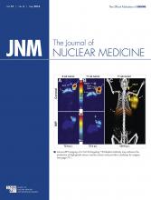
- FIGURE 1.
Examples of representative transverse slices of 18F-FDG PET scans before and after chemoradiotherapy in patient with no pathCR (i.e., non-pathCR) and in patient with a pathCR. These patients initially had comparable tumor volume, TLG, and local tumor texture (as expressed by intensity cooccurrence matrix [ICM] entropy metric). However, in the complete responder, ∆ICM entropy metric decreased and posttreatment TLG was markedly lower (underlined), which represent 2 of the important predictors in models 3 and 4. Histograms illustrate 3-dimensional 18F-FDG uptake within volumes of interest.
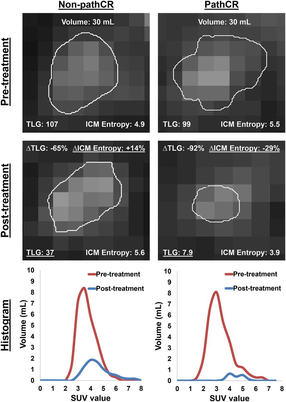
- FIGURE 2.
Receiver-operating-characteristic curve analysis of the 4 models indicating their ability to discriminate between pathCR and non-pathCR patients.
- FIGURE 3.
(A–D) Calibration plots of the 4 models demonstrating agreement between predicted probability of pathCR by model and observed incidence.
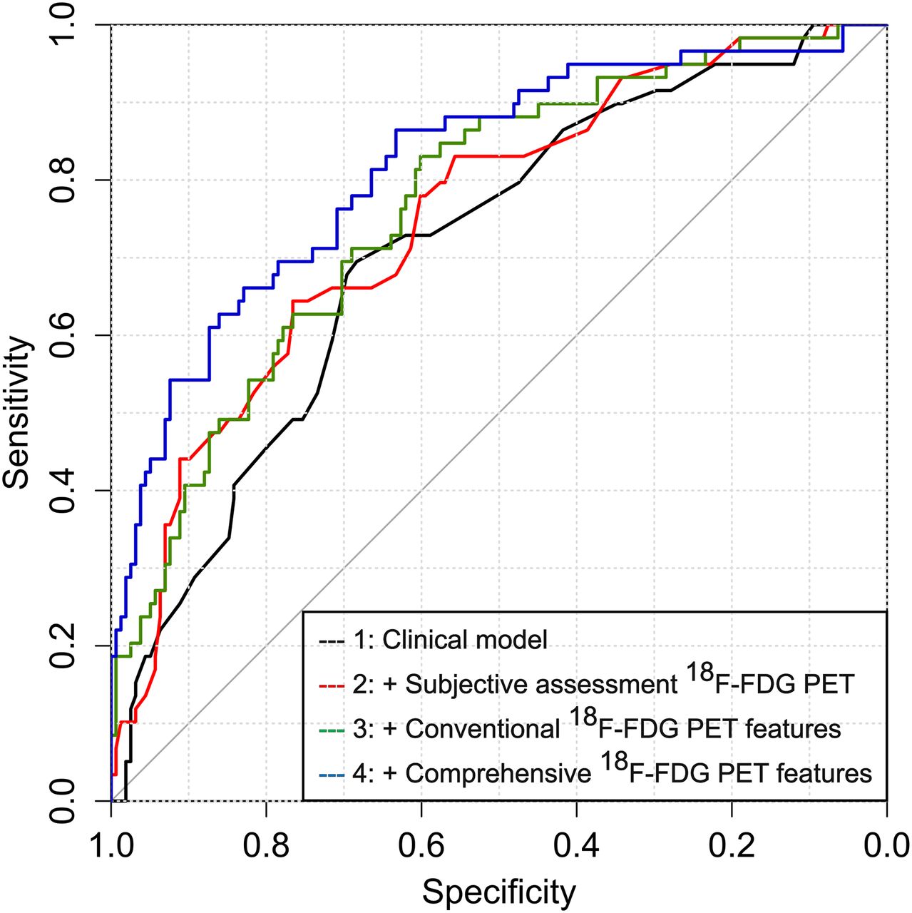
- FIGURE 4.
Decision curves graphically representing net benefit (y-axis) for the 4 models at a range of decision thresholds (i.e., minimum probabilities of pathCR at which one would be willing to change clinical decision making; x-axis). The black and gray solid lines represent making same decision in all patients (i.e., omitting surgery in none or in all of the patients, respectively).


Tables
Characteristic PathCR (n = 59) No pathCR (n = 158) P Male sex 54 (91.5) 148 (93.7) 0.558 Age (y)† 58.8 ± 12.3 60.1 ± 9.9 0.440 Body mass index (kg/m2)† 29.5 ± 5.3 29.8 ± 5.2 0.632 Hypertension 29 (49.2) 90 (57.0) 0.304 Cardiac comorbidity 14 (23.7) 24 (15.2) 0.141 Diabetes mellitus 12 (20.3) 31 (19.6) 0.906 Chronic obstructive pulmonary disease 4 (6.8) 8 (5.1) 0.739 Smoking 13 (22.0) 38 (24.1) 0.755 Karnofsky performance status† 86.4 ± 6.9 85.5 ± 6.6 0.362 Tumor location 0.324 Middle third of esophagus 2 (3.4) 1 (0.6) Distal third of esophagus 52 (88.1) 143 (90.5) Gastroesophageal junction 5 (8.5) 14 (8.9) EUS-based tumor length (cm)† 5.0 ± 2.4 5.9 ± 2.7 0.034* Histologic differentiation grade 0.055 Moderate 34 (57.6) 68 (43.0) Poor 25 (42.4) 90 (57.0) Signet ring cell adenocarcinoma 5 (8.5) 30 (19.0) 0.061 Clinical T stage 0.006* cT2 14 (23.7) 15 (9.5) cT3 45 (76.3) 143 (90.5) Clinical N stage 0.450 cN0 18 (30.5) 58 (36.7) cN+ 39 (66.1) 98 (62.0) Missing 2 (3.4) 2 (1.3) Induction chemotherapy 28 (47.5) 50 (31.6) 0.031* Total radiation dose (Gy) 0.466 45.0 4 (6.8) 6 (3.8) 50.4 55 (93.2) 152 (96.2) Radiation treatment modality 0.405 3-dimensional conformal radiation therapy 1 (1.7) 1 (0.6) Intensity-modulated radiotherapy 38 (64.4) 111 (70.3) Proton therapy 20 (33.9) 46 (29.1) Chemotherapy regimen 0.940 Oxaliplatin/5-fluorouracil 25 (42.4) 64 (40.5) Docetaxel/5-fluorouracil 25 (42.4) 67 (42.4) Other 9 (15.3) 27 (17.1) Postchemoradiation endoscopic biopsy 0.023* No residual cancer 55 (93.2) 126 (79.7) Residual cancer 4 (6.8) 32 (20.3) Days from completion chemoradiotherapy to surgery† 61.5 ± 20.4 58.3 ± 19.3 0.285 Year of patient accrual 0.072 2006–2007 10 (16.9) 47 (29.7) 2008–2010 26 (44.1) 63 (39.9) 2011–2013 23 (39.0) 48 (30.4) - TABLE 2
Univariable Analysis of Subjective and Conventional Quantitative Assessment of 18F-FDG PET for Predicting pathCR
Univariable analysis Characteristic n Odds ratio 95% confidence interval P Subjective assessment 18F-FDG PET 0.001* Clinical complete response 60 1.0 (ref) No clinical complete response 157 0.30 0.15–0.59 Baseline SUVmax (log) 217 0.63 0.37–1.07 0.087 Baseline SUVmean (log) 217 0.60 0.32–1.13 0.112 Baseline MTV (log) 217 0.85 0.57–1.26 0.408 Baseline TLG (log) 217 0.82 0.61–1.10 0.181 Postchemoradiation SUVmax (log) 217 0.32 0.13–0.80 0.015* Postchemoradiation SUVmean (log) 217 0.64 0.22–1.89 0.420 Postchemoradiation MTV (log) 217 0.34 0.21–0.53 <0.001* Postchemoradiation TLG (log) 217 0.41 0.28–0.61 <0.001* ∆SUVmax (%) 217 1.00 0.99–1.01 0.701 ∆SUVmean (%) 217 1.01 1.00–1.02 0.146 ∆MTV(%) 217 1.00 0.99–1.00 0.142 ∆TLG (%) 217 1.00 0.99–1.00 0.301 ↵* Significant difference between pathCR group and pathologic noncomplete response group (P < 0.05).
ref = reference; ∆ = relative change between baseline and postchemoradiation 18F-FDG PET scans.
- TABLE 3
Finalized Prediction Models for pathCR Using Multivariable Logistic Regression Analysis with Stepwise Backward Elimination
Model 1 Model 2 Model 3 Model 4 Characteristic Odds ratio (95% confidence interval) P Odds ratio (95% confidence interval) P Odds ratio (95% confidence interval) P Odds ratio (95% confidence interval) P EUS-based tumor length (log) 0.48 (0.24–0.95) 0.034* 0.50 (0.24–1.01) 0.054 0.55 (0.57–1.14) 0.107 0.46 (0.19–1.11) 0.085 Clinical T stage 0.077 0.046* 0.185 0.239 cT2 1.0 (ref) 1.0 (ref) 1.0 (ref) 1.0 (ref) cT3 0.45 (0.19–1.09) 0.39 (0.15–0.98) 0.53 (0.20–1.36) 0.54 (0.19–1.51) Induction chemotherapy 0.008* 0.012* 0.022* 0.008* No 1.0 (ref) 1.0 (ref) 1.0 (ref) 1.0 (ref) Yes 2.44 (1.26–4.74) 2.40 (1.21–4.77) 2.26 (1.12–4.54) 2.80 (1.31–5.98) Postchemoradiotherapy endoscopic biopsy 0.035* 0.047* 0.057 0.073 No residual cancer 1.0 (ref) 1.0 (ref) 1.0 (ref) 1.0 (ref) Residual cancer 0.30 (0.10–0.92) 0.32 (0.10–0.98) 0.32 (0.10–1.04) 0.31 (0.08–1.12) Subjective assessment 18F-FDG PET Not entered — 0.001* 0.043* 0.113 Clinical complete response 1.0 (ref) 1.0 (ref) 1.0 (ref) No clinical complete response 0.30 (0.15–0.59) 0.45 (0.21–0.98) 0.52 (0.23–1.17) Postchemoradiotherapy TLG (log) Not entered — Not entered — 0.57 (0.37–0.88) 0.011* 0.76 (0.41–1.39) 0.370 Baseline cluster shade (log) Not entered — Not entered — Not entered — 0.19 (0.03–1.03) 0.054 ∆run percentage Not entered — Not entered — Not entered — 1.07 (1.02–1.11) 0.004* ∆ICM entropy Not entered — Not entered — Not entered — 0.97 (0.94–0.99) 0.044* Postchemoradiotherapy roundness (log) Not entered — Not entered — Not entered — 0.10 (0.03–0.42) 0.001* Entered variables that were eliminated based on redundancy were year of patient accrual, histologic differentiation grade, and signet ring cell adenocarcinoma (model 1); baseline SUVmax and ∆MTV (model 3); and baseline maximum probability (log), ∆busyness, ∆cumulative histogram, postchemoradiation skewness, and postchemoradiation long-run high-intensity emphasis (log) (model 4).
ref = reference; ICM = intensity cooccurrence matrix.
Discrimination Model no Model type Apparent c-index Corrected c-index* 1 Clinical model 0.71 (0.64–0.79) 0.67 (0.60–0.75) 2 + Subjective assessment 18F-FDG PET 0.75 (0.68–0.82) 0.72 (0.65–0.79) 3 + Conventional 18F-FDG PET features 0.77 (0.70–0.84) 0.73 (0.66–0.80) 4 + Comprehensive 18F-FDG PET features 0.82 (0.75–0.88) 0.77 (0.70–0.83) ↵* Correction after internal validation for both optimism and bias from predictor selection process.
Data in parentheses are 95% confidence intervals.
- TABLE 5
Overview of Studies on 18F-FDG PET Texture Analysis in Esophageal Cancer for Treatment Response Assessment
Study, year n Tumor type Tumor stages Timing of 18F-FDG PET Outcome associated with tumor texture Tixier et al., 2011 (34)* 41 Adenocarcinoma, squamous cell carcinoma I–IV Prechemoradiotherapy Clinical response (according to RECIST) Hatt et al. 2013 (29)* 50 Adenocarcinoma, squamous cell carcinoma I–IV Prechemoradiotherapy Clinical response (according to RECIST) Tan et al., 2013 (38)† 20 Adenocarcinoma, squamous cell carcinoma II–III Pre- and postchemoradiotherapy Pathologic response (TRG 1 + 2 vs. 3 + 4) Zhang et al., 2014 (39)† 20 Adenocarcinoma, squamous cell carcinoma II–III Pre- and postchemoradiotherapy Pathologic response (TRG 1 + 2 vs. 3 + 4) Current study 217 Adenocarcinoma II–III Pre- and postchemoradiotherapy pathCR (TRG 1 vs. 2–4)
Supplemental Data
Files in this Data Supplement:


Jump to section
Related Articles
Cited By...
- A Meta-Analysis for Using Radiomics to Predict Complete Pathological Response in Esophageal Cancer Patients Receiving Neoadjuvant Chemoradiation
- A Postreconstruction Harmonization Method for Multicenter Radiomic Studies in PET
- Predicting Response to Neoadjuvant Chemoradiotherapy in Esophageal Cancer with Textural Features Derived from Pretreatment 18F-FDG PET/CT Imaging