
Main menu

  • Home
  • Content
    • Current
    • Ahead of print
    • Past Issues
    • JNM Supplement
    • SNMMI Annual Meeting Abstracts
    • Continuing Education
    • JNM Podcasts
  • Subscriptions
    • Subscribers
    • Institutional and Non-member
    • Rates
    • Journal Claims
    • Corporate & Special Sales
  • Authors
    • Submit to JNM
    • Information for Authors
    • Assignment of Copyright
    • AQARA requirements
  • Info
    • Reviewers
    • Permissions
    • Advertisers
  • About
    • About Us
    • Editorial Board
    • Contact Information
  • More
    • Alerts
    • Feedback
    • Help
    • SNMMI Journals
  • SNMMI
    • JNM
    • JNMT
    • SNMMI Journals
    • SNMMI

User menu

  • Subscribe
  • My alerts
  • Log in
  • Log out
  • My Cart

Search

  • Advanced search
Journal of Nuclear Medicine
  • SNMMI
    • JNM
    • JNMT
    • SNMMI Journals
    • SNMMI
  • Subscribe
  • My alerts
  • Log in
  • Log out
  • My Cart
Journal of Nuclear Medicine

Advanced Search

  • Home
  • Content
    • Current
    • Ahead of print
    • Past Issues
    • JNM Supplement
    • SNMMI Annual Meeting Abstracts
    • Continuing Education
    • JNM Podcasts
  • Subscriptions
    • Subscribers
    • Institutional and Non-member
    • Rates
    • Journal Claims
    • Corporate & Special Sales
  • Authors
    • Submit to JNM
    • Information for Authors
    • Assignment of Copyright
    • AQARA requirements
  • Info
    • Reviewers
    • Permissions
    • Advertisers
  • About
    • About Us
    • Editorial Board
    • Contact Information
  • More
    • Alerts
    • Feedback
    • Help
    • SNMMI Journals
  • View or Listen to JNM Podcast
  • Visit JNM on Facebook
  • Join JNM on LinkedIn
  • Follow JNM on Twitter
  • Subscribe to our RSS feeds
Research ArticleClinical Investigations

Pilot Comparison of 68Ga-RM2 PET and 68Ga-PSMA-11 PET in Patients with Biochemically Recurrent Prostate Cancer

Ryogo Minamimoto, Steven Hancock, Bernadette Schneider, Frederick T. Chin, Mehran Jamali, Andreas Loening, Shreyas Vasanawala, Sanjiv Sam Gambhir and Andrei Iagaru
Journal of Nuclear Medicine April 2016, 57 (4) 557-562; DOI: https://doi.org/10.2967/jnumed.115.168393
Ryogo Minamimoto
1Division of Nuclear Medicine and Molecular Imaging, Department of Radiology, Stanford University, Stanford, California
2Molecular Imaging Program at Stanford, Department of Radiology, Stanford University, Stanford, California
  • Find this author on Google Scholar
  • Find this author on PubMed
  • Search for this author on this site
Steven Hancock
3Department of Radiation Oncology, Stanford University, Stanford, California; and
  • Find this author on Google Scholar
  • Find this author on PubMed
  • Search for this author on this site
Bernadette Schneider
2Molecular Imaging Program at Stanford, Department of Radiology, Stanford University, Stanford, California
  • Find this author on Google Scholar
  • Find this author on PubMed
  • Search for this author on this site
Frederick T. Chin
2Molecular Imaging Program at Stanford, Department of Radiology, Stanford University, Stanford, California
  • Find this author on Google Scholar
  • Find this author on PubMed
  • Search for this author on this site
Mehran Jamali
1Division of Nuclear Medicine and Molecular Imaging, Department of Radiology, Stanford University, Stanford, California
2Molecular Imaging Program at Stanford, Department of Radiology, Stanford University, Stanford, California
  • Find this author on Google Scholar
  • Find this author on PubMed
  • Search for this author on this site
Andreas Loening
4Department of Radiology, Stanford University, Stanford, California
  • Find this author on Google Scholar
  • Find this author on PubMed
  • Search for this author on this site
Shreyas Vasanawala
4Department of Radiology, Stanford University, Stanford, California
  • Find this author on Google Scholar
  • Find this author on PubMed
  • Search for this author on this site
Sanjiv Sam Gambhir
2Molecular Imaging Program at Stanford, Department of Radiology, Stanford University, Stanford, California
  • Find this author on Google Scholar
  • Find this author on PubMed
  • Search for this author on this site
Andrei Iagaru
1Division of Nuclear Medicine and Molecular Imaging, Department of Radiology, Stanford University, Stanford, California
  • Find this author on Google Scholar
  • Find this author on PubMed
  • Search for this author on this site
  • Article
  • Figures & Data
  • Supplemental
  • Info & Metrics
  • PDF
Loading

Article Figures & Data

Figures

  • Tables
  • Additional Files
  • FIGURE 1.
    • Download figure
    • Open in new tab
    • Download powerpoint
    FIGURE 1.

    SUVmax of 68Ga-PSMA-11 and 68Ga-RM2 images for all analyzed normal tissues.

  • FIGURE 2.
    • Download figure
    • Open in new tab
    • Download powerpoint
    FIGURE 2.

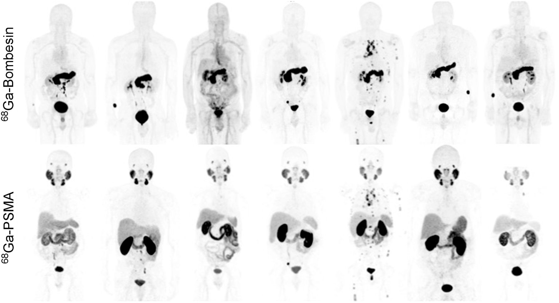
    Maximum-intensity-projection 68Ga-RM2 and 68Ga-PSMA-11 images of the 7 enrolled patients.

  • FIGURE 3.
    • Download figure
    • Open in new tab
    • Download powerpoint
    FIGURE 3.

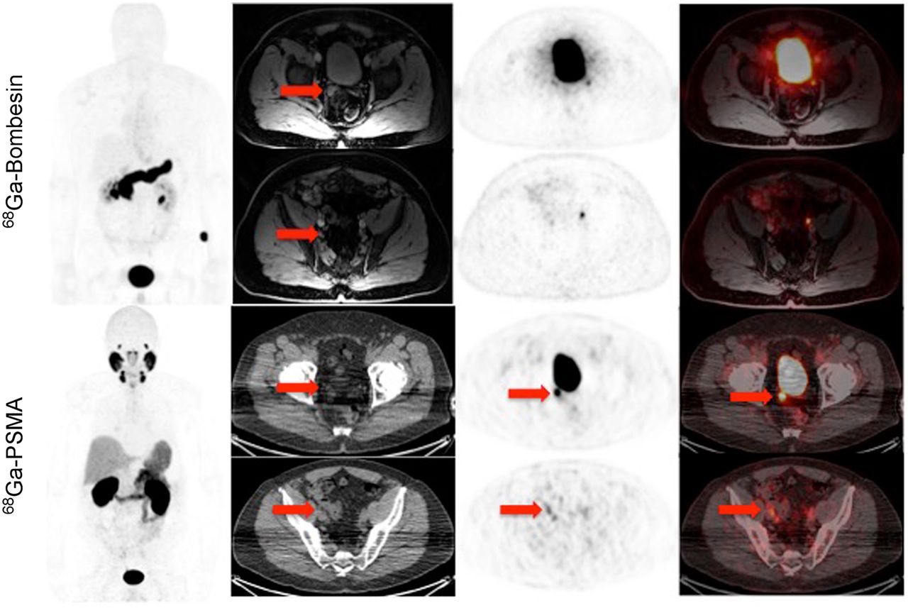
    83-y-old man with history of Gleason 5 + 4 prostate cancer treated with radiation therapy and androgen blockade, who presented with PSA level of 18.7 ng/mL and noncontributory findings on conventional imaging. Maximum-intensity-projection 68Ga-RM2 and 68Ga-PSMA-11 images, as well as axial PET images, show subcentimeter focal uptake corresponding to lymph nodes on MRI and CT, respectively.

  • FIGURE 4.
    • Download figure
    • Open in new tab
    • Download powerpoint
    FIGURE 4.

    67-y-old man with history of Gleason 3 + 3 prostate cancer treated with radiation therapy and androgen blockade, who presented with PSA level of 6.7 ng/mL and noncontributory findings on conventional imaging. 68Ga-RM2 images are negative for uptake, whereas maximum-intensity-projection 68Ga-PSMA-11 images, as well as axial PET images, show subcentimeter focal uptake corresponding to pelvic lymph node and right seminal vesicle on CT. These were biopsy-proven to represent metastatic disease.

Tables

  • Figures
  • Additional Files
    • View popup
    TABLE 1

    Patient Characteristics and Scan Results

    Patient no.Age (y)Initial cancer stageGleason scorePSA (ng/mL)Treatment68Ga-PSMA-1168Ga-RM2
    183II5 + 416.2HT + IMRTRetroperitoneal LNsRetroperitoneal LNs
    269II3 + 46.0ProstatectomyRetroperitoneal LNsRetroperitoneal LNs
    375IV4 + 536.5ProstatectomyRetroperitoneal LNs, pelvic LNRetroperitoneal LNs, pelvic LN
    467III3 + 36.7Brachytherapy + HTVas deferens, pelvic LNNegative
    572III4 + 33.5RTSupraclavicular LNSupraclavicular LN
    681I3 + 47.4ProstatectomyPelvic LNs, seminal vesiclePelvic LNs, seminal vesicle
    773III4 + 418.2ProstatectomyMediastinal LNs, retroperitoneal LNs, pelvic LNs, multiple bone lesionsMediastinal LNs, retroperitoneal LNs, pelvic LNs, multiple bone lesions
    • HT = hormone therapy; IMRT = intensity-modulated radiation therapy; LN = lymph node; RT = radiation therapy.

    • View popup
    TABLE 2

    68Ga-PSMA-11 and 68Ga-RM2 Uptake Outside Expected Physiologic Biodistribution

    Index68Ga-PSMA-1168Ga-RM2P
    SUVmax12.4 ± 7.1 (4.1–43.6)13.2 ± 8.0 (2.5–33.5)0.63
    SUVmean7.1 ± 4.0 (2.0–14.4)7.6 ± 3.8 (2.0–14.4)0.38
    F/N ratio10.4 ± 9.3 (2.3–42.8)5.9 ± 4.6 (1.2–18.8)<0.003
    F/N ratio9.2 ± 7.3 (2.4–40.4)5.2 ± 3.5 (1.6–12.8)<0.02
    • Data are mean ± SD, followed by range in parentheses.

    • View popup
    TABLE 3

    Follow-up Results After Scanning

    Patient no.Follow-up results
    1Started bicalutamide and leuprolide, and PSA decreased from 18.7 to 2.53; follow-up MRI showed decreased size and number of retroperitoneal lymph nodes
    2Chose to have no treatment, and PSA increased from 8.6 to 10.9
    3Started bicalutamide and leuprolide, and PSA decreased from 36.4 to 2.2
    4Started no treatment yet, and PSA increased from 6.7 to 12.1; on biopsy, 12 prostate cores were negative and right vas deferens showed metastatic adenocarcinoma (Gleason 4 + 3; 15% of core)
    5Started no treatment yet, and PSA increased from 8.53 to 10.2
    6Started bicalutamide, and PSA decreased from 7.36 to 0.38
    7Started bicalutamide, and PSA decreased from 18.2 to 2.5

Additional Files

  • Figures
  • Tables
  • Supplemental Data

    Files in this Data Supplement:

    • Supplemental Data
PreviousNext
Back to top

In this issue

Journal of Nuclear Medicine: 57 (4)
Journal of Nuclear Medicine
Vol. 57, Issue 4
April 1, 2016
  • Table of Contents
  • Table of Contents (PDF)
  • About the Cover
  • Index by author
Print
Download PDF
Article Alerts
Sign In to Email Alerts with your Email Address
Email Article

Thank you for your interest in spreading the word on Journal of Nuclear Medicine.

NOTE: We only request your email address so that the person you are recommending the page to knows that you wanted them to see it, and that it is not junk mail. We do not capture any email address.

Enter multiple addresses on separate lines or separate them with commas.
Pilot Comparison of 68Ga-RM2 PET and 68Ga-PSMA-11 PET in Patients with Biochemically Recurrent Prostate Cancer
(Your Name) has sent you a message from Journal of Nuclear Medicine
(Your Name) thought you would like to see the Journal of Nuclear Medicine web site.
Citation Tools
Pilot Comparison of 68Ga-RM2 PET and 68Ga-PSMA-11 PET in Patients with Biochemically Recurrent Prostate Cancer
Ryogo Minamimoto, Steven Hancock, Bernadette Schneider, Frederick T. Chin, Mehran Jamali, Andreas Loening, Shreyas Vasanawala, Sanjiv Sam Gambhir, Andrei Iagaru
Journal of Nuclear Medicine Apr 2016, 57 (4) 557-562; DOI: 10.2967/jnumed.115.168393

Citation Manager Formats

  • BibTeX
  • Bookends
  • EasyBib
  • EndNote (tagged)
  • EndNote 8 (xml)
  • Medlars
  • Mendeley
  • Papers
  • RefWorks Tagged
  • Ref Manager
  • RIS
  • Zotero
Share
Pilot Comparison of 68Ga-RM2 PET and 68Ga-PSMA-11 PET in Patients with Biochemically Recurrent Prostate Cancer
Ryogo Minamimoto, Steven Hancock, Bernadette Schneider, Frederick T. Chin, Mehran Jamali, Andreas Loening, Shreyas Vasanawala, Sanjiv Sam Gambhir, Andrei Iagaru
Journal of Nuclear Medicine Apr 2016, 57 (4) 557-562; DOI: 10.2967/jnumed.115.168393
Twitter logo Facebook logo LinkedIn logo Mendeley logo
  • Tweet Widget
  • Facebook Like
  • Google Plus One
Bookmark this article

Jump to section

  • Article
    • Abstract
    • MATERIALS AND METHODS
    • RESULTS
    • DISCUSSION
    • CONCLUSION
    • Acknowledgments
    • Footnotes
    • REFERENCES
  • Figures & Data
  • Supplemental
  • Info & Metrics
  • PDF

Related Articles

  • This Month in JNM
  • PubMed
  • Google Scholar

Cited By...

  • Utility of 64Cu-Sarcophagine-Bombesin PET/CT in Men with Biochemically Recurrent Prostate Cancer and Negative or Equivocal Findings on 68Ga-PSMA-11 PET/CT
  • Prospective Comparison of 68Ga-NeoB and 68Ga-PSMA-R2 PET/MRI in Patients with Biochemically Recurrent Prostate Cancer
  • A Pilot Study of 68Ga-PSMA11 and 68Ga-RM2 PET/MRI for Biopsy Guidance in Patients with Suspected Prostate Cancer
  • A Pilot Study of 68Ga-PSMA11 and 68Ga-RM2 PET/MRI for Evaluation of Prostate Cancer Response to High-Intensity Focused Ultrasound Therapy
  • Comparison of 68Ga-PSMA-617 PET/CT and 68Ga-RM2 PET/CT in Patients with Localized Prostate Cancer Who Are Candidates for Radical Prostatectomy: A Prospective, Single-Arm, Single-Center, Phase II Study
  • Correlation of 68Ga-RM2 PET with Postsurgery Histopathology Findings in Patients with Newly Diagnosed Intermediate- or High-Risk Prostate Cancer
  • GRPr Antagonist 68Ga-SB3 PET/CT Imaging of Primary Prostate Cancer in Therapy-Naive Patients
  • PSMA- and GRPR-Targeted PET: Results from 50 Patients with Biochemically Recurrent Prostate Cancer
  • PET Imaging Quantifying 68Ga-PSMA-11 Uptake in Metastatic Colorectal Cancer
  • Clinical Evaluation of (4S)-4-(3-[18F]Fluoropropyl)-L-glutamate (18F-FSPG) for PET/CT Imaging in Patients with Newly Diagnosed and Recurrent Prostate Cancer
  • Development of Improved Tumor-Residualizing, GRPR-Targeted Agents: Preclinical Comparison of an Endolysosomal Trapping Approach in Agonistic and Antagonistic Constructs
  • Simultaneous transrectal ultrasound and photoacoustic human prostate imaging
  • Healthy Tissue Uptake of 68Ga-Prostate-Specific Membrane Antigen, 18F-DCFPyL, 18F-Fluoromethylcholine, and 18F-Dihydrotestosterone
  • New Developments in Peptide Receptor Radionuclide Therapy
  • PET Using a GRPR Antagonist 68Ga-RM26 in Healthy Volunteers and Prostate Cancer Patients
  • Prospective Evaluation of 68Ga-RM2 PET/MRI in Patients with Biochemical Recurrence of Prostate Cancer and Negative Findings on Conventional Imaging
  • Will GRPR Compete with PSMA as a Target in Prostate Cancer?
  • Approaches to Multireceptor Targeting: Hybrid Radioligands, Radioligand Cocktails, and Sequential Radioligand Applications
  • Glu-Ureido-Based Inhibitors of Prostate-Specific Membrane Antigen: Lessons Learned During the Development of a Novel Class of Low-Molecular-Weight Theranostic Radiotracers
  • 68Ga/177Lu-NeoBOMB1, a Novel Radiolabeled GRPR Antagonist for Theranostic Use in Oncology
  • Theranostic Perspectives in Prostate Cancer with the Gastrin-Releasing Peptide Receptor Antagonist NeoBOMB1: Preclinical and First Clinical Results
  • What Medical, Urologic, and Radiation Oncologists Want from Molecular Imaging of Prostate Cancer
  • Molecular Imaging and Targeted Radionuclide Therapy of Prostate Cancer
  • Bombesin-Targeted PET of Prostate Cancer
  • Google Scholar

More in this TOC Section

  • Feasibility of Ultra-Low-Activity 18F-FDG PET/CT Imaging Using a Long–Axial-Field-of-View PET/CT System
  • Cardiac Presynaptic Sympathetic Nervous Function Evaluated by Cardiac PET in Patients with Chronotropic Incompetence Without Heart Failure
  • Validation and Evaluation of a Vendor-Provided Head Motion Correction Algorithm on the uMI Panorama PET/CT System
Show more Clinical Investigations

Similar Articles

Keywords

  • prostate cancer
  • 68Ga
  • RM2
  • PSMA
  • PET/MRI
  • PET/CT
SNMMI

© 2025 SNMMI

Powered by HighWire