
Main menu

  • Home
  • Content
    • Current
    • Ahead of print
    • Past Issues
    • JNM Supplement
    • SNMMI Annual Meeting Abstracts
    • Continuing Education
    • JNM Podcasts
  • Subscriptions
    • Subscribers
    • Institutional and Non-member
    • Rates
    • Journal Claims
    • Corporate & Special Sales
  • Authors
    • Submit to JNM
    • Information for Authors
    • Assignment of Copyright
    • AQARA requirements
  • Info
    • Reviewers
    • Permissions
    • Advertisers
  • About
    • About Us
    • Editorial Board
    • Contact Information
  • More
    • Alerts
    • Feedback
    • Help
    • SNMMI Journals
  • SNMMI
    • JNM
    • JNMT
    • SNMMI Journals
    • SNMMI

User menu

  • Subscribe
  • My alerts
  • Log in
  • My Cart

Search

  • Advanced search
Journal of Nuclear Medicine
  • SNMMI
    • JNM
    • JNMT
    • SNMMI Journals
    • SNMMI
  • Subscribe
  • My alerts
  • Log in
  • My Cart
Journal of Nuclear Medicine

Advanced Search

  • Home
  • Content
    • Current
    • Ahead of print
    • Past Issues
    • JNM Supplement
    • SNMMI Annual Meeting Abstracts
    • Continuing Education
    • JNM Podcasts
  • Subscriptions
    • Subscribers
    • Institutional and Non-member
    • Rates
    • Journal Claims
    • Corporate & Special Sales
  • Authors
    • Submit to JNM
    • Information for Authors
    • Assignment of Copyright
    • AQARA requirements
  • Info
    • Reviewers
    • Permissions
    • Advertisers
  • About
    • About Us
    • Editorial Board
    • Contact Information
  • More
    • Alerts
    • Feedback
    • Help
    • SNMMI Journals
  • View or Listen to JNM Podcast
  • Visit JNM on Facebook
  • Join JNM on LinkedIn
  • Follow JNM on Twitter
  • Subscribe to our RSS feeds
Research ArticleClinical Investigations

Partial-Volume Effect Correction Improves Quantitative Analysis of 18F-Florbetaben β-Amyloid PET Scans

Michael Rullmann, Juergen Dukart, Karl-Titus Hoffmann, Julia Luthardt, Solveig Tiepolt, Marianne Patt, Hermann-Josef Gertz, Matthias L. Schroeter, John Seibyl, Walter J. Schulz-Schaeffer, Osama Sabri and Henryk Barthel
Journal of Nuclear Medicine February 2016, 57 (2) 198-203; DOI: https://doi.org/10.2967/jnumed.115.161893
Michael Rullmann
1Department of Nuclear Medicine, University of Leipzig, Leipzig, Germany
  • Find this author on Google Scholar
  • Find this author on PubMed
  • Search for this author on this site
Juergen Dukart
2LREN, Département des Neurosciences Cliniques, CHUV, Université de Lausanne, Lausanne, Switzerland
3Max Planck Institute for Human Cognitive and Brain Sciences, Leipzig, Germany
  • Find this author on Google Scholar
  • Find this author on PubMed
  • Search for this author on this site
Karl-Titus Hoffmann
4Department of Neuroradiology, University of Leipzig, Leipzig, Germany
  • Find this author on Google Scholar
  • Find this author on PubMed
  • Search for this author on this site
Julia Luthardt
1Department of Nuclear Medicine, University of Leipzig, Leipzig, Germany
  • Find this author on Google Scholar
  • Find this author on PubMed
  • Search for this author on this site
Solveig Tiepolt
1Department of Nuclear Medicine, University of Leipzig, Leipzig, Germany
  • Find this author on Google Scholar
  • Find this author on PubMed
  • Search for this author on this site
Marianne Patt
1Department of Nuclear Medicine, University of Leipzig, Leipzig, Germany
  • Find this author on Google Scholar
  • Find this author on PubMed
  • Search for this author on this site
Hermann-Josef Gertz
5Department of Psychiatry, University of Leipzig, Leipzig, Germany
  • Find this author on Google Scholar
  • Find this author on PubMed
  • Search for this author on this site
Matthias L. Schroeter
3Max Planck Institute for Human Cognitive and Brain Sciences, Leipzig, Germany
6Clinic for Cognitive Neurology, University Hospital Leipzig, Leipzig, Germany
  • Find this author on Google Scholar
  • Find this author on PubMed
  • Search for this author on this site
John Seibyl
7Molecular NeuroImaging, L.L.C., New Haven, Connecticut; and
  • Find this author on Google Scholar
  • Find this author on PubMed
  • Search for this author on this site
Walter J. Schulz-Schaeffer
8Department of Neuropathology, University Medical Center, Goettingen, Germany
  • Find this author on Google Scholar
  • Find this author on PubMed
  • Search for this author on this site
Osama Sabri
1Department of Nuclear Medicine, University of Leipzig, Leipzig, Germany
  • Find this author on Google Scholar
  • Find this author on PubMed
  • Search for this author on this site
Henryk Barthel
1Department of Nuclear Medicine, University of Leipzig, Leipzig, Germany
  • Find this author on Google Scholar
  • Find this author on PubMed
  • Search for this author on this site
  • Article
  • Figures & Data
  • Info & Metrics
  • PDF
Loading

Article Figures & Data

Figures

  • Tables
  • FIGURE 1.
    • Download figure
    • Open in new tab
    • Download powerpoint
    FIGURE 1.

    Workflow chart of processing pipeline. PET and MRI data were provided as input to PVELab. Main output of PVELab was PVE-corrected data, which were masked by corresponding gray matter. PVELab additionally generated gray matter tissue probability map (TPMGM), which was used to mask uncorrected PET data. To interpret gray matter–masked PET data (GM-PET) and PVEC GM-PET data, hand-drawn VOIs were used for cohorts 1 and 2 and anatomic automatic-labeling (AAL) VOIs for cohort 3.

  • FIGURE 2.
    • Download figure
    • Open in new tab
    • Download powerpoint
    FIGURE 2.

    PVEC-related changes in differential regional and composite SUVR in AD patients and HCs. Groups were compared using Student t test.
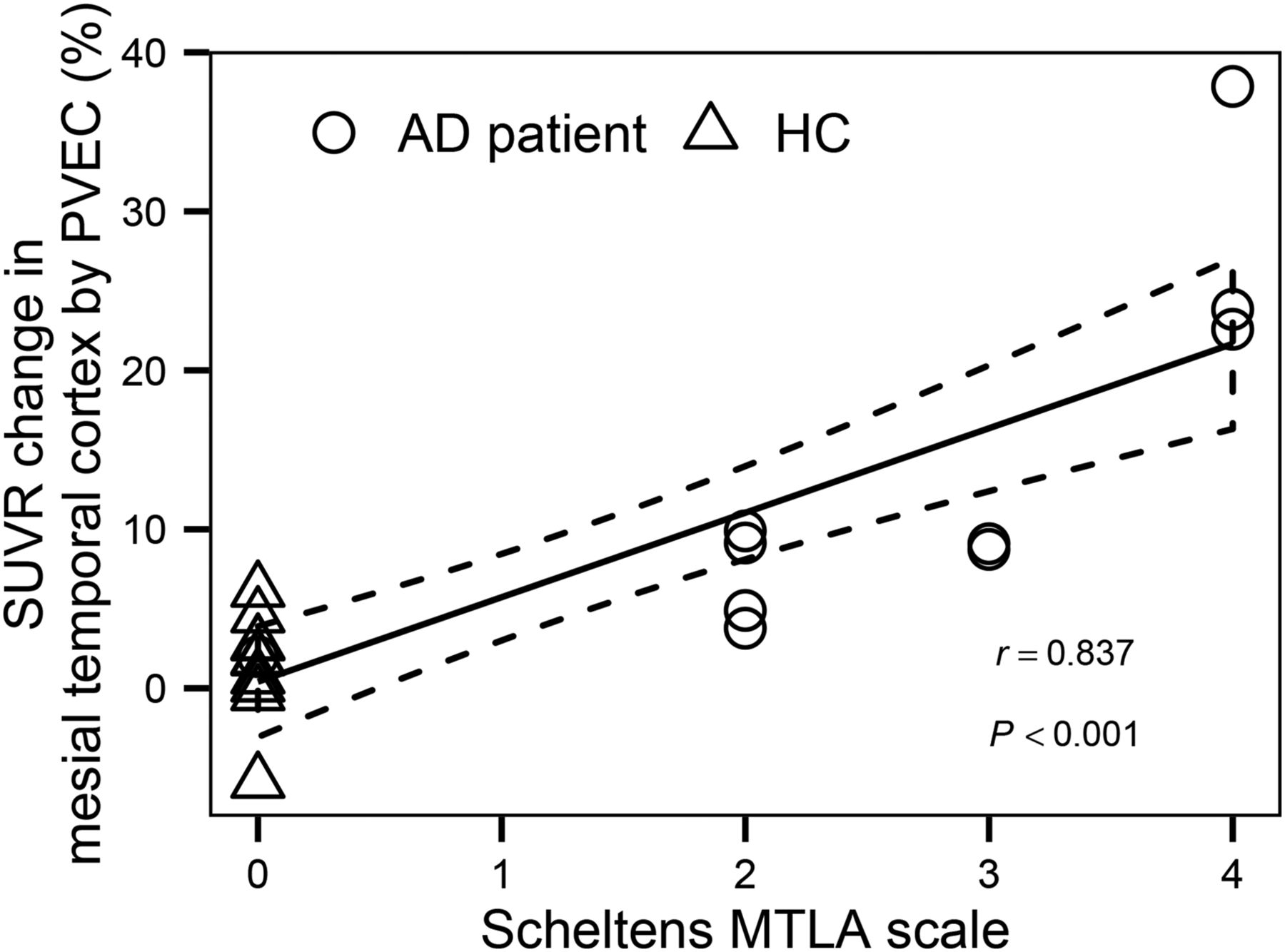
  • FIGURE 3.
    • Download figure
    • Open in new tab
    • Download powerpoint
    FIGURE 3.

    Correlation between PVEC-related SUVR change in mesial temporal cortex (mean of left and right) and degree of mesial temporal lobe atrophy as scored by Scheltens scale.

  • FIGURE 4.
    • Download figure
    • Open in new tab
    • Download powerpoint
    FIGURE 4.

    Correlation between PVEC-related composite SUVR change and relative volume of gray matter in composite regions.

  • FIGURE 5.
    • Download figure
    • Open in new tab
    • Download powerpoint
    FIGURE 5.

    T1-weighted magnetization-prepared rapid-acquisition gradient echo MRI data (left), gray matter–masked 18F-florbetaben PET data (middle), and PVE-corrected gray matter 18F-florbetaben PET data (right) of 2 AD patients with different degrees of mesial temporal lobe atrophy as scored by Scheltens scale. Tracer uptake increase by PVEC was higher in more atrophic brain than in less atrophic brain. GM = gray matter; MTLA = mesial temporal lobe atrophy.
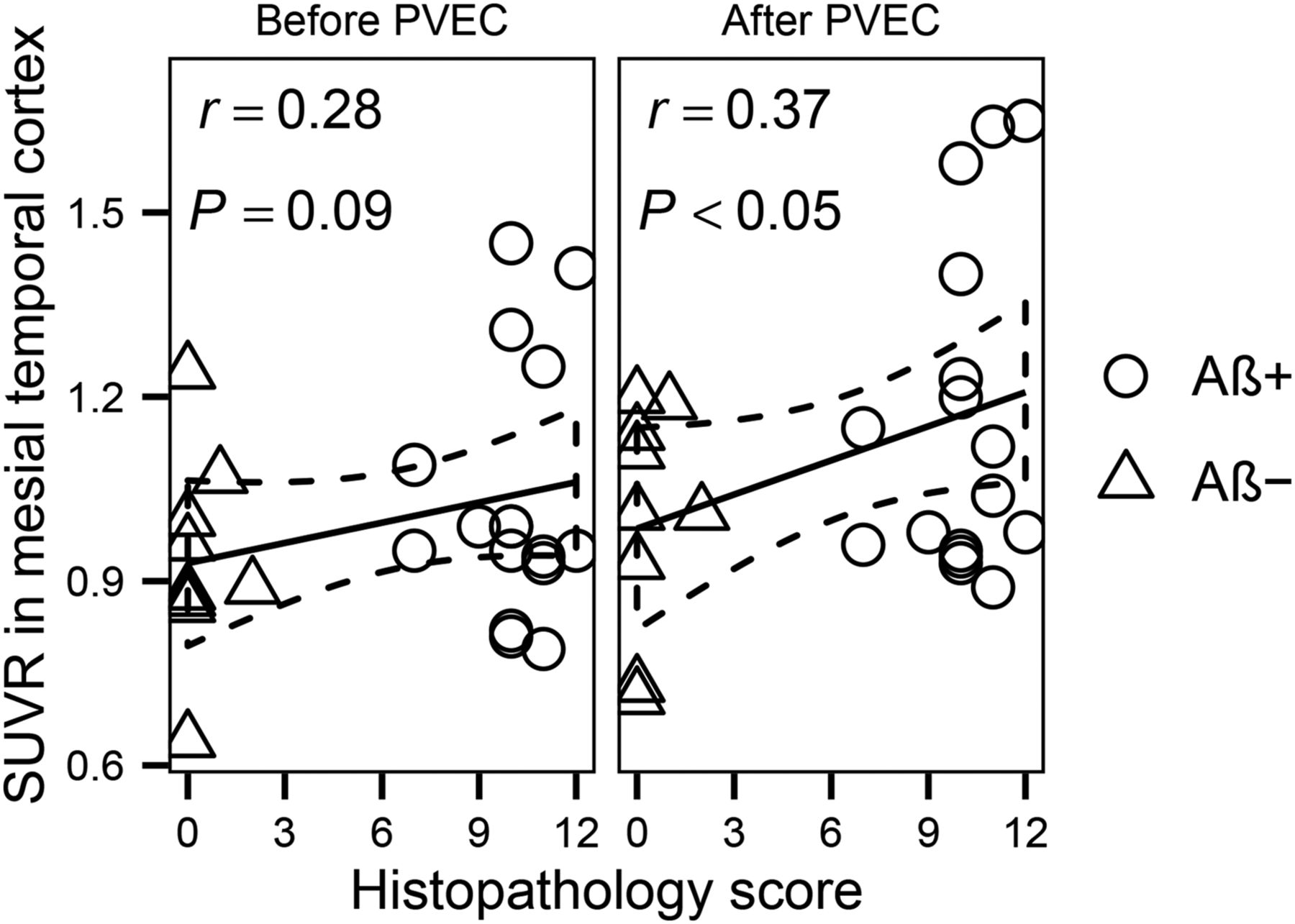
  • FIGURE 6.
    • Download figure
    • Open in new tab
    • Download powerpoint
    FIGURE 6.

    Influence of PVEC on correlation between SUVRs in mesial temporal cortex and histopathology score. Aβ− = Aβ-negative; Aβ+ = Aβ-positive.
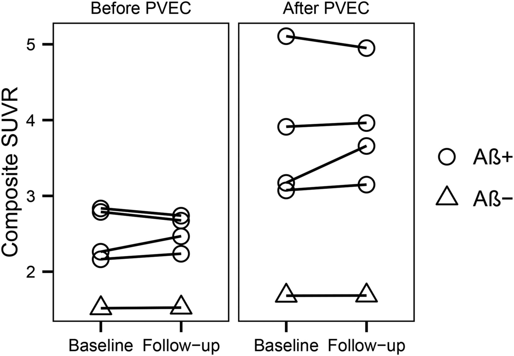
  • FIGURE 7.
    • Download figure
    • Open in new tab
    • Download powerpoint
    FIGURE 7.

    Influence of PVEC on composite SUVRs over time. Aβ− = Aβ-negative; Aβ+ = Aβ-positive.

Tables

  • Figures
    • View popup
    TABLE 1

    Spatial Resolutions of the Different PET Scanners Used on Cohort 2

    ScannerManufacturerTransaxialAxial
    Discovery LSGE Healthcare5.27.5
    Discovery STGE Healthcare6.75.8
    AllegroPhilips Healthcare5.76.7
    SET-2400WShimadzu5.06.7
    Biograph 2Siemens Healthcare7.17.0
    Biograph 16Siemens Healthcare4.85.5
    ECAT Exact HR+Siemens Healthcare5.45.3
    Sensation 16Siemens Healthcare6.87.1
    • Data are full width at half maximum at 10 cm according to National Electrical Manufacturers Association NU 2-2001 standards as implemented in PVEC approach used in this study.

    • View popup
    TABLE 2

    Influence of PVEC on 18F-Florbetaben SUVR Discrimination Between AD Patients and HCs in Cohort 1

    Before PVECAfter PVEC
    ROIADHCPCohen dADHCPCohen d
    Frontal cortex*1.46 ± 0.221.13 ± 0.220.0041.481.85 ± 0.401.15 ± 0.280.00032.02
    Parietal cortex*1.45 ± 0.151.31 ± 0.150.0360.771.84 ± 0.341.40 ± 0.200.0021.59
    Mesial temporal cortex*1.24 ± 0.111.23 ± 0.150.930.041.39 ± 0.131.25 ± 0.150.031.04
    Composite1.55 ± 0.181.27 ± 0.160.0011.681.92 ± 0.361.34 ± 0.190.00032.00
    • ↵* Mean of left and right.

    • Data are mean ± SD SUVR.

PreviousNext
Back to top

In this issue

Journal of Nuclear Medicine: 57 (2)
Journal of Nuclear Medicine
Vol. 57, Issue 2
February 1, 2016
  • Table of Contents
  • Table of Contents (PDF)
  • About the Cover
  • Index by author
Print
Download PDF
Article Alerts
Sign In to Email Alerts with your Email Address
Email Article

Thank you for your interest in spreading the word on Journal of Nuclear Medicine.

NOTE: We only request your email address so that the person you are recommending the page to knows that you wanted them to see it, and that it is not junk mail. We do not capture any email address.

Enter multiple addresses on separate lines or separate them with commas.
Partial-Volume Effect Correction Improves Quantitative Analysis of 18F-Florbetaben β-Amyloid PET Scans
(Your Name) has sent you a message from Journal of Nuclear Medicine
(Your Name) thought you would like to see the Journal of Nuclear Medicine web site.
Citation Tools
Partial-Volume Effect Correction Improves Quantitative Analysis of 18F-Florbetaben β-Amyloid PET Scans
Michael Rullmann, Juergen Dukart, Karl-Titus Hoffmann, Julia Luthardt, Solveig Tiepolt, Marianne Patt, Hermann-Josef Gertz, Matthias L. Schroeter, John Seibyl, Walter J. Schulz-Schaeffer, Osama Sabri, Henryk Barthel
Journal of Nuclear Medicine Feb 2016, 57 (2) 198-203; DOI: 10.2967/jnumed.115.161893

Citation Manager Formats

  • BibTeX
  • Bookends
  • EasyBib
  • EndNote (tagged)
  • EndNote 8 (xml)
  • Medlars
  • Mendeley
  • Papers
  • RefWorks Tagged
  • Ref Manager
  • RIS
  • Zotero
Share
Partial-Volume Effect Correction Improves Quantitative Analysis of 18F-Florbetaben β-Amyloid PET Scans
Michael Rullmann, Juergen Dukart, Karl-Titus Hoffmann, Julia Luthardt, Solveig Tiepolt, Marianne Patt, Hermann-Josef Gertz, Matthias L. Schroeter, John Seibyl, Walter J. Schulz-Schaeffer, Osama Sabri, Henryk Barthel
Journal of Nuclear Medicine Feb 2016, 57 (2) 198-203; DOI: 10.2967/jnumed.115.161893
Twitter logo Facebook logo LinkedIn logo Mendeley logo
  • Tweet Widget
  • Facebook Like
  • Google Plus One
Bookmark this article

Jump to section

  • Article
    • Abstract
    • MATERIALS AND METHODS
    • RESULTS
    • DISCUSSION
    • CONCLUSION
    • DISCLOSURE
    • Acknowledgments
    • Footnotes
    • REFERENCES
  • Figures & Data
  • Info & Metrics
  • PDF

Related Articles

  • This Month in JNM
  • PubMed
  • Google Scholar

Cited By...

  • Traumatic brain injury and Alzheimers Disease biomarkers: A systematic review of findings from amyloid and tau positron emission tomography (PET)
  • Reshaping the Amyloid Buildup Curve in Alzheimer Disease? Partial-Volume Effect Correction of Longitudinal Amyloid PET Data
  • Optimal Reference Region to Measure Longitudinal Amyloid-{beta} Change with 18F-Florbetaben PET
  • Google Scholar

More in this TOC Section

  • Cardiac Presynaptic Sympathetic Nervous Function Evaluated by Cardiac PET in Patients with Chronotropic Incompetence Without Heart Failure
  • Validation and Evaluation of a Vendor-Provided Head Motion Correction Algorithm on the uMI Panorama PET/CT System
  • Prognostic Role of 68Ga-PSMA11 PET–Based Response in Patients with Prostate Cancer Undergoing Taxane-Based Chemotherapy
Show more Clinical Investigations

Similar Articles

Keywords

  • partial-volume effect correction
  • PET
  • positron emission tomography
  • β-amyloid
  • florbetaben
SNMMI

© 2025 SNMMI

Powered by HighWire