


Article Figures & Data
Figures
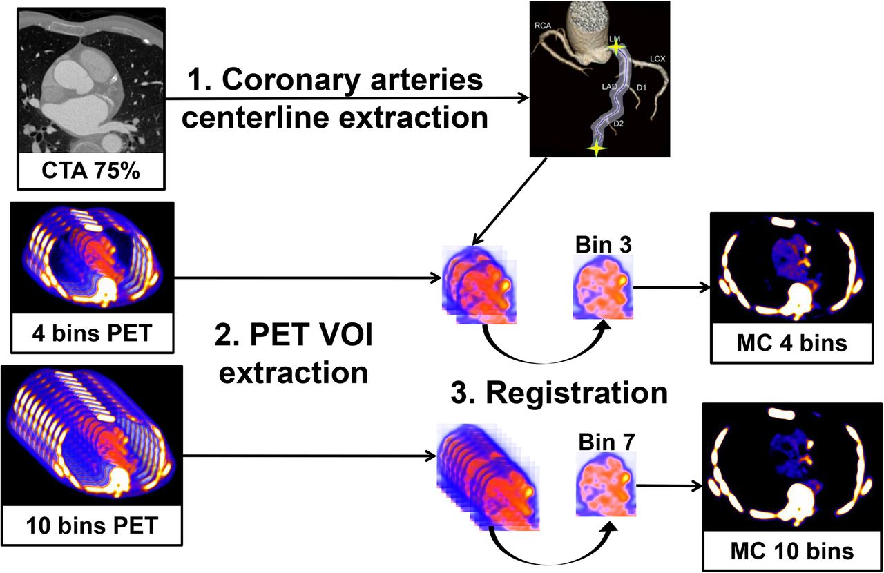
- FIGURE 1.
Overview of the motion correction method. (1) Coronary artery centerlines are extracted from CCTA in end-diastolic phase using CCTA analysis software. (2) Volumes of interest surrounding coronary arteries are extracted from 4- and 10-bin PET data using previously extracted CCTA centerlines. (3) All bins of data are registered to the common end-diastolic reference bin (bins 3 and 7 for 4- and 10-bin data, respectively) by nonlinear level-set registration restricted to coronary regions. Then, registered VOIs are inserted back into their original PET volumes, and all registered PET images are summed into a single volume to obtain motion-corrected 4- and 10-bin data. MC = motion-corrected; VOI = volume of interest.
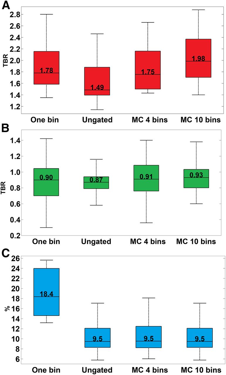
- FIGURE 2.
TBRs in positive (A) and negative (B) lesions and noise (C) are shown for different PET datasets (1-bin data, ungated data, motion-corrected data created from 4 bins, and motion-corrected data created from 10 bins). (A) TBR in positive lesions increases with motion correction for 10-bin data, as compared with 1-bin data and ungated data. (B) TBR in negative lesions remains below 1 using motion correction. (C) Noise is almost halved using motion correction, as compared with 1-bin data. MC = motion-corrected.
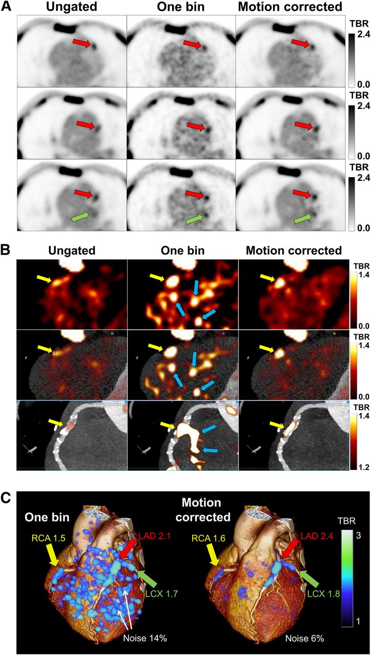
- FIGURE 3.
Noise decrease and TBR improvement in 18F-NaF PET images of 65-y-old man. (A) On linear grayscale transaxial slices, 18F-NaF plaque uptake is seen in left anterior descending (LAD) (red arrows) and left circumflex (LCX) (green arrows) coronary arteries. The images show blurred lesion signal in ungated images, significant noise in 1-bin images, and high lesion signal with reduced noise in motion-corrected images. (B) On PET (top), PET/CCTA (middle), and multiplanar-reformatted PET/CCTA (bottom) images with exponential color table and same window and level settings, vulnerable plaque is seen in right coronary artery (RCA) (yellow arrows). Low lesion signal is seen in ungated images, significant noise in 1-bin images (blue arrows), and high signal with less noise in motion-corrected images. (C) 3-dimensional rendering of 1-bin image (25% of PET counts) as in study of Joshi et al. (2) (left) and motion-corrected image (right) superimposed on rendered CCTA volume. Increased uptake is seen in RCA, LAD, and LCX coronary arteries in high-noise 1-bin image and remains clear in motion-corrected image.

Tables
Parameter Stable angina (n = 10) MI (n = 7) Mean age ± SD (y) 67 ± 9 66 ± 7 Men (n) 9 (90%) 7 (100%) Mean BMI ± SD (kg/m2) 29 ± 5 28 ± 8 Agatston score Median 1,010 498 IQR 560–2,175 355–771 History (n) MI 4 (40%) 0 PCI 5 (50%) 1 (14%) CABG 5 (50%) 0 Lesions (n) 31 (61%) 20 (39%) Positive 19 (68%) 9 (32%) Negative 12 (52%) 11 (48%) BMI = body mass index; MI = myocardial infarction; PCI = percutaneous coronary intervention; CABG = coronary artery bypass graft.
Parameter 18F-NaF CCTA dose AC CT dose PET/CT + CCTA Prospective Range 2.8–3.1 1.3–5.8 0.5–1.8 4.7–9.9 Mean ± SD 2.9 ± 0.1 3.4 ± 1.2 1.0 ± 0.4 7.3 ± 1.4 Retrospective Range 2.8–3.0 7.8–12.7 0.9–1.1 11.6–16.7 Mean ± SD 2.9 ± 0.1 9.7 ± 2.7 1.0 ± 0.1 13.6 ± 2.7 Conversion factors are 0.014 mSv/mGy⋅cm for CCTA and attenuation-corrected (AC) CT and 0.024 mSv/MBq for 18F-NaF.


Jump to section
Related Articles
Cited By...
- Machine Learning with 18F-Sodium Fluoride PET and Quantitative Plaque Analysis on CT Angiography for the Future Risk of Myocardial Infarction
- The Changing Face of Nuclear Cardiology: Guiding Cardiovascular Care Toward Molecular Medicine
- Coronary 18F-Sodium Fluoride Uptake Predicts Outcomes in Patients With Coronary Artery Disease
- In vivo alpha-V beta-3 integrin expression in human aortic atherosclerosis
- Peri-Coronary Adipose Tissue Density Is Associated With 18F-Sodium Fluoride Coronary Uptake in Stable Patients With High-Risk Plaques
- Data-Driven Gross Patient Motion Detection and Compensation: Implications for Coronary 18F-NaF PET Imaging
- Three-Hour Delayed Imaging Improves Assessment of Coronary 18F-Sodium Fluoride PET
- Motion-Corrected Imaging of the Aortic Valve with 18F-NaF PET/CT and PET/MRI: A Feasibility Study
- MR/PET Imaging of the Cardiovascular System
- Coronary Artery PET/MR Imaging: Feasibility, Limitations, and Solutions
- Detection of Atherosclerotic Inflammation by 68Ga-DOTATATE PET Compared to [18F]FDG PET Imaging
- Cardiac {alpha}V{beta}3 integrin expression following acute myocardial infarction in humans
- Molecular Imaging of Vulnerable Coronary Plaque: A Pathophysiologic Perspective
- Optimization and Reproducibility of Aortic Valve 18F-Fluoride Positron Emission Tomography in Patients With Aortic Stenosis
- Imaging Atherosclerosis