


Article Figures & Data
Figures
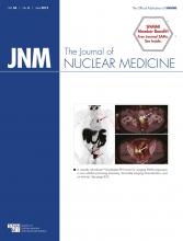
- FIGURE 1.
Sequential scan of 70-y-old patient (P3; PSA level, 101.2 ng/mL) with initial diagnosis of prostate cancer (dotted arrow) showing high tumor-to-background ratio. Maximum-intensity projections (upper row) and axial slices (middle and lower rows) at different time points are displayed ([A] early rapid scan, [B] 1-h scan, [C] 2-h scan, [D] 4-h scan). Primary prostate cancer (middle row; 1-h SUVmax, 55.0) as well as numerous iliacal (lower row; 1-h SUVmax, 57.0) and mediastinal lymph node metastases (upper row; 1-h SUVmax, 31.4) can be depicted immediately after injection and up to 4 h.
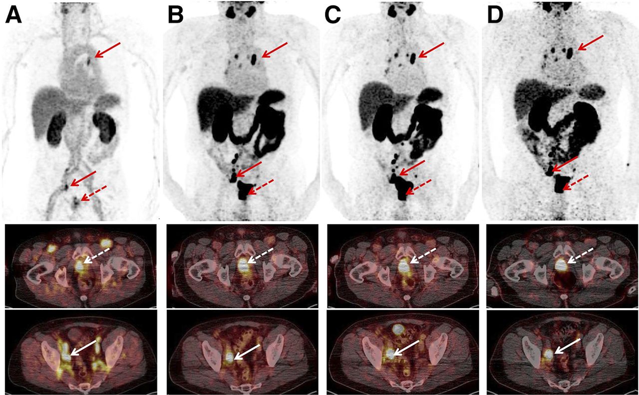
- FIGURE 2.
Sequential patient scan (P1) of 67-y-old patient with biochemical relapse (PSA level, 6.7 ng/mL) 1.8 y after curative radiotherapy. Maximum-intensity projections (upper row) and axial slices (middle and lower row) of early rapid scan (A) and scans after 1 h (B), 2 h (C), and 4 h (D) show increased uptake in rib metastasis (A–D, arrows) and physiologic uptake in lacrimal glands, salivary glands, liver, spleen, kidneys, and slightly in bowel (A–D). Red dotted arrow in A depicts unspecific uptake in left subclavian vein.
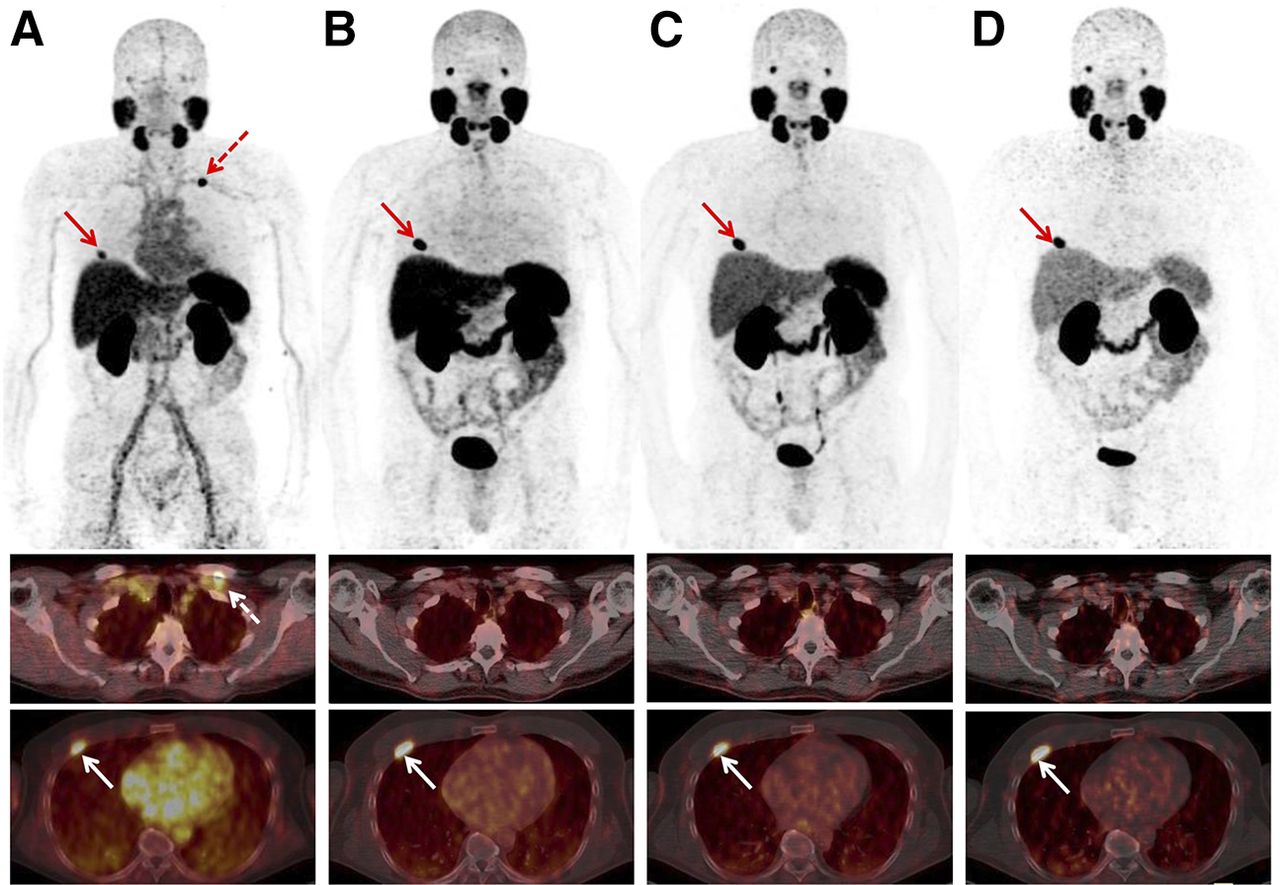
- FIGURE 3.
Time–activity curves for P1 for all organs showing uptake, for whole body, and for blood. For blood, percentage of activity is given per liter of blood.
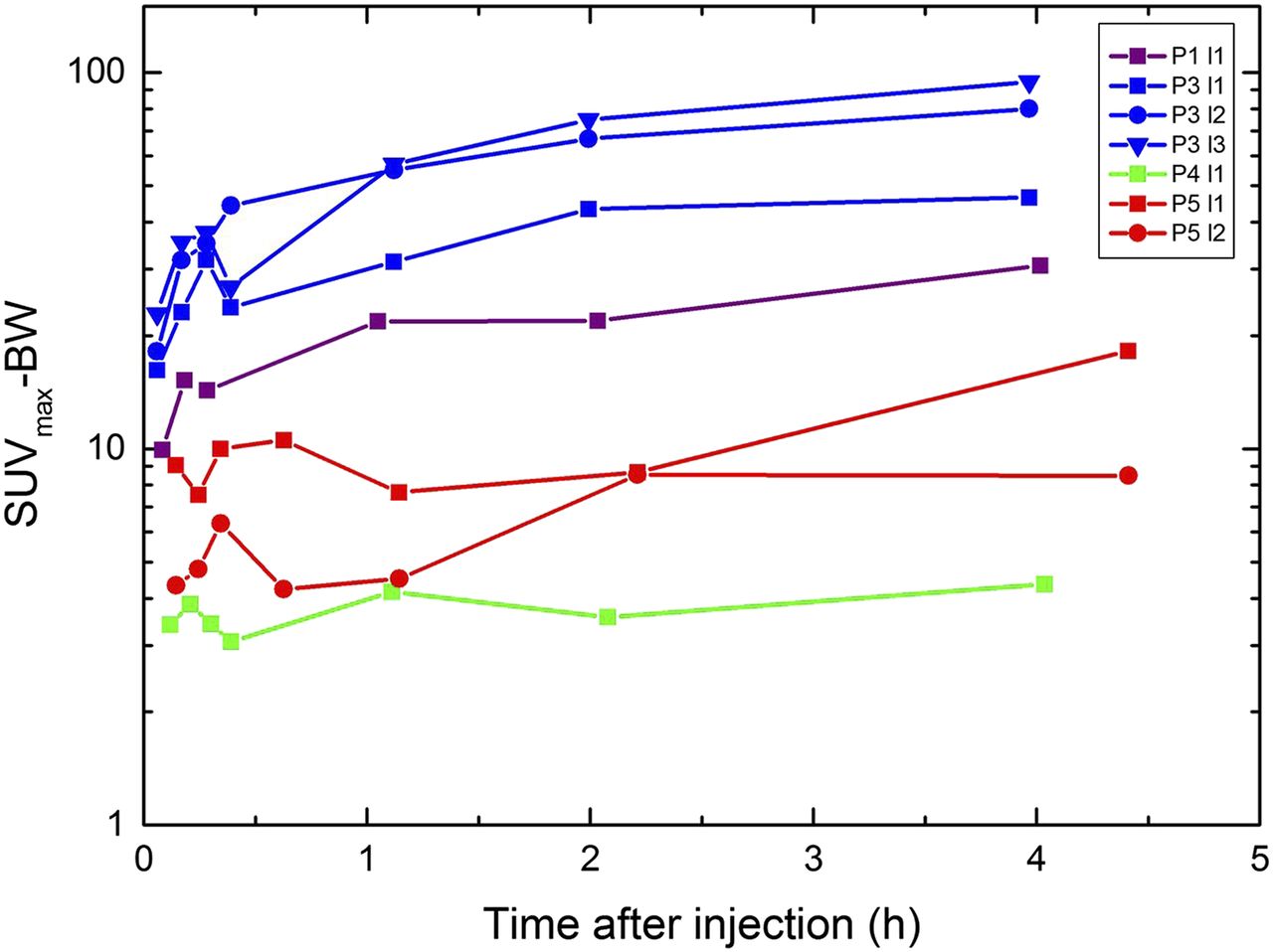
- FIGURE 4.
Temporal variation of SUVmax–body weight in visible lesions in P1 (red), P3 (light blue), P4 (green), and P5 (blue). P1-l1 = bone lesion; P3-l1 = lymph node mediastinal; P3-l2 = tumor tissue prostate; P3-l3 = lymph node iliacal; P4-l4 = lymph node; P5-l1 = tumor tissue prostate; P5-l2 = bone lesion.


Tables
Patient no. Age (y) Height (cm) Weight (kg) Activity (MBq) PD PET indication PSA level (ng/mL) Gleason Score Previous treatment PET findings 1 66.8 170 74 148 09/12 Relapse 6.7 7 RTx B 2 66.3 178 95 124 01/13 Relapse 0.5 8 RPT None 3 69.9 172 81 146 07/14 Staging 101.2 7 — Loc, B, LNs 4 59.1 185 110 133 07/11 Relapse 1.6 8 RPT and RTx LNs 5 63.5 177 70 91 07/14 Follow-up 1.4 8 ADT B, Loc PD = primary diagnosis; RTx = radiotherapy; B = bone metastasis; RPT = radical prostatectomy; Loc = tumor tissue in the prostate; LN = lymph node metastasis; ADT = androgen-deprivation therapy.
Time-integrated activity coefficients (h) Source organ P1 P2 P3 P4 P5 Mean SD Remainder 1.023 1.029 0.952 1.098 0.964 1.013 0.059 Liver 0.181 0.141 0.100 0.142 0.173 0.147 0.032 Right kidney 0.080 0.068 0.075 0.078 0.038 0.068 0.018 Left kidney 0.084 0.076 0.087 0.073 0.042 0.073 0.018 Kidneys, sum 0.164 0.144 0.163 0.151 0.080 0.140 0.035 Heart 0.021 0.018 0.015 0.024 0.017 0.019 0.003 Bladder contents 0.042 0.029 0.055 0.039 0.088 0.051 0.023 Gallbladder 0.002 0.001 0.001 0.002 0.002 0.002 0.001 Spleen 0.042 0.010 0.011 0.019 0.032 0.023 0.014 Parotid glands 0.010 0.004 0.005 0.006 0.003 Submandibular glands 0.005 0.003 0.004 0.002 0.003 0.004 0.001 LV2–4 0.002 0.001 0.001 0.001 0.001 0.001 0.000 Red marrow lumbar vertebrae 0.027 0.013 0.009 0.018 0.016 0.017 0.007 Red marrow blood 0.028 0.026 0.019 0.024 0.022 0.024 0.003 Calculation methods for red marrow lumbar vertebrae and red marrow blood are presented in “Material and Methods” section.
P = patient; LV2–4 = lumbar vertebrae 2–4.
Target organ β (mGy/MBq) Photon (mGy/MBq) Total (mGy/MBq) Absorbed dose (mGy) (150 MBq) Adrenals 5.87E–03 7.62E–03 1.35E–02 2.0 Brain 5.87E–03 2.43E–03 8.29E–03 1.2 Breasts 5.87E–03 2.58E–03 8.44E–03 1.3 Gallbladder wall 1.16E–02 7.99E–03 1.96E–02 2.9 Lower large intestine wall 5.87E–03 4.68E–03 1.06E–02 1.6 Small intestine 5.87E–03 5.24E–03 1.11E–02 1.7 Stomach wall 5.87E–03 5.24E–03 1.11E–02 1.7 Upper large intestine wall 5.87E–03 5.25E–03 1.11E–02 1.7 Heart wall 1.48E–02 5.36E–03 2.02E–02 3.0 Kidneys 1.96E–01 2.31E–02 2.20E–01 33.0 Liver 3.30E–02 1.01E–02 4.31E–02 6.5 Lungs 5.87E–03 3.91E–03 9.78E–03 1.5 Muscle 5.87E–03 3.65E–03 9.52E–03 1.4 Ovaries 5.87E–03 4.94E–03 1.08E–02 1.6 Pancreas 5.87E–03 7.38E–03 1.32E–02 2.0 Red marrow 8.07E–03 4.35E–03 1.24E–02 1.9 Osteogenic cells 1.16E–02 4.04E–03 1.57E–02 2.4 Skin 5.87E–03 2.28E–03 8.15E–03 1.2 Spleen 5.26E–02 1.09E–02 6.34E–02 9.5 Testes 5.87E–03 3.28E–03 9.14E–03 1.4 Thymus 5.87E–03 3.62E–03 9.49E–03 1.4 Thyroid 5.87E–03 3.15E–03 9.01E–03 1.4 Urinary bladder wall 5.70E–02 1.03E–02 6.74E–02 10.1 Uterus 5.87E–03 5.72E–03 1.16E–02 1.7 Salivary glands 6.07E–02 — 6.07E–02 9.1 Total body 7.88E–03 3.85E–03 1.17E–02 1.8 Effective dose coefficient (mSv/MBq) 1.99E–02 Effective dose coefficient (mSv/MBq)* 1.93E–02 ± 0.09E–02 Effective dose (mSv) 3.0 ↵* Mean effective dose coefficient of P1–P5.
- TABLE 4
Comparison of Absorbed Dose Coefficients and Absorbed Doses for Several Prostate-Specific Compounds
124I-PSMA 123I-MIP-1072 123I-MIP-1095 Pentixafor DOTATOC DOTATATE 18F-FDG PSMA-IT Target organ Unit Zechmann (33) Barrett (36) Barrett (36) Herrmann (25) Sandstrom (34) Sandstrom (34) ICRP 106 (35) This work Kidneys mSv/MBq 1.39E+00 5.4E–02 1.10E–2 3.50E–02 8.20E–02 9.30E–02 1.70E–02 2.20E–01 Liver mSv/MBq 1.66E–00 2.4E–02 5.8E–2 1.75E–02 4.10E–02 5.00E–02 2.10E–02 4.31E–02 Spleen mSv/MBq 7.7E–01 2.3E–2 4.7E–2 5.38E–02 1.08E–01 1.09E–01 1.10E–02 6.34E–02 Urinary bladder wall mSv/MBq 5.7E–01 9.2E–2 2.1E–2 8.14E–02 1.19E–01 9.80E–02 1.30E–01 6.74E–02 Effective dose coefficient mSv/MBq 5.8E–01 2.5E–2 3.2E–2 1.56E–02 2.10E–02 2.10E–02 1.90E–02 1.99E–02 Typical injected activity MBq 67 370 370 150 185 150 370 150 Effective dose mSv 38.9 9.3 11.8 2.3 3.9 3.2 7.0 3.0


Jump to section
Related Articles
Cited By...
- Delayed Imaging Improves Lesion Detectability in [99mTc]Tc-PSMA-I&S SPECT/CT in Recurrent Prostate Cancer
- Matched-Pair Comparison of 18F-DCFPyL PET/CT and 18F-PSMA-1007 PET/CT in 240 Prostate Cancer Patients: Interreader Agreement and Lesion Detection Rate of Suspected Lesions
- Diagnostic Performance of 18F-DCFPyL-PET/CT in Men with Biochemically Recurrent Prostate Cancer: Results from the CONDOR Phase III, Multicenter Study
- Therapeutic Efficacy of a Bivalent Inhibitor of Prostate-Specific Membrane Antigen Labeled with 67Cu
- Current and Emerging Clinical Applications of PSMA PET Diagnostic Imaging for Prostate Cancer
- Quantitative and Qualitative Analyses of Biodistribution and PET Image Quality of a Novel Radiohybrid PSMA, 18F-rhPSMA-7, in Patients with Prostate Cancer
- Impact of 18F-PSMA-1007 Uptake in Prostate Cancer Using Different Peptide Concentrations: Preclinical PET/CT Study on Mice
- First Clinicopathologic Evidence of a Non-PSMA-Related Uptake Mechanism for 68Ga-PSMA-11 in Salivary Glands
- Phase I Study of CTT1057, an 18F-Labeled Imaging Agent with Phosphoramidate Core Targeting Prostate-Specific Membrane Antigen in Prostate Cancer
- Initial Experience with Volumetric 68Ga-PSMA I&T PET/CT for Assessment of Whole-Body Tumor Burden as a Quantitative Imaging Biomarker in Patients with Prostate Cancer
- Seduction by Sensitivity: Reality, Illusion, or Delusion? The Challenge of Assessing Outcomes after PSMA Imaging Selection of Patients for Treatment
- 18F-DCFPyL PET/CT in the Detection of Prostate Cancer at 60 and 120 Minutes: Detection Rate, Image Quality, Activity Kinetics, and Biodistribution
- Glu-Ureido-Based Inhibitors of Prostate-Specific Membrane Antigen: Lessons Learned During the Development of a Novel Class of Low-Molecular-Weight Theranostic Radiotracers
- The Clinical Impact of Additional Late PET/CT Imaging with 68Ga-PSMA-11 (HBED-CC) in the Diagnosis of Prostate Cancer
- Safety, Dosimetry, and Tumor Detection Ability of 68Ga-NOTA-AE105: First-in-Human Study of a Novel Radioligand for uPAR PET Imaging
- 68Ga-PSMA Ligand PET/CT-based Radiotherapy for Lymph Node Relapse of Prostate Cancer After Primary Therapy Delays Initiation of Systemic Therapy
- Evaluation of 68Ga-Glutamate Carboxypeptidase II Ligand Positron Emission Tomography for Clinical Molecular Imaging of Atherosclerotic Plaque Neovascularization
- The Rise of PSMA Ligands for Diagnosis and Therapy of Prostate Cancer
- 177Lu-Labeled Prostate-Specific Membrane Antigen Radioligand Therapy of Metastatic Castration-Resistant Prostate Cancer: Safety and Efficacy
- New Strategies in Prostate Cancer: Prostate-Specific Membrane Antigen (PSMA) Ligands for Diagnosis and Therapy
- The Theranostic PSMA Ligand PSMA-617 in the Diagnosis of Prostate Cancer by PET/CT: Biodistribution in Humans, Radiation Dosimetry, and First Evaluation of Tumor Lesions