


Article Figures & Data
Figures
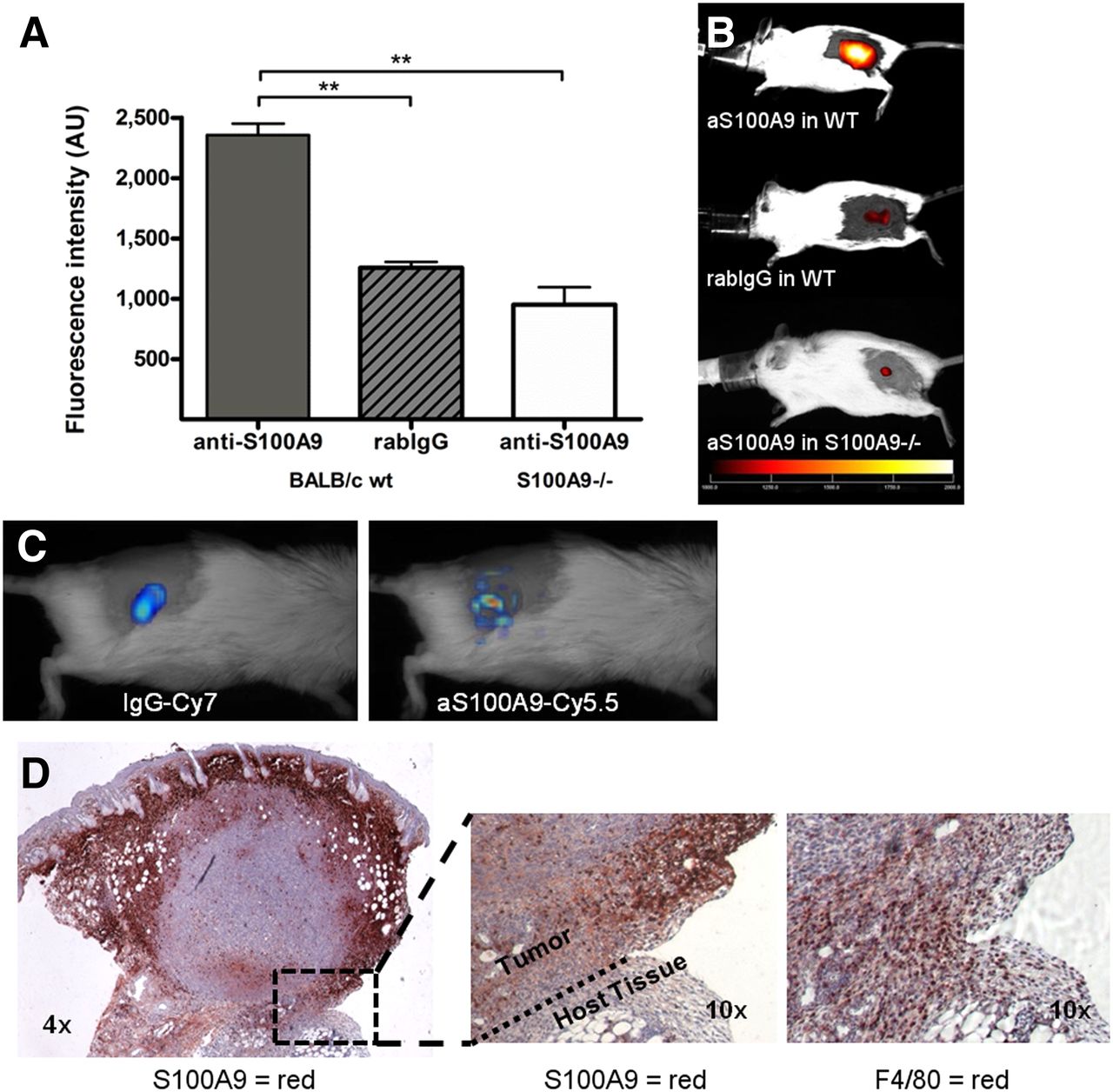
- FIGURE 1.
(A and B) S100A9 imaging specifically identifies tumor-associated immune cells. aS100A9-Cy5.5 injection in 4T1 tumor–bearing wild-type (wt) animals results in significantly higher specific fluorescence than unspecific rabIgG-Cy5.5 or injection of specific aS100A9-Cy5.5 in S100A9−/− knock-out mice. (C) FMT after parallel injection of rabIgG-Cy7 and aS100A9-Cy5.5 showed homogeneous distribution of rabIgG-Cy7 in tumor region, reflecting perfusion, whereas aS100A9-Cy5.5 accumulated in delineated regions only. (D) Histology confirmed S100A9+ cells (red) in corresponding, peripheral areas of tumor (left) with F4/80+ TAM detectable within clusters of S100A8/A9+ active monocytes (right).
- FIGURE 2.
Correlation between tumor growth and S100A9 fluorescence intensity. S100A9 imaging at early time points during tumor development (lesion size < 5 mm; A) shows only low fluorescence signals in animals with subsequently only moderate tumor growth (left). Ex vivo, only few S100A9+ cells (red/brown) could be found in tumor specimens of these animals (B, left). High initial S100A9 signal (A, right) predicted more rapid tumor growth and reflected pronounced infiltration of tumor by S100A9+ cells (B, right). Developmental variation among 4T1 tumor lesions could be predicted by S100A9 imaging, correlating relative growth rate over 10 d, after imaging (C; r2 = 0.86; P < 0.0001; n = 11).
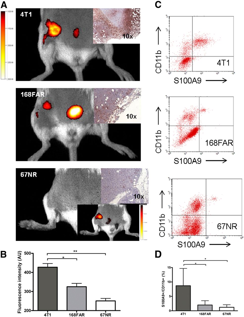
- FIGURE 3.
S100A9 imaging reflects metastatic capability. S100A9 in vivo imaging reveals significant differences between tumor entities of different metastatic capabilities (exemplary images in A; corresponding S100A9 histology; data in C). In vivo signal in 67NR tumors was visible only after threshold was reduced (insert). Ex vivo analysis of cellular tumor infiltrate revealed CD11b+ S100A9+ cells in all 3 tumors in amounts, confirming in vivo imaging results (B and D). Individual isotype controls and resulting gating are elaborated in Supplemental Figure 2.
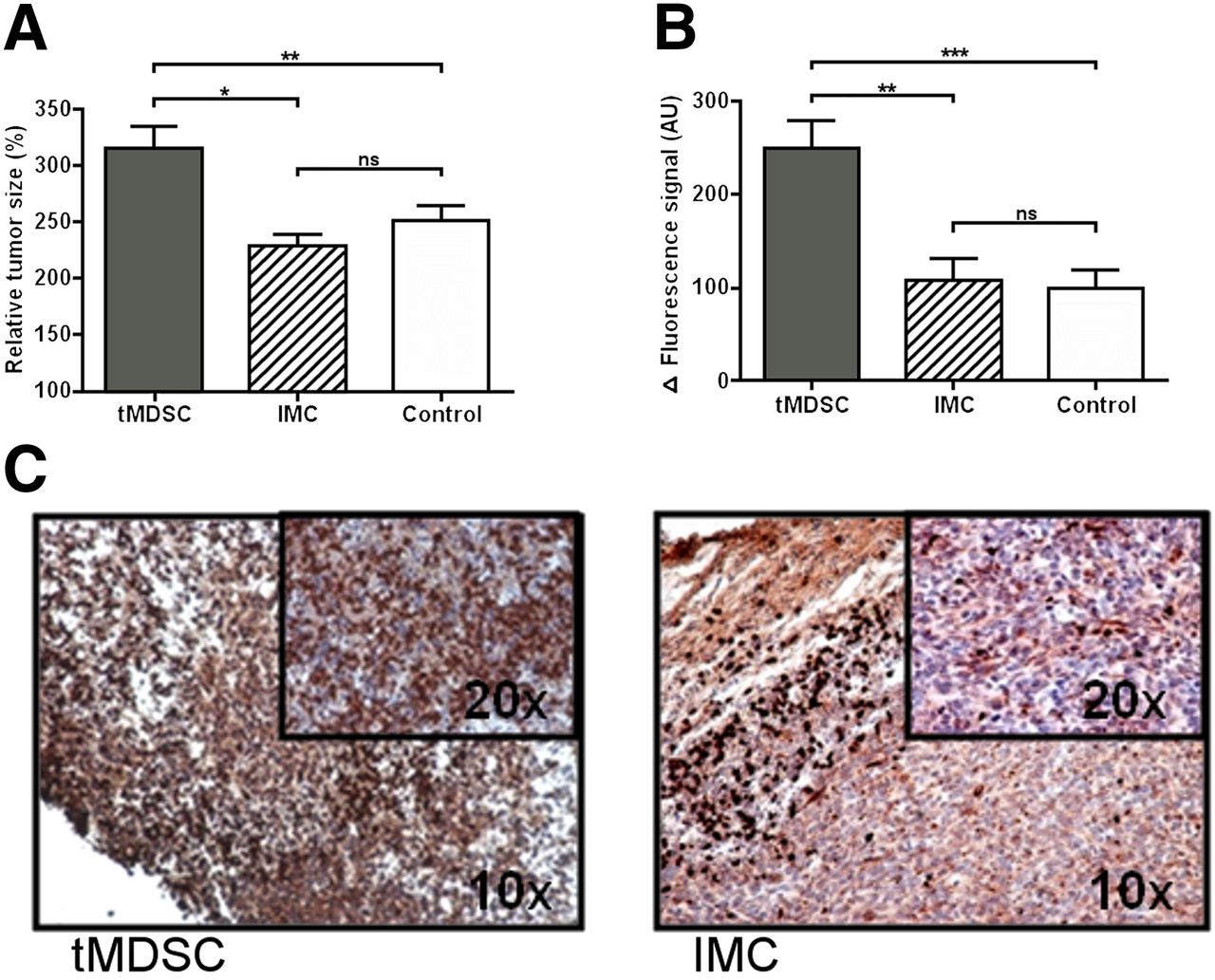
- FIGURE 4.
(A) tMDSC promote tumor growth. Splenic monocytes (1 × 10⁶) from either tumor-bearing (tMDSC) or healthy animals (IMC) were injected intravenously into mice, inoculated with 4T1 (scheme in Supplemental Fig. 3). Accelerated growth resulted in relatively increased tumor size (% of size compared with d1) in tMDSC-treated animals. (B) S100A9-specific imaging revealed significantly higher immune cell activity in tMDSC-treated tumors as compared with controls. (C) Immunohistochemistry for S100A9 confirmed increased infiltration of S100A9+ cells into tMDSC-treated tumors as compared with IMC-treated controls.

Tables
Experiment Mouse strain Tumor type n Tracer Purpose Fig. I BALB/c wt 4T1 28 1, 2 Proof of principle 1 II S100A9−/− 4T1 5 1 Proof of specificity 1 III BALB/c wt 4T1 6 1, 3 Parallel injection (tracer 2 labeled with Cy7) 1 IV BALB/c wt 4T1 11 1 Correlation signal/growth 2 V BALB/c wt 4T1; 67NR, 168FAR 32 1 Correlation signal/malignancy 3 VI BALB/c wt 4T1 20 1 Cell transfer 4 wt = wild-type; 1 = tracer aS100A9-Cy5.5; 2 = tracer rabIgG-Cy5.5; 3 = tracer rabIgG-Cy7.
Characteristic 4T1 168FAR 67NR Shed cells ++ + — Invasion + — — Regional lymph node — + — Solid metastasis ++ — — ++ = very strong; + = detectable; — = not detectable.
TC supernatant TC cell lysates Resected tumor lysates Parameter 4T1 168FAR 67NR 4T1 168FAR 67NR 4T1 168FAR 67NR S100A8/A9 (ng/mL) 0 0 0 0 0 0 86,897 4,665 1,927 SD Not applicable Not applicable Not applicable Not applicable Not applicable Not applicable 50,044 3,513 1,348
Supplemental Data
Files in this Data Supplement:


