


Article Figures & Data
Figures
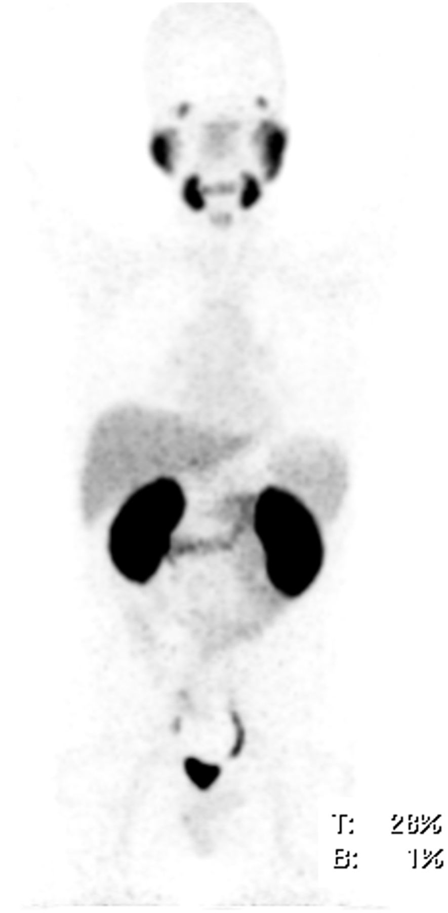
- FIGURE 1.
Maximum-intensity projections of patient 4 with normal distribution of 68Ga-PSMA-617 at 3 h after injection. Physiologic accumulation is seen in lacrimal and salivary glands, nasal mucosa, liver, spleen, bowels, and kidneys. Surplus tracer is excreted via urinary tract and urinary bladder. No pathologic tracer uptake was found.
- FIGURE 2.
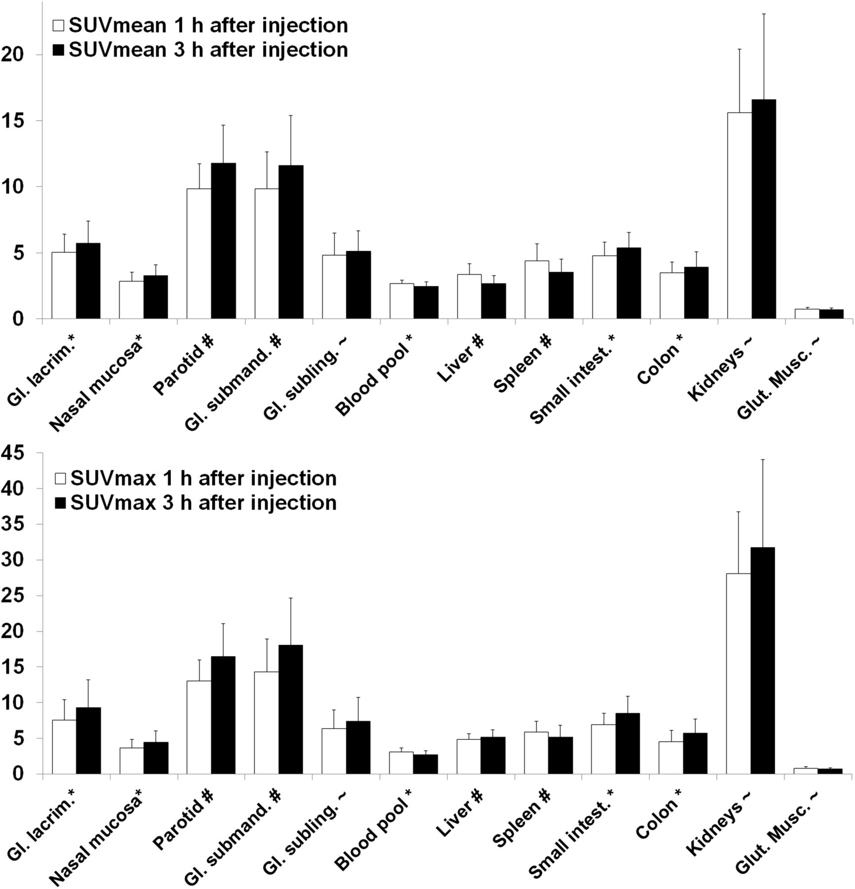
Average SUVmean and SUVmax in different organs 1 and 3 h after injection. Significance of differences between 1 and 3 h (t test): #P < 0.001; *P < 0.05; ∼P > 0.05. Gl. = glandular; Glut. Musc. = gluteal musculature.
- FIGURE 3.
(A-C) 68Ga-PSMA-617 PET/CT of patient 11 at 1 h after injection. Red arrows point to a bone metastasis with SUVmax of 21.7 at 1 h and 32.6 at 3 h after injection. (A) Low dose CT. (B) Fusion of PET and CT. (C) MIP of PET/CT. MIP = maximum-intensity projection.
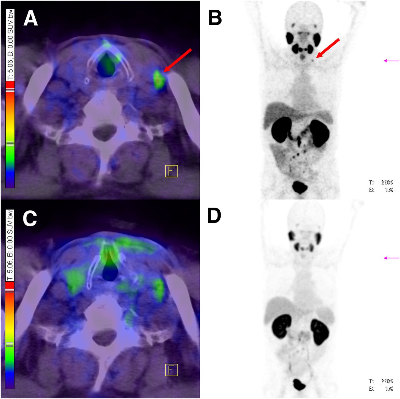
- FIGURE 4.
68Ga-PSMA-617-PET/CT of patient 3 at 1 h (C and D) and 3 h after injection (A and B). Red arrows point to LN (Virchow’s node), which was identified as metastasis in images at 3 h after injection only. In images at 1 h after injection, tracer accumulation was rated as artifact. In this patient, also 4 other LNs (retroperitoneal) were identified as metastases in images at 3 h after injection only. (A) Fusion of PET and CT 3 h after injection. (B) Maximum-intensity projection of PET/CT 3 h after injection. (C) Fusion of PET and CT 1 h after injection. (D) Maximum-intensity projection of PET/CT 1 h after injection.
- FIGURE 5.
Average SUVmax in 53 representative PCa lesions and their ratio to background (contrast) 1 and 3 h after injection. Five of overall 8 lesions (lesions 11, 12, 14, 17, and 18) in patient 3 and 1 of overall 2 lesions (lesion 31) patient 16 were not visible in images at 1 h after injection because of low contrast.
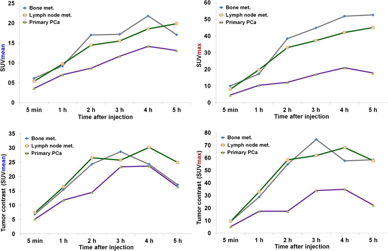
- FIGURE 6.
Development of SUVs and tumor contrast over time in all bone metastases (n = 7), LN metastases (n = 15), and primary tumors (n = 2) in patients 16–19. met. = metastases.
- FIGURE 7.
(Upper) Maximum-intensity projections of sequential PET/CT scan of patient 18 at different times after injection of 213 MBq of 68Ga-PSMA-617. Blue and red arrows point to 2 primary PCa lesions. (Lower) SUVmax of the 2 primary PCa lesions at the different times. p.i. = after injection.


Tables
Patient no. Age (y) 68Ga-PSMA-617 (MBq) Gleason score PSA (ng/mL) Previous treatment LN metastases Bone metastases Local relapse Soft-tissue metastases Primary tumor 1* 66 237 7 0.40 RPx + RT 0 0 0 0 0 2 55 295 8 6.40 RPx >10 0 0 0 0 3 73 286 7 10.30 RPx 8 0 0 0 0 4* 49 255 7 0.10 RPx + RT 0 0 0 0 0 5 69 262 5 1.30 RPx + RT 0 0 1 0 0 6 71 178 7 4.90 RPx 3 0 0 0 0 7* 71 68 7 1.30 RPx + RT 0 0 0 0 0 8 47 138 6 2.81 RPx 2 0 0 0 0 9* 64 238 8 0.40 RPx 0 0 0 0 0 10 60 194 7 2.90 RPx + RT + ADT 0 0 1 0 0 11 69 177 8 5.16 RPx + RT + ADT 0 1 0 0 0 12 60 151 6 24.30 Biopsy 0 0 0 0 1 13* 70 149 7 0.22 RPx 0 0 0 0 0 14 75 164 7 1.40 RPx 0 0 1 0 0 15 59 132 7 1.30 RPx + RT + ADT 1 0 0 0 0 Dosimetry 16 67 260 7 6.60 RPx + RT 2 0 0 0 0 17 75 230 9 14.91 RPx + ADT >10 0 0 0 0 18 61 213 NA 13.70 ADT 0 0 0 0 2 19 60 221 NA 2.86 RT >10 >10 0 0 0 ↵* Patients without pathologic tracer uptake in 68Ga-PSMA-617 PET/CT (n = 5).
RPx = radical prostatectomy; RT = radiation therapy; ADT = androgen-deprivation therapy; NA = not available.
68Ga-PSMA-617 PET/CT at 1 h after injection 68Ga-PSMA-617 PET/CT at 3 h after injection Different types of PCa SUV Range Median SUV Range Median LN metastases (n = 39) SUVmean 7.1 ± 4.3 1.4–22.0 6.4 SUVmean 9.9 ± 2.4 2.5–34.1 7.7 Bone metastases (n = 8) SUVmean 9.4 ± 7.5 3.3–25.6 5.9 SUVmean 16.9 ± 12.5 4.5–44.9 12.5 Local relapses (n = 3) SUVmean 6.5 ± 0.9 5.9–7.5 6.2 SUVmean 7.7 ± 0.6 7.2–8.3 7.7 Primary tumors (n = 3) SUVmean 7.1 ± 0.4 6.8–7.3 7.1 SUVmean 11.7 ± 1.1 10.9–12.5 11.7 LN metastases (n = 39) SUVmax 12.3 ± 10.3 1.4–50.4 9.3 SUVmax 20.4 ± 22.5 2.5–84.4 12.3 Bone metastases (n = 8) SUVmax 17.8 ± 12.0 4.7–43.0 13.9 SUVmax 43.3 ± 24.8 12.1–92.6 38.6 Local relapses (n = 3) SUVmax 11.1 ± 2.1 9.6–13.5 10.1 SUVmax 16.3 ± 0.9 15.4–17.1 16.4 Primary tumors (n = 3) SUVmax 8.6 ± 3.4 4.8–11.4 9.5 SUVmax 12.7 ± 7.5 4.1–18.2 15.8 Data are SUVmean or SUVmax ± SD.
Residence time (Bq × h/Bq) for patient Source organ 16 17 18 19 Liver 0.110 0.068 0.102 0.090 Spleen 0.014 0.006 0.003 0.013 Kidneys 0.153 0.053 0.196 0.125 Urinary bladder 0.076 0.095 0.045 0.059 Small intestine 0.011 0.002 0.018 0.004 Colon 0.042 0.033 0.052 0.004 Remainder 1.190 1.320 1.190 1.290 Absorbed organ dose (mGy/MBq) for patient Target organ 16 17 18 19 Adrenals 0.015 0.014 0.015 0.015 Brain 0.010 0.011 0.010 0.011 Breasts 0.010 0.011 0.010 0.010 Gallbladder 0.015 0.014 0.016 0.015 Lower colon 0.013 0.014 0.013 0.013 Small intestine 0.019 0.015 0.024 0.015 Stomach 0.013 0.013 0.013 0.013 Upper colon 0.053 0.045 0.064 0.017 Heart 0.012 0.012 0.012 0.012 Kidneys 0.239 0.085 0.305 0.196 Liver 0.033 0.022 0.032 0.028 Lungs 0.011 0.012 0.011 0.012 Muscle 0.011 0.012 0.011 0.012 Pancreas 0.014 0.014 0.015 0.015 Red marrow 0.010 0.010 0.010 0.010 Osteogenic cells 0.015 0.017 0.015 0.016 Skin 0.009 0.010 0.009 0.010 Spleen 0.040 0.020 0.015 0.039 Testes 0.011 0.012 0.011 0.012 Thymus 0.011 0.012 0.011 0.012 Thyroid 0.011 0.012 0.011 0.011 Urinary bladder 0.098 0.121 0.062 0.080 Total body 0.013 0.013 0.013 0.013 Effective dose (mSv/MBq) 0.023 0.018 0.022 0.020
Supplemental Data
Files in this Data Supplement:


Jump to section
Related Articles
Cited By...
- Renal and Multiorgan Safety of 177Lu-PSMA-617 in Patients with Metastatic Castration-Resistant Prostate Cancer in the VISION Dosimetry Substudy
- The Hierarchy of SUVs: From Diagnostics to Therapeutics and the Pathway to Effective Theranostics
- Evaluation of 134Ce/134La as a PET Imaging Theranostic Pair for 225Ac {alpha}-Radiotherapeutics
- Comparison of 68Ga-PSMA-617 PET/CT and 68Ga-RM2 PET/CT in Patients with Localized Prostate Cancer Who Are Candidates for Radical Prostatectomy: A Prospective, Single-Arm, Single-Center, Phase II Study
- From Concept to Regulatory Drug Approval: Lessons for Theranostics
- Antitumor efficacy of 90Y-NM600 targeted radionuclide therapy and PD-1 blockade is limited by regulatory T cells in murine prostate tumors
- Bringing VISION to Nuclear Medicine: Accelerating Evidence and Changing Paradigms with Theranostics
- Matched-Pair Comparison of 18F-DCFPyL PET/CT and 18F-PSMA-1007 PET/CT in 240 Prostate Cancer Patients: Interreader Agreement and Lesion Detection Rate of Suspected Lesions
- Diagnostic Performance of 18F-DCFPyL-PET/CT in Men with Biochemically Recurrent Prostate Cancer: Results from the CONDOR Phase III, Multicenter Study
- Radiolabeling of PSMA-617 with 89Zr: A Novel Use of DMSO for Radiochemical Yield Enhancement and Preliminary Small-Animal PET Results
- Current and Emerging Clinical Applications of PSMA PET Diagnostic Imaging for Prostate Cancer
- Harnessing 64Cu/67Cu for a theranostic approach to pretargeted radioimmunotherapy
- 68Ga-PSMA PET/CT Combined with PET/Ultrasound-Guided Prostate Biopsy Can Diagnose Clinically Significant Prostate Cancer in Men with Previous Negative Biopsy Results
- Quantitative and Qualitative Analyses of Biodistribution and PET Image Quality of a Novel Radiohybrid PSMA, 18F-rhPSMA-7, in Patients with Prostate Cancer
- Theranostics: Leveraging Molecular Imaging and Therapy to Impact Patient Management and Secure the Future of Nuclear Medicine
- Prospective Comparison of PET Imaging with PSMA-Targeted 18F-DCFPyL Versus Na18F for Bone Lesion Detection in Patients with Metastatic Prostate Cancer
- Development of Novel PSMA Ligands for Imaging and Therapy with Copper Isotopes
- Radiation Dosimetry and Biodistribution of 18F-PSMA-11 for PET Imaging of Prostate Cancer
- Is the Vision of Radioligand Therapy for Prostate Cancer Becoming a Reality? An Overview of the Phase III VISION Trial and Its Importance for the Future of Theranostics
- Effect of External Cooling on 177Lu-PSMA Uptake by the Parotid Glands
- Rapid Modulation of PSMA Expression by Androgen Deprivation: Serial 68Ga-PSMA-11 PET in Men with Hormone-Sensitive and Castrate-Resistant Prostate Cancer Commencing Androgen Blockade
- Albumin-Binding PSMA Ligands: Implications for Expanding the Therapeutic Window
- PET-CT Cancer Imaging
- Why Targeting PSMA Is a Game Changer in the Management of Prostate Cancer
- 18F-DCFPyL PET/CT in the Detection of Prostate Cancer at 60 and 120 Minutes: Detection Rate, Image Quality, Activity Kinetics, and Biodistribution
- PSMA Ligands for PET Imaging of Prostate Cancer
- Dual-Target Binding Ligands with Modulated Pharmacokinetics for Endoradiotherapy of Prostate Cancer
- Prostate-Specific Membrane Antigen Ligands for Imaging and Therapy
- Glu-Ureido-Based Inhibitors of Prostate-Specific Membrane Antigen: Lessons Learned During the Development of a Novel Class of Low-Molecular-Weight Theranostic Radiotracers
- 68Ga-PSMA-PET/CT Has a Role in Detecting Prostate Cancer Lesions in Patients with Recurrent Disease
- 68Ga or 18F for Prostate Cancer Imaging?
- Type I collagen-targeted PET probe for pulmonary fibrosis detection and staging in preclinical models
- Preclinical Evaluation and First Patient Application of 99mTc-PSMA-I&S for SPECT Imaging and Radioguided Surgery in Prostate Cancer
- Predictors of Response to Radioligand Therapy of Metastatic Castrate-Resistant Prostate Cancer with 177Lu-PSMA-617
- Synthesis and Biologic Evaluation of Novel 18F-Labeled Probes Targeting Prostate-Specific Membrane Antigen for PET of Prostate Cancer
- The Rise of PSMA Ligands for Diagnosis and Therapy of Prostate Cancer
- Response and Tolerability of a Single Dose of 177Lu-PSMA-617 in Patients with Metastatic Castration-Resistant Prostate Cancer: A Multicenter Retrospective Analysis