
Main menu

  • Home
  • Content
    • Current
    • Ahead of print
    • Past Issues
    • JNM Supplement
    • SNMMI Annual Meeting Abstracts
    • Continuing Education
    • JNM Podcasts
  • Subscriptions
    • Subscribers
    • Institutional and Non-member
    • Rates
    • Journal Claims
    • Corporate & Special Sales
  • Authors
    • Submit to JNM
    • Information for Authors
    • Assignment of Copyright
    • AQARA requirements
  • Info
    • Reviewers
    • Permissions
    • Advertisers
  • About
    • About Us
    • Editorial Board
    • Contact Information
  • More
    • Alerts
    • Feedback
    • Help
    • SNMMI Journals
  • SNMMI
    • JNM
    • JNMT
    • SNMMI Journals
    • SNMMI

User menu

  • Subscribe
  • My alerts
  • Log in
  • My Cart

Search

  • Advanced search
Journal of Nuclear Medicine
  • SNMMI
    • JNM
    • JNMT
    • SNMMI Journals
    • SNMMI
  • Subscribe
  • My alerts
  • Log in
  • My Cart
Journal of Nuclear Medicine

Advanced Search

  • Home
  • Content
    • Current
    • Ahead of print
    • Past Issues
    • JNM Supplement
    • SNMMI Annual Meeting Abstracts
    • Continuing Education
    • JNM Podcasts
  • Subscriptions
    • Subscribers
    • Institutional and Non-member
    • Rates
    • Journal Claims
    • Corporate & Special Sales
  • Authors
    • Submit to JNM
    • Information for Authors
    • Assignment of Copyright
    • AQARA requirements
  • Info
    • Reviewers
    • Permissions
    • Advertisers
  • About
    • About Us
    • Editorial Board
    • Contact Information
  • More
    • Alerts
    • Feedback
    • Help
    • SNMMI Journals
  • View or Listen to JNM Podcast
  • Visit JNM on Facebook
  • Join JNM on LinkedIn
  • Follow JNM on Twitter
  • Subscribe to our RSS feeds
Research ArticleClinical Investigations

Comparison of 18F-FDG PET/CT for Systemic Staging of Newly Diagnosed Invasive Lobular Carcinoma Versus Invasive Ductal Carcinoma

Molly P. Hogan, Debra A. Goldman, Brittany Dashevsky, Christopher C. Riedl, Mithat Gönen, Joseph R. Osborne, Maxine Jochelson, Clifford Hudis, Monica Morrow and Gary A. Ulaner
Journal of Nuclear Medicine November 2015, 56 (11) 1674-1680; DOI: https://doi.org/10.2967/jnumed.115.161455
Molly P. Hogan
1Department of Radiology, Memorial Sloan Kettering Cancer Center, New York, New York
2Department of Radiology, Weill Cornell Medical College, New York, New York
  • Find this author on Google Scholar
  • Find this author on PubMed
  • Search for this author on this site
Debra A. Goldman
3Department of Epidemiology and Biostatistics, Memorial Sloan Kettering Cancer Center, New York, New York
  • Find this author on Google Scholar
  • Find this author on PubMed
  • Search for this author on this site
Brittany Dashevsky
1Department of Radiology, Memorial Sloan Kettering Cancer Center, New York, New York
2Department of Radiology, Weill Cornell Medical College, New York, New York
  • Find this author on Google Scholar
  • Find this author on PubMed
  • Search for this author on this site
Christopher C. Riedl
1Department of Radiology, Memorial Sloan Kettering Cancer Center, New York, New York
  • Find this author on Google Scholar
  • Find this author on PubMed
  • Search for this author on this site
Mithat Gönen
3Department of Epidemiology and Biostatistics, Memorial Sloan Kettering Cancer Center, New York, New York
  • Find this author on Google Scholar
  • Find this author on PubMed
  • Search for this author on this site
Joseph R. Osborne
1Department of Radiology, Memorial Sloan Kettering Cancer Center, New York, New York
2Department of Radiology, Weill Cornell Medical College, New York, New York
  • Find this author on Google Scholar
  • Find this author on PubMed
  • Search for this author on this site
Maxine Jochelson
1Department of Radiology, Memorial Sloan Kettering Cancer Center, New York, New York
2Department of Radiology, Weill Cornell Medical College, New York, New York
  • Find this author on Google Scholar
  • Find this author on PubMed
  • Search for this author on this site
Clifford Hudis
4Department of Medicine, Memorial Sloan Kettering Cancer Center, New York, New York; and
  • Find this author on Google Scholar
  • Find this author on PubMed
  • Search for this author on this site
Monica Morrow
5Department of Surgery, Memorial Sloan Kettering Cancer Center, New York, New York
  • Find this author on Google Scholar
  • Find this author on PubMed
  • Search for this author on this site
Gary A. Ulaner
1Department of Radiology, Memorial Sloan Kettering Cancer Center, New York, New York
2Department of Radiology, Weill Cornell Medical College, New York, New York
  • Find this author on Google Scholar
  • Find this author on PubMed
  • Search for this author on this site
  • Article
  • Figures & Data
  • Info & Metrics
  • PDF
Loading

Article Figures & Data

Figures

  • Tables
  • FIGURE 1.
    • Download figure
    • Open in new tab
    • Download powerpoint
    FIGURE 1.

    A 62-y-old woman with initial stage III ILC upstaged to IV by 18F-FDG PET and CT. (A) Axial fused 18F-FDG PET/CT image demonstrates previously unknown right humeral head metastases as 18F-FDG–avid osseous lesion (arrow). (B) Metastatic lesion is apparent as sclerotic osseous lesion on CT (arrow). Biopsy confirmed osseous metastasis.
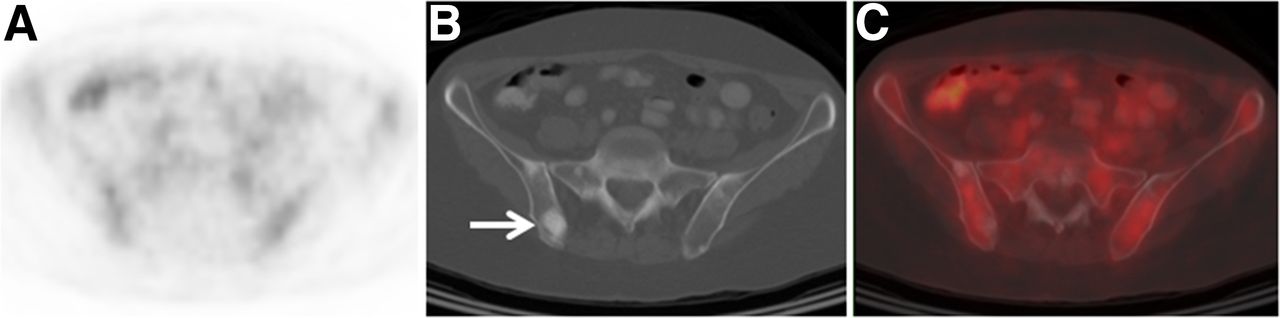
  • FIGURE 2.
    • Download figure
    • Open in new tab
    • Download powerpoint
    FIGURE 2.

    A 56-y-old woman with initial stage III ILC upstaged to IV on CT component of 18F-FDG PET/CT. (A) Axial 18F-FDG PET does not demonstrate suggestive foci. (B) Axial CT component of PET/CT demonstrates multiple osseous sclerotic lesions suggestive of metastases (arrow). (C) Axial fused 18F-FDG PET/CT image confirms that osseous sclerotic lesions demonstrate background 18F-FDG avidity. Biopsy confirmed osseous metastasis.
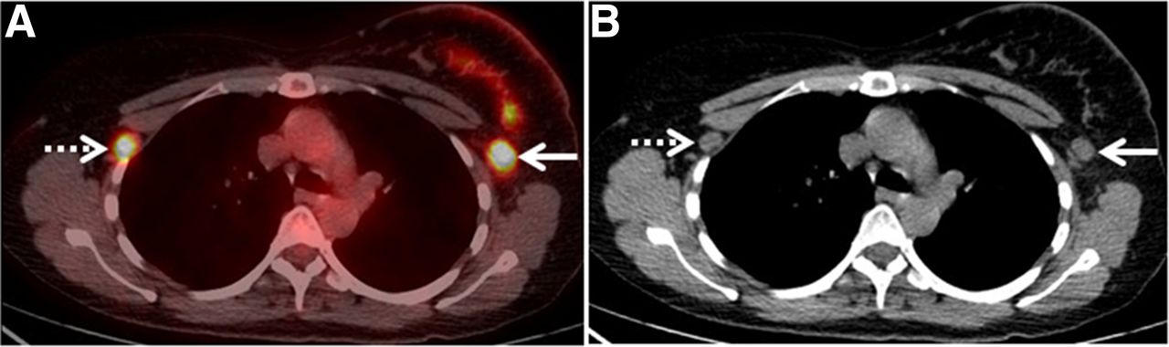
  • FIGURE 3.
    • Download figure
    • Open in new tab
    • Download powerpoint
    FIGURE 3.

    A 52-y-old woman with initial stage III left breast ILC upstaged to IV by 18F-FDG PET and CT. (A) Axial fused 18F-FDG PET/CT image demonstrates previously known ipsilateral left axillary nodal metastasis as 18F-FDG–avid lesion (solid arrow), as well as previously unknown contralateral right axillary node (dashed arrow). (B) Both ipsilateral and contralateral axillary nodal lesions are apparent as enlarged and rounded nodes on CT. Biopsy of contralateral right axillary node demonstrated nodal metastasis. Contralateral axillary nodal metastases are distant metastases (M1 disease) as classified by American Joint Committee on Cancer (18).
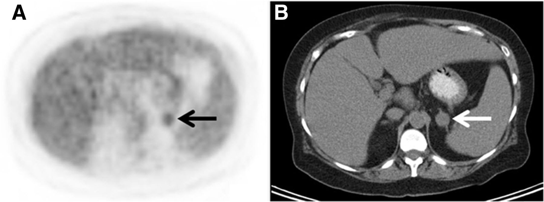
  • FIGURE 4.
    • Download figure
    • Open in new tab
    • Download powerpoint
    FIGURE 4.

    A 64-y-old woman with initial stage III ILC false-positive for distant metastasis on 18F-FDG PET/CT. (A) Axial 18F-FDG PET demonstrates 18F-FDG–avid (SUV, 2.9) focus in left abdomen (arrow). (B) Axial CT component of PET/CT demonstrates 2.1-cm adrenal nodule with average Hounsfield units of 29 (arrow). Biopsy resulted in diagnosis of benign adrenal adenoma.
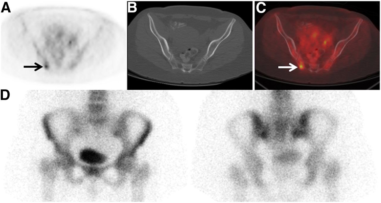
  • FIGURE 5.
    • Download figure
    • Open in new tab
    • Download powerpoint
    FIGURE 5.

    A 42-y-old woman with initial stage III IDC upstaged to IV by 18F-FDG PET. (A) Axial fused 18F-FDG PET/CT image demonstrates previously unknown right ilium metastases as 18F-FDG–avid osseous lesion (arrow). (B) No definite corresponding lesion is seen on axial CT component of PET/CT. (C) Axial fused 18F-FDG PET/CT image confirms osseous localization of 18F-FDG–avid focus (arrow). (D) No corresponding focus is seen on 99mTc-MDP bone scan (anterior [left] and posterior [right] spot views of pelvis shown). Biopsy confirmed osseous metastasis.
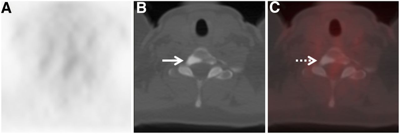
  • FIGURE 6.
    • Download figure
    • Open in new tab
    • Download powerpoint
    FIGURE 6.

    A 46-y-old woman with initial stage III IDC false-positive for distant metastasis on CT component of 18F-FDG PET/CT. (A) Axial 18F-FDG PET does not demonstrate suggestive foci. (B) Axial CT component of PET/CT demonstrates osseous sclerotic lesion (arrow) in T1 vertebra. (C) Axial fused 18F-FDG PET/CT image confirms that osseous sclerotic lesion (arrow) demonstrates background 18F-FDG avidity. Biopsy of sclerotic lesion yielded dense cortical bone without evidence of malignancy, consistent with bone island.

Tables

  • Figures
    • View popup
    TABLE 1

    Characteristics of Patients and Tumors

    CharacteristicILC cohortComparison IDC cohort
    Age (y)
     Median5759
     Range34–9233–90
    Number of patients146 (100%)89 (100%)
    AJCC stage before PET/CT* (n)
     I8 (5%)0 (0%)
     II50 (35%)0 (0%)
     III88 (60%)89 (100%)
    Race (n)
     Caucasian129 (88.4%)69 (77.5%)
     African American9 (6.2%)13 (14.6%)
     Asian8 (5.5%)6 (6.7%)
     Other0 (0.0%)1 (1.1%)
    Receptor phenotype (n)
     ER+/HER2−132 (90.4%)46 (51.7%)
     HER2+8 (5.5%)19 (21.3%)
     Triple-negative5 (3.4%)23 (25.8%)
     Other/unspecified1 (0.7%)1 (1.1%)
    • ↵* Clinical classification is according to seventh edition of AJCC Cancer Staging Manual (18). Inclusion criteria for IDC cohort specified initial stage III disease, to allow comparison to stage III ILC patients.

PreviousNext
Back to top

In this issue

Journal of Nuclear Medicine: 56 (11)
Journal of Nuclear Medicine
Vol. 56, Issue 11
November 1, 2015
  • Table of Contents
  • Table of Contents (PDF)
  • About the Cover
  • Index by author
Print
Download PDF
Article Alerts
Sign In to Email Alerts with your Email Address
Email Article

Thank you for your interest in spreading the word on Journal of Nuclear Medicine.

NOTE: We only request your email address so that the person you are recommending the page to knows that you wanted them to see it, and that it is not junk mail. We do not capture any email address.

Enter multiple addresses on separate lines or separate them with commas.
Comparison of 18F-FDG PET/CT for Systemic Staging of Newly Diagnosed Invasive Lobular Carcinoma Versus Invasive Ductal Carcinoma
(Your Name) has sent you a message from Journal of Nuclear Medicine
(Your Name) thought you would like to see the Journal of Nuclear Medicine web site.
Citation Tools
Comparison of 18F-FDG PET/CT for Systemic Staging of Newly Diagnosed Invasive Lobular Carcinoma Versus Invasive Ductal Carcinoma
Molly P. Hogan, Debra A. Goldman, Brittany Dashevsky, Christopher C. Riedl, Mithat Gönen, Joseph R. Osborne, Maxine Jochelson, Clifford Hudis, Monica Morrow, Gary A. Ulaner
Journal of Nuclear Medicine Nov 2015, 56 (11) 1674-1680; DOI: 10.2967/jnumed.115.161455

Citation Manager Formats

  • BibTeX
  • Bookends
  • EasyBib
  • EndNote (tagged)
  • EndNote 8 (xml)
  • Medlars
  • Mendeley
  • Papers
  • RefWorks Tagged
  • Ref Manager
  • RIS
  • Zotero
Share
Comparison of 18F-FDG PET/CT for Systemic Staging of Newly Diagnosed Invasive Lobular Carcinoma Versus Invasive Ductal Carcinoma
Molly P. Hogan, Debra A. Goldman, Brittany Dashevsky, Christopher C. Riedl, Mithat Gönen, Joseph R. Osborne, Maxine Jochelson, Clifford Hudis, Monica Morrow, Gary A. Ulaner
Journal of Nuclear Medicine Nov 2015, 56 (11) 1674-1680; DOI: 10.2967/jnumed.115.161455
Twitter logo Facebook logo LinkedIn logo Mendeley logo
  • Tweet Widget
  • Facebook Like
  • Google Plus One
Bookmark this article

Jump to section

  • Article
    • Abstract
    • MATERIALS AND METHODS
    • RESULTS
    • DISCUSSION
    • CONCLUSION
    • DISCLOSURE
    • Acknowledgments
    • Footnotes
    • REFERENCES
  • Figures & Data
  • Info & Metrics
  • PDF

Related Articles

  • This Month in JNM
  • PubMed
  • Google Scholar

Cited By...

  • Diagnostic Potential of 68Ga-NeoB PET/CT with Estrogen Receptor- and Progesterone Receptor-Positive Breast Cancer Undergoing Staging or Restaging for Metastatic Disease
  • Oncogenic NOVA1 expression dysregulates alternative splicing in breast cancer
  • 68Ga-FAP-2286 PET of Solid Tumors: Biodistribution, Dosimetry, and Comparison with 18F-FDG
  • 68Ga-FAPI PET/CT as an Alternative to 18F-FDG PET/CT in the Imaging of Invasive Lobular Breast Carcinoma
  • WNT4 regulates cellular metabolism via intracellular activity at the mitochondria in breast and gynecologic cancers
  • Summary: Appropriate Use Criteria for Estrogen Receptor-Targeted PET Imaging with 16{alpha}-18F-Fluoro-17{beta}-Fluoroestradiol
  • WNT4 executes estrogen regulation of cellular metabolism via intracellular activity at the mitochondria
  • Head-to-Head Evaluation of 18F-FES and 18F-FDG PET/CT in Metastatic Invasive Lobular Breast Cancer
  • Mars Shot for Nuclear Medicine, Molecular Imaging, and Molecularly Targeted Radiopharmaceutical Therapy
  • 18F-FDG PET/CT for Systemic Staging of Newly Diagnosed Breast Cancer in Men
  • Expression of Gastrin-Releasing Peptide Receptor in Breast Cancer and Its Association with Pathologic, Biologic, and Clinical Parameters: A Study of 1,432 Primary Tumors
  • Prospective Clinical Trial of 18F-Fluciclovine PET/CT for Determining the Response to Neoadjuvant Therapy in Invasive Ductal and Invasive Lobular Breast Cancers
  • Initial Results of a Prospective Clinical Trial of 18F-Fluciclovine PET/CT in Newly Diagnosed Invasive Ductal and Invasive Lobular Breast Cancers
  • Google Scholar

More in this TOC Section

  • Feasibility of Ultra-Low-Activity 18F-FDG PET/CT Imaging Using a Long–Axial-Field-of-View PET/CT System
  • Cardiac Presynaptic Sympathetic Nervous Function Evaluated by Cardiac PET in Patients with Chronotropic Incompetence Without Heart Failure
  • Validation and Evaluation of a Vendor-Provided Head Motion Correction Algorithm on the uMI Panorama PET/CT System
Show more Clinical Investigations

Similar Articles

Keywords

  • lobular
  • ductal
  • breast cancer
  • 18F-FDG
  • PET/CT
  • staging
SNMMI

© 2025 SNMMI

Powered by HighWire