
Main menu

  • Home
  • Content
    • Current
    • Ahead of print
    • Past Issues
    • JNM Supplement
    • SNMMI Annual Meeting Abstracts
    • Continuing Education
    • JNM Podcasts
  • Subscriptions
    • Subscribers
    • Institutional and Non-member
    • Rates
    • Journal Claims
    • Corporate & Special Sales
  • Authors
    • Submit to JNM
    • Information for Authors
    • Assignment of Copyright
    • AQARA requirements
  • Info
    • Reviewers
    • Permissions
    • Advertisers
  • About
    • About Us
    • Editorial Board
    • Contact Information
  • More
    • Alerts
    • Feedback
    • Help
    • SNMMI Journals
  • SNMMI
    • JNM
    • JNMT
    • SNMMI Journals
    • SNMMI

User menu

  • Subscribe
  • My alerts
  • Log in
  • Log out
  • My Cart

Search

  • Advanced search
Journal of Nuclear Medicine
  • SNMMI
    • JNM
    • JNMT
    • SNMMI Journals
    • SNMMI
  • Subscribe
  • My alerts
  • Log in
  • Log out
  • My Cart
Journal of Nuclear Medicine

Advanced Search

  • Home
  • Content
    • Current
    • Ahead of print
    • Past Issues
    • JNM Supplement
    • SNMMI Annual Meeting Abstracts
    • Continuing Education
    • JNM Podcasts
  • Subscriptions
    • Subscribers
    • Institutional and Non-member
    • Rates
    • Journal Claims
    • Corporate & Special Sales
  • Authors
    • Submit to JNM
    • Information for Authors
    • Assignment of Copyright
    • AQARA requirements
  • Info
    • Reviewers
    • Permissions
    • Advertisers
  • About
    • About Us
    • Editorial Board
    • Contact Information
  • More
    • Alerts
    • Feedback
    • Help
    • SNMMI Journals
  • View or Listen to JNM Podcast
  • Visit JNM on Facebook
  • Join JNM on LinkedIn
  • Follow JNM on Twitter
  • Subscribe to our RSS feeds
Review ArticleContinuing Education

Dynamic Bone Imaging with 99mTc-Labeled Diphosphonates and 18F-NaF: Mechanisms and Applications

Ka Kit Wong and Morand Piert
Journal of Nuclear Medicine April 2013, 54 (4) 590-599; DOI: https://doi.org/10.2967/jnumed.112.114298
Ka Kit Wong
1Nuclear Medicine/Radiology Department, University of Michigan Hospital, Ann Arbor, Michigan; and
2Nuclear Medicine Service, Department of Veterans Affairs Health System, Ann Arbor, Michigan
  • Find this author on Google Scholar
  • Find this author on PubMed
  • Search for this author on this site
Morand Piert
1Nuclear Medicine/Radiology Department, University of Michigan Hospital, Ann Arbor, Michigan; and
  • Find this author on Google Scholar
  • Find this author on PubMed
  • Search for this author on this site
  • Article
  • Figures & Data
  • Info & Metrics
  • PDF
Loading
  1. FIGURE 1.
    • Download figure
    • Open in new tab
    • Download powerpoint
    FIGURE 1.

    Diffusely increased 99mTc-MDP uptake of left lower extremity on delayed-phase whole-body bone scan (anterior and posterior projections) as result of increased blood flow due to osteosarcoma in proximal tibia. INJ = injection site.

  2. FIGURE 2.
    • Download figure
    • Open in new tab
    • Download powerpoint
    FIGURE 2.

    An 18F-NaF 2-tissue-compartment model (3 compartments and 4 parameters).
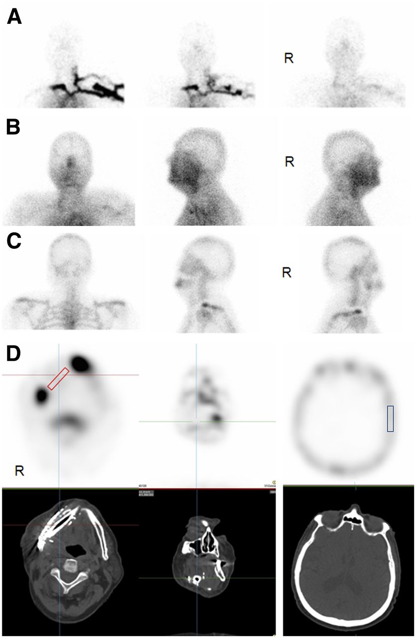
  3. FIGURE 3.
    • Download figure
    • Open in new tab
    • Download powerpoint
    FIGURE 3.

    99mTc-MDP bone scan for viability assessment of vascularized fibula graft in patient with osteonecrosis of jaw. On anterior, left, and right planar projections, angiographic (A) and soft-tissue (B) phases show nonspecific diffuse hyperemia overlying mandible due to surgical trauma. Delayed planar images (C) display focal increased uptake at genuine mandible near union sites of fibula graft. Transaxial and coronal SPECT/CT reveals photopenia in graft region (D). Regions of interest are defined in fibula graft (red) and calvarium (blue) on 3 consecutive slices to calculate graft-to-calvarium uptake ratio (41). Visual inspection and ratio result (0.72) are suggestive of nonviable graft.
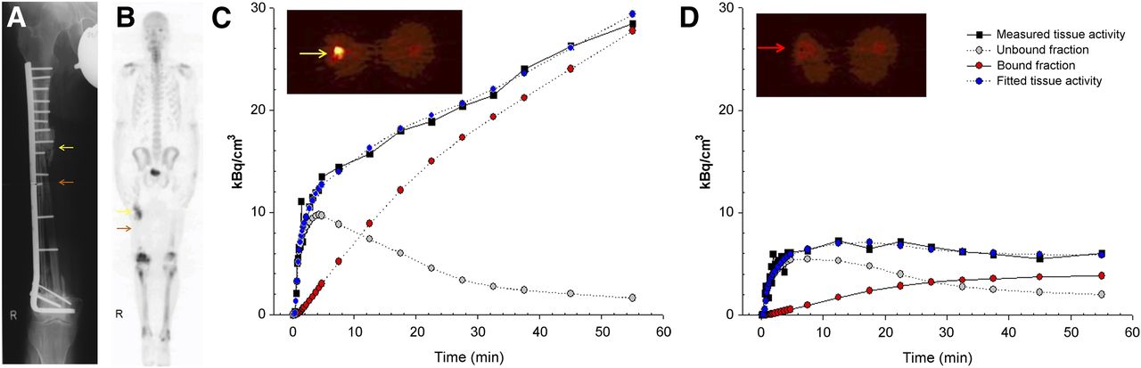
  4. FIGURE 4.
    • Download figure
    • Open in new tab
    • Download powerpoint
    FIGURE 4.

    Plate fixation of vascularized fibula transplant (red arrow) after resection of osteosarcoma of right femur (A). Revision plate fixation was performed after fracture due to nonunion (yellow arrow). After injection of 370 MBq of 18F-NaF, dynamic PET study of mid thigh was performed for 60 min, followed by non–attenuation-corrected whole-body scan (B). Attenuation-corrected and axial slices (50–60 min) of hypertrophic nonunion (C) and fibular graft (D) areas show markedly increased uptake at nonunion and decreased uptake at level of fibula graft. Tissue time–activity curves (black squares) demonstrate rapid and sustained net-accumulation of radiotracer in nonunion but poor accumulation in fibula graft. Compartmental modeling was performed, providing estimates for total tissue activity (blue dots) and fraction of bound (red dots) and unbound (gray dots) radiotracer in tissue. At later time points, relationship of bound to unbound radiotracer is much higher in hypertrophic nonunion (C) than in failing graft (D).

  5. FIGURE 5.
    • Download figure
    • Open in new tab
    • Download powerpoint
    FIGURE 5.

    An 8-y-old boy with right hip pain and avascular necrosis of right femur head (Legg-Calve-Perthes disease). Anterior and posterior projection soft-tissue (A) and delayed-phase images (B) show focally decreased 99mTc-MDP uptake in right femoral head.

  6. FIGURE 6.
    • Download figure
    • Open in new tab
    • Download powerpoint
    FIGURE 6.

    Breast cancer bone metastases identified on anterior and posterior projection whole-body bone scan in ribs, spine, pelvis, and left femur shaft (A). Treatment with denosumab results in near-normalization of scan findings (B), although with continued rise of tumor marker CA15-3. Three months after treatment, bone scan again identifies apparent disease progression (C).

  • © 2013 by the Society of Nuclear Medicine and Molecular Imaging, Inc.
PreviousNext
Back to top

In this issue

Journal of Nuclear Medicine: 54 (4)
Journal of Nuclear Medicine
Vol. 54, Issue 4
April 1, 2013
  • Table of Contents
  • Table of Contents (PDF)
  • About the Cover
  • Index by author
Print
Download PDF
Article Alerts
Sign In to Email Alerts with your Email Address
Email Article

Thank you for your interest in spreading the word on Journal of Nuclear Medicine.

NOTE: We only request your email address so that the person you are recommending the page to knows that you wanted them to see it, and that it is not junk mail. We do not capture any email address.

Enter multiple addresses on separate lines or separate them with commas.
Dynamic Bone Imaging with 99mTc-Labeled Diphosphonates and 18F-NaF: Mechanisms and Applications
(Your Name) has sent you a message from Journal of Nuclear Medicine
(Your Name) thought you would like to see the Journal of Nuclear Medicine web site.
Citation Tools
Dynamic Bone Imaging with 99mTc-Labeled Diphosphonates and 18F-NaF: Mechanisms and Applications
Ka Kit Wong, Morand Piert
Journal of Nuclear Medicine Apr 2013, 54 (4) 590-599; DOI: 10.2967/jnumed.112.114298

Citation Manager Formats

  • BibTeX
  • Bookends
  • EasyBib
  • EndNote (tagged)
  • EndNote 8 (xml)
  • Medlars
  • Mendeley
  • Papers
  • RefWorks Tagged
  • Ref Manager
  • RIS
  • Zotero
Share
Dynamic Bone Imaging with 99mTc-Labeled Diphosphonates and 18F-NaF: Mechanisms and Applications
Ka Kit Wong, Morand Piert
Journal of Nuclear Medicine Apr 2013, 54 (4) 590-599; DOI: 10.2967/jnumed.112.114298
Twitter logo Facebook logo LinkedIn logo Mendeley logo
  • Tweet Widget
  • Facebook Like
  • Google Plus One
Bookmark this article

Jump to section

  • Article
    • Abstract
    • UPTAKE MECHANISMS OF BONE-SEEKING RADIOPHARMACEUTICALS
    • CLINICAL APPLICATIONS
    • CONCLUSION
    • Footnotes
    • REFERENCES
  • Figures & Data
  • Info & Metrics
  • PDF

Related Articles

  • This Month in JNM
  • PubMed
  • Google Scholar

Cited By...

  • Feasibility of dual phase 99mTc-MDP SPECT/CT imaging in rheumatoid arthritis evaluation
  • 11C-Choline Pharmacokinetics in Recurrent Prostate Cancer
  • Evaluation of 18F-Fluoride PET/MR and PET/CT in Patients with Foot Pain of Unclear Cause
  • Regarding Dynamic Bone Imaging with 99mTc-Labeled Diphosphonates and 18F-NaF: Mechanisms and Applications
  • Reply: Regarding Dynamic Bone Imaging with 99mTc-Labeled Diphosphonates and 18F-NaF: Mechanisms and Applications
  • Google Scholar

More in this TOC Section

  • Approaches to Imaging Immune Activation Using PET
  • Large Language Models and Large Multimodal Models in Medical Imaging: A Primer for Physicians
  • Precision Oncology in Melanoma: Changing Practices
Show more Continuing Education

Similar Articles

Keywords

  • 99mTc-methyl-diphosphonate
  • 18F-NaF
  • dynamic imaging
SNMMI

© 2025 SNMMI

Powered by HighWire