
Main menu

  • Home
  • Content
    • Current
    • Ahead of print
    • Past Issues
    • JNM Supplement
    • SNMMI Annual Meeting Abstracts
    • Continuing Education
    • JNM Podcasts
  • Subscriptions
    • Subscribers
    • Institutional and Non-member
    • Rates
    • Journal Claims
    • Corporate & Special Sales
  • Authors
    • Submit to JNM
    • Information for Authors
    • Assignment of Copyright
    • AQARA requirements
  • Info
    • Reviewers
    • Permissions
    • Advertisers
  • About
    • About Us
    • Editorial Board
    • Contact Information
  • More
    • Alerts
    • Feedback
    • Help
    • SNMMI Journals
  • SNMMI
    • JNM
    • JNMT
    • SNMMI Journals
    • SNMMI

User menu

  • Subscribe
  • My alerts
  • Log in
  • My Cart

Search

  • Advanced search
Journal of Nuclear Medicine
  • SNMMI
    • JNM
    • JNMT
    • SNMMI Journals
    • SNMMI
  • Subscribe
  • My alerts
  • Log in
  • My Cart
Journal of Nuclear Medicine

Advanced Search

  • Home
  • Content
    • Current
    • Ahead of print
    • Past Issues
    • JNM Supplement
    • SNMMI Annual Meeting Abstracts
    • Continuing Education
    • JNM Podcasts
  • Subscriptions
    • Subscribers
    • Institutional and Non-member
    • Rates
    • Journal Claims
    • Corporate & Special Sales
  • Authors
    • Submit to JNM
    • Information for Authors
    • Assignment of Copyright
    • AQARA requirements
  • Info
    • Reviewers
    • Permissions
    • Advertisers
  • About
    • About Us
    • Editorial Board
    • Contact Information
  • More
    • Alerts
    • Feedback
    • Help
    • SNMMI Journals
  • View or Listen to JNM Podcast
  • Visit JNM on Facebook
  • Join JNM on LinkedIn
  • Follow JNM on Twitter
  • Subscribe to our RSS feeds
Research ArticleClinical Investigations

N-(4-18F-Fluorobenzoyl)Interleukin-2 for PET of Human-Activated T Lymphocytes

Valentina Di Gialleonardo, Alberto Signore, Andor W.J.M. Glaudemans, Rudi A.J.O. Dierckx and Erik F.J. De Vries
Journal of Nuclear Medicine May 2012, 53 (5) 679-686; DOI: https://doi.org/10.2967/jnumed.111.091306
Valentina Di Gialleonardo
  • Find this author on Google Scholar
  • Find this author on PubMed
  • Search for this author on this site
Alberto Signore
  • Find this author on Google Scholar
  • Find this author on PubMed
  • Search for this author on this site
Andor W.J.M. Glaudemans
  • Find this author on Google Scholar
  • Find this author on PubMed
  • Search for this author on this site
Rudi A.J.O. Dierckx
  • Find this author on Google Scholar
  • Find this author on PubMed
  • Search for this author on this site
Erik F.J. De Vries
  • Find this author on Google Scholar
  • Find this author on PubMed
  • Search for this author on this site
  • Article
  • Figures & Data
  • Info & Metrics
  • PDF
Loading

Article Figures & Data

Figures

  • Tables
  • FIGURE 1.
    • Download figure
    • Open in new tab
    • Download powerpoint
    FIGURE 1.

    MALDI-TOF profile of 18F-FB-IL2.
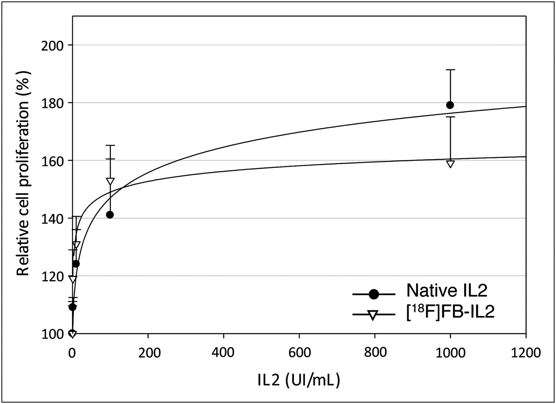
  • FIGURE 2.
    • Download figure
    • Open in new tab
    • Download powerpoint
    FIGURE 2.

    MTT cell proliferation assay of phytohemoagglutinin-activated hPBMc stimulated by native IL2 or 18F-FB-IL2. Results are expressed as percentage of increase in cellular proliferation as compared with untreated cells (mean ± SD of 3 independent experiments, each performed in triplicate). There is no significant difference between labeled and unlabeled IL2 at any tested concentration.
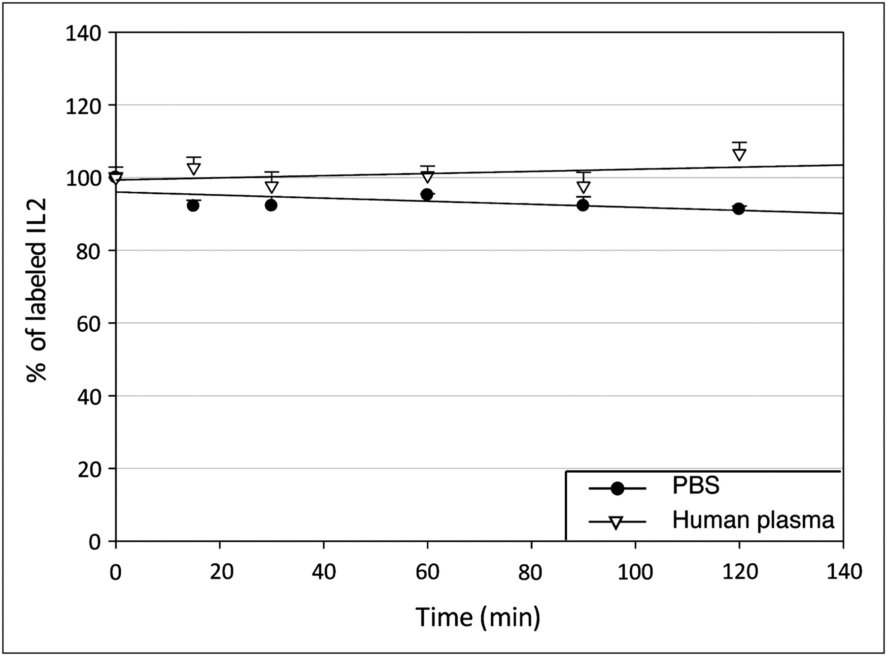
  • FIGURE 3.
    • Download figure
    • Open in new tab
    • Download powerpoint
    FIGURE 3.

    3-chloroacetic acid precipitation assay as percentage of labeled IL2 at different times in phosphate-buffered saline and human plasma. PBS = phosphate-buffered saline.
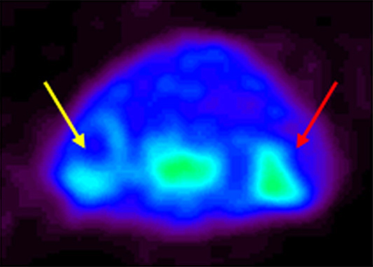
  • FIGURE 4.
    • Download figure
    • Open in new tab
    • Download powerpoint
    FIGURE 4.

    Small-animal PET images of SCID mice inoculated with phytohemoagglutinin-activated T lymphocytes. Small-animal PET image (transaxial section of mouse shoulders) shows 18F-FB-IL2 uptake in right shoulder (red arrow) and, to lesser extent, in contralateral, control shoulder (yellow arrow) due to migration of lymphocytes from injection site to contralateral site.
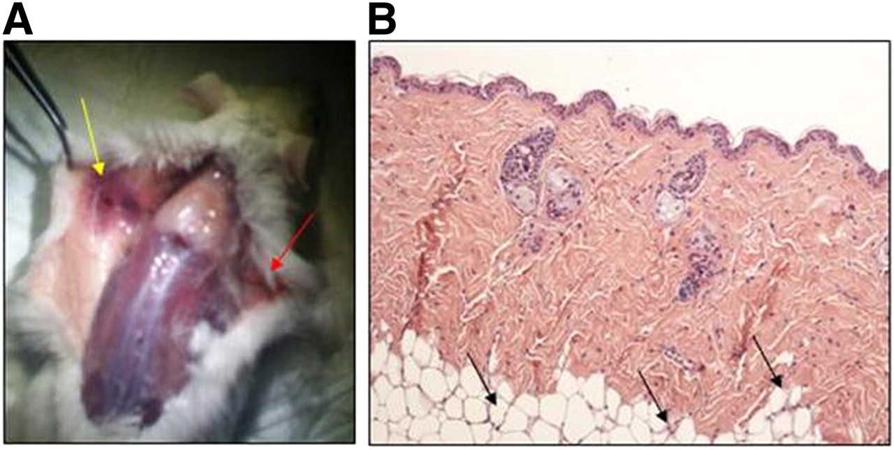
  • FIGURE 5.
    • Download figure
    • Open in new tab
    • Download powerpoint
    FIGURE 5.

    (A) Visual inspection of derma of inoculated mouse, of which PET image is presented in Figure 4. Red arrow highlights site at which lymphocytes were inoculated with Matrigel in right shoulder, and yellow arrow indicates inflammatory reaction after inoculation of Matrigel only in left shoulder. (B) Hematoxylin and eosin staining of skin from left shoulder showing presence of lymphocytes at border of Matrigel (black arrows).
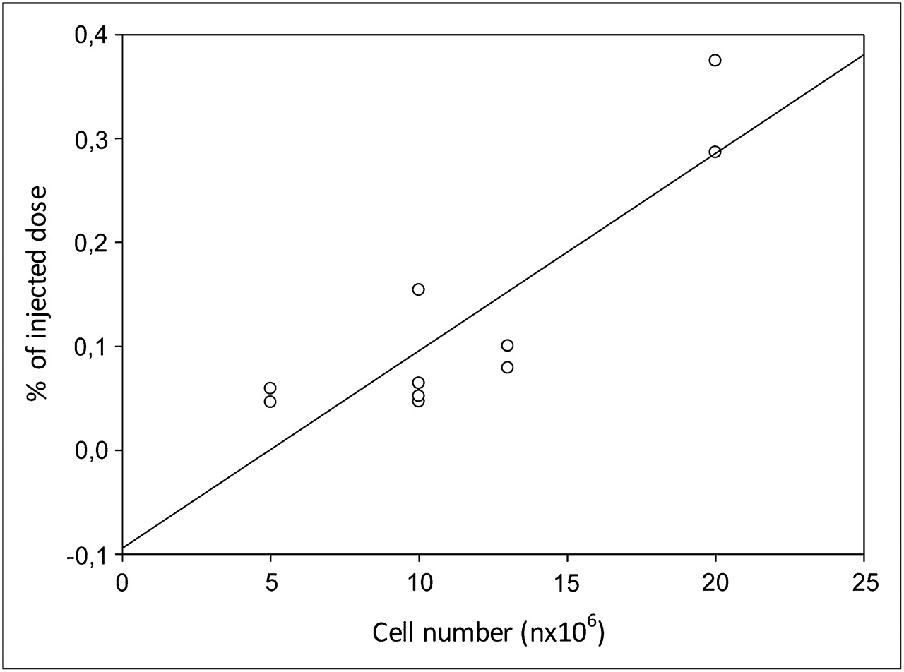
  • FIGURE 6.
    • Download figure
    • Open in new tab
    • Download powerpoint
    FIGURE 6.

    Correlation between number of inoculated lymphocytes and 18F-FB-IL2 uptake in both shoulders (R2 = 0.768, P = 0.0012).

  • FIGURE 7.
    • Download figure
    • Open in new tab
    • Download powerpoint
    FIGURE 7.

    In vivo binding of 18F-FB-IL2 to 10 × 106 phytohemoagglutinin-activated T lymphocytes inoculated in shoulder of SCID mice (n = 4) (left bar) and in mice (n = 4) pretreated with 100-fold excess of unlabeled IL2 (right bar). Data are mean ± SD of standardized uptake value calculated 30 min after radiopharmaceutical injection.

Tables

  • Figures
    • View popup
    TABLE 1

    Biodistribution of Radiotracer 18F-FB-IL2 in 12 BALB/c Mice at Different Time Points

    Organ15 min (n = 4)60 min (n = 4)90 min (n = 4)
    Bone8.35 ± 7.792.86 ± 4.053.48 ± 3.01
    Cerebellum1.40 ± 0.950.53 ± 0.780.55 ± 0.22
    Cerebrum0.93 ± 0.680.51 ± 0.750.37 ± 0.09
    Colon3.09 ± 2.501.38 ± 1.792.95 ± 1.65
    Duodenum30.87 ± 23.9612.49 ± 6.107.56 ± 11.82
    Heart3.01 ± 2.871.78 ± 2.441.01 ± 0.13
    Ileum9.46 ± 13.281.54 ± 2.172.93 ± 1.36
    Kidney52.68 ± 55.3921.64 ± 28.7030.82 ± 17.87
    Liver5.76 ± 4.913.51 ± 5.262.67 ± 0.87
    Lung4.37 ± 3.203.75 ± 4.414.89 ± 5.65
    Muscle5.11 ± 3.210.89 ± 1.195.05 ± 4.42
    Pancreas2.06 ± 1.591.41 ± 1.961.04 ± 0.25
    Plasma12.18 ± 11.446.96 ± 9.493.44 ± 1.14
    Red blood cells4.20 ± 3.922.80 ± 3.951.85 ± 0.35
    Spleen1.26 ± 0.551.46 ± 2.101.40 ± 0.53
    Stomach2.44 ± 1.190.26 ± 0.152.67 ± 2.81
    • Data are %ID/g, as mean ± SD.

PreviousNext
Back to top

In this issue

Journal of Nuclear Medicine: 53 (5)
Journal of Nuclear Medicine
Vol. 53, Issue 5
May 1, 2012
  • Table of Contents
  • Table of Contents (PDF)
  • About the Cover
  • Index by author
Print
Download PDF
Article Alerts
Sign In to Email Alerts with your Email Address
Email Article

Thank you for your interest in spreading the word on Journal of Nuclear Medicine.

NOTE: We only request your email address so that the person you are recommending the page to knows that you wanted them to see it, and that it is not junk mail. We do not capture any email address.

Enter multiple addresses on separate lines or separate them with commas.
N-(4-18F-Fluorobenzoyl)Interleukin-2 for PET of Human-Activated T Lymphocytes
(Your Name) has sent you a message from Journal of Nuclear Medicine
(Your Name) thought you would like to see the Journal of Nuclear Medicine web site.
Citation Tools
N-(4-18F-Fluorobenzoyl)Interleukin-2 for PET of Human-Activated T Lymphocytes
Valentina Di Gialleonardo, Alberto Signore, Andor W.J.M. Glaudemans, Rudi A.J.O. Dierckx, Erik F.J. De Vries
Journal of Nuclear Medicine May 2012, 53 (5) 679-686; DOI: 10.2967/jnumed.111.091306

Citation Manager Formats

  • BibTeX
  • Bookends
  • EasyBib
  • EndNote (tagged)
  • EndNote 8 (xml)
  • Medlars
  • Mendeley
  • Papers
  • RefWorks Tagged
  • Ref Manager
  • RIS
  • Zotero
Share
N-(4-18F-Fluorobenzoyl)Interleukin-2 for PET of Human-Activated T Lymphocytes
Valentina Di Gialleonardo, Alberto Signore, Andor W.J.M. Glaudemans, Rudi A.J.O. Dierckx, Erik F.J. De Vries
Journal of Nuclear Medicine May 2012, 53 (5) 679-686; DOI: 10.2967/jnumed.111.091306
Twitter logo Facebook logo LinkedIn logo Mendeley logo
  • Tweet Widget
  • Facebook Like
  • Google Plus One
Bookmark this article

Jump to section

  • Article
    • Abstract
    • MATERIALS AND METHODS
    • RESULTS
    • DISCUSSION
    • CONCLUSION
    • DISCLOSURE STATEMENT
    • Acknowledgments
    • Footnotes
    • REFERENCES
  • Figures & Data
  • Info & Metrics
  • PDF

Related Articles

  • This Month in JNM
  • PubMed
  • Google Scholar

Cited By...

  • Imaging of Activated T Cells
  • Imaging Calreticulin for Early Detection of Immunogenic Cell Death During Anticancer Treatment
  • Molecular Imaging of Chimeric Antigen Receptor T Cells by ICOS-ImmunoPET
  • Development and Evaluation of Interleukin-2-Derived Radiotracers for PET Imaging of T Cells in Mice
  • Combination treatment with hypofractionated radiotherapy plus IL-2/anti-IL-2 complexes and its theranostic evaluation
  • A PET Imaging Strategy to Visualize Activated T Cells in Acute Graft-versus-Host Disease Elicited by Allogenic Hematopoietic Cell Transplant
  • Synthesis and Characterization of 18F-Interleukin-8 Using a Cell-Free Translation System and 4-18F-Fluoro-L-Proline
  • PET Imaging of Macrophage Mannose Receptor-Expressing Macrophages in Tumor Stroma Using 18F-Radiolabeled Camelid Single-Domain Antibody Fragments
  • Detection of Insulitis by Pancreatic Scintigraphy With 99mTc-Labeled IL-2 and MRI in Patients With LADA (Action LADA 10)
  • Google Scholar

More in this TOC Section

  • Feasibility of Ultra-Low-Activity 18F-FDG PET/CT Imaging Using a Long–Axial-Field-of-View PET/CT System
  • Cardiac Presynaptic Sympathetic Nervous Function Evaluated by Cardiac PET in Patients with Chronotropic Incompetence Without Heart Failure
  • Validation and Evaluation of a Vendor-Provided Head Motion Correction Algorithm on the uMI Panorama PET/CT System
Show more Clinical Investigations

Similar Articles

SNMMI

© 2025 SNMMI

Powered by HighWire