


Abstract
Recently, N-(4-18F-fluorobenzoyl)-interleukin-2 (18F-FB-IL2) was introduced as a PET tracer for T cell imaging. However, production is complex and time-consuming. Therefore, we developed 2 radiolabeled IL2 variants, namely aluminum 18F-fluoride-(restrained complexing agent)-IL2 (18F-AlF-RESCA-IL2) and 68Ga-gallium-(1,4,7-triazacyclononane-4,7-diacetic acid-1-glutaric acid)-IL2 (68Ga-Ga-NODAGA-IL2), and compared their in vitro and in vivo characteristics with 18F-FB-IL2. Methods: Radiolabeling of 18F-AlF-RESCA-IL2 and 68Ga-Ga-NODAGA-IL2 was optimized, and stability was evaluated in human serum. Receptor binding was studied with activated human peripheral blood mononuclear cells (hPBMCs). Ex vivo tracer biodistribution in immunocompetent BALB/cOlaHsd (BALB/c) mice was performed at 15, 60, and 90 min after tracer injection. In vivo binding characteristics were studied in severe combined immunodeficient (SCID) mice inoculated with activated hPBMCs in Matrigel. Tracer was injected 15 min after hPBMC inoculation, and a 60-min dynamic PET scan was acquired, followed by ex vivo biodistribution studies. Specific uptake was determined by coinjection of tracer with unlabeled IL2 and by evaluating uptake in a control group inoculated with Matrigel only. Results: 68Ga-Ga-NODAGA-IL2 and 18F-AlF-RESCA-IL2 were produced with radiochemical purity of more than 95% and radiochemical yield of 13.1% ± 4.7% and 2.4% ± 1.6% within 60 and 90 min, respectively. Both tracers were stable in serum, with more than 90% being intact tracer after 1 h. In vitro, both tracers displayed preferential binding to activated hPBMCs. Ex vivo biodistribution studies on BALB/c mice showed higher uptake of 18F-AlF-RESCA-IL2 than of 18F-FB-IL2 in liver, kidney, spleen, bone, and bone marrow. 68Ga-Ga-NODAGA-IL2 uptake in liver and kidney was higher than 18F-FB-IL2 uptake. In vivo, all tracers revealed uptake in activated hPBMCs in SCID mice. Low uptake was seen after a blocking dose of IL2 and in the Matrigel control group. In addition, 18F-AlF-RESCA-IL2 yielded the highest-contrast PET images of target lymph nodes. Conclusion: Production of 18F-AlF-RESCA-IL2 and 68Ga-Ga-NODAGA-IL2 is simpler and faster than that of 18F-FB-IL2. Both tracers showed good in vitro and in vivo characteristics, with high uptake in lymphoid tissue and hPBMC xenografts.
Molecular imaging of immune cells for diagnosis and therapy evaluation in inflammatory and infectious diseases has been performed for decades (1,2). Now, interest in visualizing the immune response in cancer is also growing, as a result of the introduction of cancer immunotherapies. Immune checkpoint inhibitors have antitumor effects across several tumor types. However, not all patients respond, and some patients experience serious immune-related side effects. Therefore, strategies to select patients and optimize therapy are needed. Molecular imaging of immune cells might be a suitable method for patient selection, response prediction, and treatment evaluation in this context (2).
T cells are major players in both immune-related diseases and the tumor-immune response. During an immune response, T cells secrete the protein interleukin-2 (IL2). IL2 binds to the IL2 receptor (IL2-R), consisting of 3 subunits: IL2-Rα (CD25), IL2-Rβ (CD122), and IL2-Rγ (CD132) (3–6). Binding of IL2 to this complex leads to T-cell activation and differentiation (3). The IL2-R is expressed not only on activated cytotoxic T cells but also on other subpopulations, such as regulatory T cells. Specific binding of radiolabeled IL2 to T cells could be exploited for molecular imaging and might provide insight on immune responses.
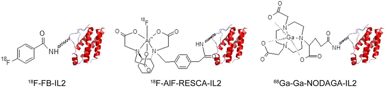
Recombinant human IL2 binds to human and murine IL2-R and has been radiolabeled previously with SPECT isotopes such as 99mTc and 123I (7–9). As PET offers better resolution than SPECT, the 18F-labeled PET tracer N-(4-18F-fluorobenzoyl)-IL2 (18F-FB-IL2) was developed. 18F-FB-IL2 PET imaging detected subcutaneously injected activated T cells and tumor infiltration of T cells in response to radiotherapy and immunization (10–12). Clinical trials with 18F-FB-IL2 are ongoing. However, radiotracer production is complex, requiring 2.5 h and several synthesis modules. Moreover, the production yields sufficient radiotracer for only 1 or 2 patients (13). Therefore, we aimed to develop other radiolabeled IL2 PET tracers. First, we developed a simplified method for radiolabeling with 18F, requiring fewer synthesis steps. Second, we used another PET isotope, 68Ga, which is a generator-produced radioisotope. Here, we present the development of aluminum 18F-fluoride-(restrained complexing agent)-IL2 (18F-AlF-RESCA-IL2) and 68Ga-gallium-(1,4,7-triazacyclononane-4,7-diacetic acid-1-glutaric acid)-IL2 (68Ga-Ga-NODAGA-IL2) (Fig. 1). To determine their potential for clinical use, in vitro and in vivo characteristics were evaluated and compared with 18F-FB-IL2. For this purpose, in vitro binding assays with activated human peripheral blood mononuclear cells (hPBMCs) and ex vivo biodistribution studies on immunocompetent BALB/cOlaHsd (BALB/c) mice were performed. In addition, PET imaging and ex vivo biodistribution studies were performed on severe-combined immunodeficient (SCID) mice subcutaneously inoculated with activated hPBMCs.

Structures of radiolabeled IL2 PET tracers.
MATERIALS AND METHODS
Production of 18F-FB-IL2
The production method for 18F-FB-IL2 has been described previously (10). This method was adapted to improve tracer yields (13).
Production of 18F-AlF-RESCA-IL2
We developed a simplified method to label IL2 with 18F, using the indirect 18F-AlF-RESCA methodology, combining the chemical advantages of a chelator-based radiolabeling method with the unique physical properties of 18F (14–16). 18F-AlF was allowed to react with the RESCA-tetrafluorophenol ester ((±)-H3RESCA-TFP) (Leuven University). Subsequently, the 18F-AlF-RESCA complex was conjugated with IL2. The complete synthesis is described in the supplemental materials (available at http://jnm.snmjournals.org).
Production of 68Ga-Ga-NODAGA-IL2
Our second strategy was radiolabeling of IL2 with 68Ga. Because of the short half-life of 68Ga, fewer synthesis steps and less synthesis time are required. First, 2,2′-(7-(1-carboxy-4-((2,5-dioxopyrrolidin-1-yl)oxy)-4-oxobutyl)-1,4,7-triazonane1,4-diyl)diacetic-acid (NODAGA-NHS) was conjugated to IL2, followed by radiolabeling with 68Ga. We used 2 synthesis methods. In method 1, 68Ga was eluted from the PS-H+ cartridge in the cationic form (0.1 M HCl). However, large amounts of HCl are needed to efficiently elute all 68Ga, leading to low concentrations of 68Ga. Thus, for obtaining high concentrations of 68Ga in a nonfractionated elution, we implemented method 2, in which 68Ga was eluted from a PS-HCO3 cartridge with deionized water (17). The complete synthesis methods are described in the supplemental materials.
In Vitro Binding Assays
In vitro binding to IL2-Rs was determined by performing binding assays with hPBMCs, isolated from peripheral blood from healthy volunteers by Ficoll-Paque Plus separation (GE Healthcare). Cells were kept in RPMI-1640 medium (Gibco) supplemented with 10% fetal calf serum. Isolated hPBMCs were activated by incubation with a 5 μg/mL concentration of phytohemagglutinin (Sigma-Aldrich) in a 5% CO2 atmosphere at 37°C for 48 h (10). As a control, nonactivated hPBMCs were incubated for 48 h at 37°C and 5% CO2. On the day of the experiment, another batch of hPBMCs was isolated, to compare CD25 (IL2-Rα) expression on these freshly isolated cells with expression on cells that had been incubated for 48 h.
Approximately 5 × 10⁵ cells were incubated with 50 μL of tracer solution at 37°C for 30 min. The cells were washed twice with 1 mL of ice-cold phosphate-buffered saline (PBS) containing 1% human serum albumin (Sanquin). Activity in the cell fraction was measured in a γ-counter (Wizard2 2480-0019, SW 2.1; PerkinElmer). Tracer uptake was corrected for the number of viable cells, which were counted manually using trypan blue. Uptake was expressed as percentage of cell-associated radioactivity per 500,000 cells. For 18F-FB-IL2 and 18F-AlF-RESCA-IL2, 4 independent experiments were performed, each experiment in triplicate. For 68Ga-Ga-NODAGA-IL2, 2 independent experiments were performed, each experiment in triplicate.
In Vitro Stability Studies
The stability of 18F-AlF-RESCA-IL2 and 68Ga-Ga-NODAGA-IL2 was evaluated by adding 50 μL of tracer to 200 μL of human serum. The mixture was vortexed and incubated at 37°C for 60 and 120 min. After incubation, radiochemical purity was determined by a trichloroacetic acid precipitation assay (18,19).
Animal Studies
Animal studies were performed according to Dutch Regulations for Animal Welfare (IvD number 16395-01-007). The protocol was approved by the animal ethical committee of the University of Groningen. Animals were randomly assigned to different groups. A power calculation was performed to calculate the experimental group size. With an expected variation coefficient of 10%, a meaningful effect size of 25%, a confidence interval of 95%, and a power of 90%, a minimum of 5 animals per group is required. Therefore 5–6 animals were included in each group. Exclusion criteria were abnormal behavior, signs of sickness, more than a 15% reduction in body weight, and death.
Biodistribution Studies and In Vivo Stability in BALB/c Mice
Ex vivo biodistribution studies were performed on immunocompetent BALB/c mice 5–8 wk old (Envigo). The weights of the animals were comparable. Tracer (150 μL) was injected via the penile vein: 4.20 ± 0.8 MBq (0.75 ± 0.22 μg) of 18F-FB-IL2; 1.21 ± 0.95 MBq (0.25 ± 0.24 μg) of 18F-AlF-RESCA-IL2; and 0.55 ± 0.13 MBq (0.32 ± 0.18 μg) of 68Ga-Ga-NODAGA-IL2 (method 1). The mice were sacrificed at 15, 60. and 90 min after injection (6 mice per time point). Organs were dissected and counted in a γ-counter. Bone marrow was isolated from the hind limb long bones (femurs and tibias) via centrifugation. Uptake in organs was calculated as percentage injected dose per gram of tissue. Total injected dose was determined by measuring activity in the syringe before and after injection. In vivo stability was determined in plasma samples by a trichloroacetic acid precipitation assay.
PET Imaging and Ex Vivo Biodistribution in SCID Mice with a hPBMC Xenograft
PET studies were performed on Fox Chase severe combined immunodeficient (SCID) beige mutant mice 5–8 wk old (Envigo). The mice were subcutaneously implanted with 10 × 106 48-h phytohemagglutinin–activated hPBMCs in 300 μL of 1:1 PBS:Matrigel (Corning) in the right shoulder. As a negative control group, mice were subcutaneously inoculated with Matrigel only. Tracer was injected via the penile vein 15 min after inoculation: 1.17 ± 0.62 MBq (0.15 ± 0.11 μg) of 18F-FB-IL2; 2.10 ± 2.41 MBq (0.18 ± 0.12 μg) of 18F-AlF-RESCA-IL2; and 0.44 ± 0.18 MBq (0.26 ± 0.13 μg) of 68Ga-Ga-NODAGA-IL2 (method 2). For blocking experiments, a third group inoculated with 10 × 106 phytohemagglutinin-activated hPBMCs received a coinjection of tracer with a blocking dose of unlabeled IL2 (100 μg). Directly after tracer injection, a 60-min dynamic PET scan was made using a Focus 220 PET scanner (CTI Siemens), followed by a 15-min transmission scan with a 57Co point source to correct for tissue attenuation, random coincidences, and scatter. After the scan, the mice were sacrificed and organs were dissected and counted in a γ-counter. During all scans and invasive procedures, the mice were anesthetized with isoflurane and medical air inhalation anesthesia (5% induction, 2.5% maintenance).
PET Reconstruction
PET data were reconstructed into 6 frames and corrected for radioactive decay, random coincidences, scatter, and attenuation. Reconstructed images were analyzed using PMOD software (version 3.9; PMOD Technologies LCC). Three-dimensional regions of interest were drawn around the site of cell inoculation. For other organs, a fixed-size sphere was drawn on representative parts of the organs. PET data are presented as percentage injected dose per gram of tissue.
FACS Analysis
The CD25 (IL2-Rα) expression on hPBMCs was determined by fluorescence-activated cell sorting (FACS) analysis. For in vitro experiments, hPBMCs were washed once with 3 mL of ice-cold PBS and resuspended in 500 μL of PBS. To select viable cells, 5 μL of Zombie Aqua (Biolegend) were added and incubated in the dark at room temperature for 15 min. The cells were washed with PBS containing 5% fetal calf serum and were resuspended in PBS containing 2% fetal calf serum (FACS buffer), to a concentration of 1 × 10⁶ cells/mL. To 0.1 mL of cell suspension, 5 μL of either fluorescein isothiocyanate–conjugated mouse antihuman antibody CD25 (ImmunoTools) or mouse fluorescein isothiocyanate–IgG (BD Biosciences) as a control were added. Then, 5 μL of phycoerythrin-conjugated antihuman CD3 antibody (eBioscience) were added and the cells were incubated for 45 min on ice. Afterward, the cells were washed twice with 3 mL of cold FACS buffer and resuspended in 0.1 mL of FACS buffer. FACS measurements were performed on a BD FACSVerse apparatus (BD Biosciences). FACS data were analyzed using Flow-Jo software (version 10). Cells were gated for living cells, followed by CD3-positive cells. In this population, the percentage of CD25-positive cells was selected. For in vivo experiments, hPBMC activation was confirmed by FACS analysis of CD25 only.
Statistical Analysis
Data are presented as mean ± SD. Groups were compared using an unpaired 2-tailed t test (in vitro binding data and stability data) or a Bonferroni-corrected Mann–Whitney U test (PET imaging and biodistribution data) (GraphPad Prism, version 7.0). P values of 0.05 or less were considered statistically significant.
RESULTS
Production of 18F-FB-IL2
18F-FB-IL2 was obtained with a radiochemical yield of 1.0% ± 0.4%, a molar activity of 342 ± 385 GBq/μmol, and a radiochemical purity of more than 95% within 150 min.
Production of 18F-AlF-RESCA-IL2
In the first step, the 18F-AlF-RESCA-TFP complex was formed at room temperature with high yield (radiochemical yield > 80%). The subsequent conjugation of IL2 with 18F-AlF-RESCA-TFP provided the final product, 18F-AlF-RESCA-IL2, with a radiochemical yield of 2.4% ± 1.6%, a molar activity of 910 ± 927 GBq/μmol, and a radiochemical purity of more than 95% within 90 min.
Production of 68Ga-Ga-NODAGA-IL2
The conversion of NODAGA-IL2 into 68Ga-Ga-NODAGA-IL2 according to method 1 resulted in almost quantitative yields (85% ± 29%) when small volumes (100–200 μL, 20–100 MBq) of freshly eluted 68Ga-Cl3 were used. When large volumes (1 mL, 200–600 MBq) were used, the yields dropped to 6.0% ± 6.6%.
Labeling of NODAGA-IL2 with small volumes (200 μL, 200–600 MBq) of freshly eluted 68Ga-Cl3 according to method 2 resulted in 68Ga-NODAGA-IL2 with a radiochemical yield of 13.1% ± 4.7%, a molar activity of 76 ± 34 GBq/μmol, and a radiochemical purity of more than 91% within 60 min.
In Vitro Binding Assays
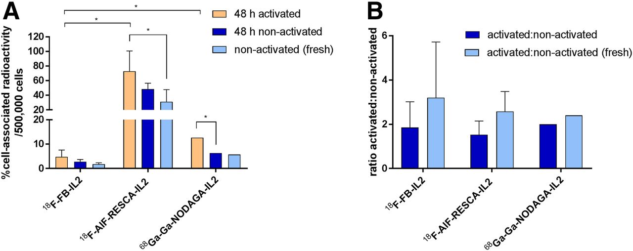
18F-AlF-RESCA-IL2 uptake in activated hPBMCs (73% ± 27.6%) was substantially higher than uptake of 18F-FB-IL2 (4.8% ± 2.8%, P = 0.015) or 68Ga-Ga-NODAGA-IL2 (12.7% ± 0.1%, P = 0.019) (Fig. 2A). When we compared activated-to-nonactivated ratios, we found that the new tracers were just as selective as 18F-FB-IL2 (Fig. 2B). All tracers showed a reduction in cell binding in fresh and nonactivated hPBMCs, compared with uptake in activated hPBMCs. More CD3-positive T cells expressed CD25 in the activated hPBMCs than in the incubated nonactivated hPBMCs (32.1% ± 6.0% vs. 3.4% ± 2.1%, P = 0.002) or in the freshly isolated hPBMCs (3.1% ± 1.2%, P = 0.001) (Supplemental Fig. 1).

In vitro binding assay in human PBMCs for 18F-FB-IL2, 18F-AlF-RESCA-IL2, and 68Ga-Ga-NODAGA-IL2. Four independent experiments were performed for 18F-FB-IL2 and 18F-AlF-RESCA-IL2, in triplicate; For 68Ga-Ga-NODAGA-IL2, 2 independent experiments were performed, in triplicate. (A) Data expressed as percentage of cell-associated radioactivity per 500,000 cells. (B) Data expressed as ratio of activated to nonactivated cells. Data are mean ± SD. *P ≤ 0.05.
In Vitro Stability Studies
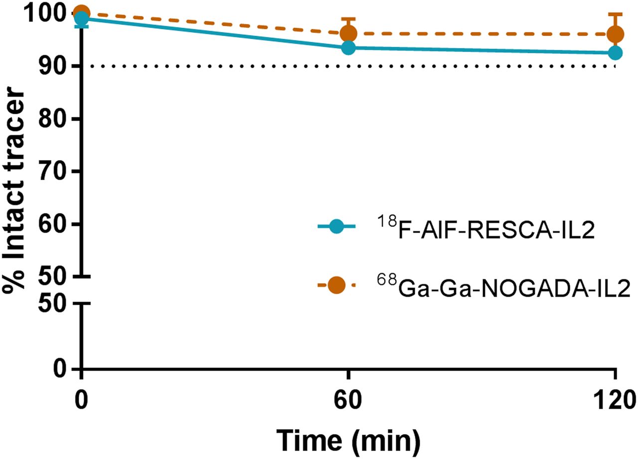
18F-AlF-RESCA-IL2 and 68Ga-Ga-NODAGA-IL2 showed comparable high stability in human serum, with more than 90% of both tracers remaining intact after 1 and 2 h (Fig. 3). For comparison, 18F-FB-IL2 showed slightly higher stability (100% at 1 and 2 h, as described earlier (10,11)).

In vitro stability studies in human serum for 18F-AlF-RESCA-IL2 and 68Ga-Ga-NODAGA-IL2. Data are expressed as percentage of intact tracer after 60 and 120 min of incubation (mean ± SD).
Animal Studies
Ex Vivo Biodistribution Studies in Immunocompetent Mice
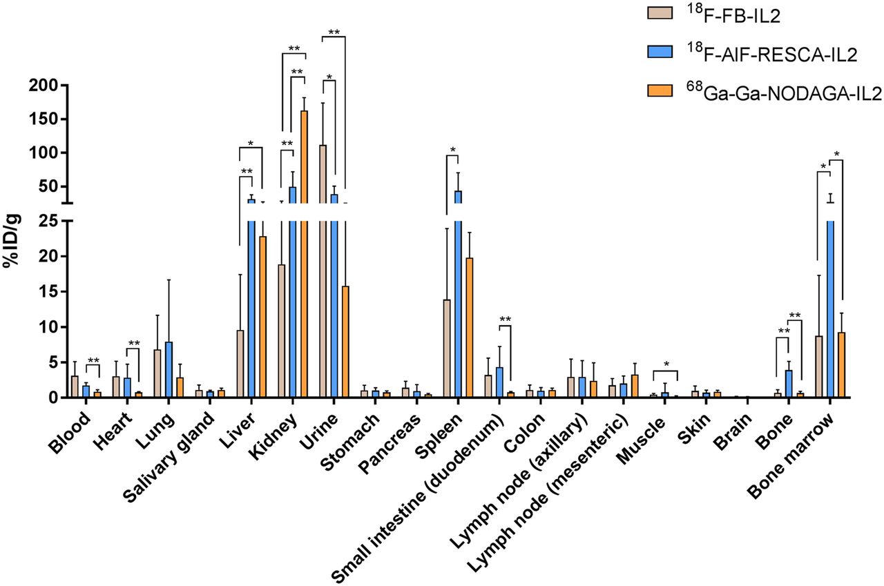
Ex vivo biodistribution studies showed high uptake of 18F-FB-IL2 at sites of renal excretion (Fig. 4; Supplemental Table 1). After 15 min, kidney uptake of 18F-AlF-RESCA-IL2 was higher than that of 18F-FB-IL2 (P = 0.009) (Supplemental Table 1). However, activity in urine was higher for 18F-FB-IL2 (P = 0.004), indicating faster renal excretion. At 60 and 90 min after injection, 18F-AlF-RESCA-IL2 showed higher uptake than 18F-FB-IL2 in liver, kidney, spleen, bone, and bone marrow. 68Ga-Ga-NODAGA-IL2 showed a biodistribution pattern comparable to that of 18F-AlF-RESCA-IL2, with high uptake in liver and kidney. Moreover, 68Ga-Ga-NODAGA-IL2 uptake in the kidney was higher than either 18F-FB-IL2 or 18F-AlF-RESCA-IL2 uptake at all time points (Fig. 4). 18F-AlF-RESCA-IL2 showed the highest uptake in lymphoid organs, such as spleen, lymph nodes, and bone marrow, at 60 min and 90 min.

Comparison of ex vivo biodistribution in immunocompetent BALB/c mice 60 min after injection between 18F-FB-IL2, 18F-AlF-RESCA-IL2, and 68Ga-Ga-NODAGA-IL2 (n = 6 per tracer). Uptake is expressed as percentage injected dose per gram of tissue (%ID/g, mean ± SD). *P ≤ 0.05. **P ≤ 0.01.
In vivo stability of 18F-AlF-RESCA-IL2 was comparable to that of 18F-FB-IL2 at 90 min after injection (81% ± 9% for 18F-FB-IL2 vs. 72% ± 9% for 18F-AlF-RESCA-IL2, P = 0.058) (Supplemental Fig. 2). 68Ga-Ga-NODAGA-IL2 was less stable than 18F-FB-IL2 at 90 min after injection (68Ga-Ga-NODAGA-IL2, 65% ± 5%; P = 0.003).
PET Imaging and Ex Vivo Biodistribution in SCID Mice with a hPBMC Xenograft
Activated hPBMC xenografts inoculated in SCID mice were visualized with all tracers by dynamic PET imaging. No uptake was observed in the Matrigel control group or after coinjection of tracer with a blocking dose of unlabeled IL2. PET images of all tracers showed high uptake in liver and kidney, which did not substantially change over time.
With 18F-FB-IL2 PET, lymph nodes could be detected in only 3 of 5 mice (Fig. 5A), whereas 18F-AlF-RESCA-IL2 PET could clearly visualize lymph nodes in all mice at 60 min after injection (Fig. 5B). With 68Ga-Ga-NODOGA-IL2 PET, lymph nodes could be detected as well, although not as clearly as with 18F-AlF-RESCA-IL2 PET (Fig. 5C).
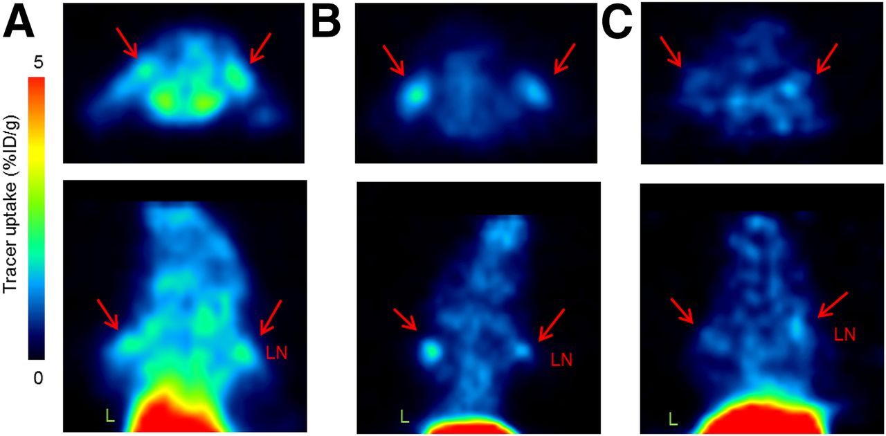
Representative PET images in immunodeficient SCID mice inoculated with activated PBMCs or Matrigel control in right shoulder 15 min before tracer injection for 18F-FB-IL2 (A), 18F-AlF-RESCA-IL2 (B), and 68Ga-Ga-NODAGA-IL2 (C). Clear lymph node uptake is depicted by arrows. Upper panel shows transaxial view; lower panel shows coronal view. L = liver; LN = lymph node; %ID/g = percentage injected dose per gram of tissue.
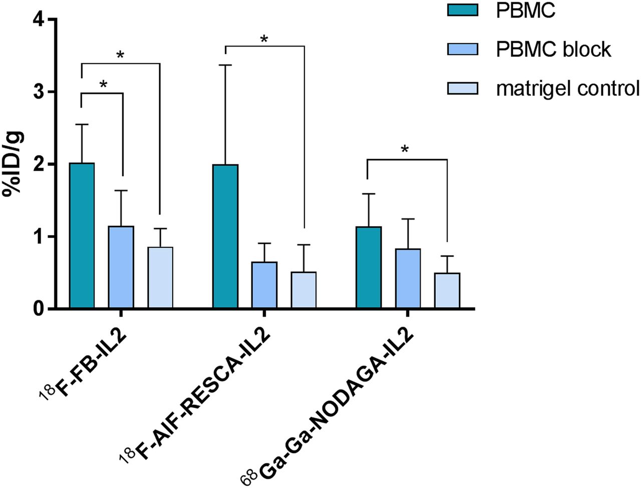
The PET results were confirmed by ex vivo studies showing higher uptake in activated PBMCs than in Matrigel control for all tracers (Fig. 6; Supplemental Table 2). A blocking dose of unlabeled IL2 reduced tracer uptake in PBMCs, with the largest reduction being for 18F-AlF-RESCA-IL2 (18F-FB-IL2, 43.2%; 18F-AlF-RESCA-IL2, 67.5%; 68Ga-Ga-NODAGA-IL2, 26.9%).

Ex vivo uptake (percentage injected dose per gram of tissue [%ID/g]) of 3 radiolabeled IL2 variants in mice inoculated with activated PBMCs or Matrigel control in right shoulder 15 min before tracer injection. For blocking, excess of unlabeled IL2 was coinjected with radiolabeled IL2 (PBMC block). 18F-FB-IL2: PBMC, n = 4; PBMC block, n = 6; Matrigel control, n = 5. 18F-AlF-RESCA-IL2: PBMC, n = 5; PBMC block, n = 6; Matrigel control, n = 6. 68Ga-Ga-NODAGA-IL2: PBMC, n = 5; PBMC block, n = 6; Matrigel control, n = 5. Data are expressed as mean ± SD. *P ≤ 0.05.
DISCUSSION
We developed 2 radiolabeled IL2 tracers, namely 18F-AlF-RESCA-IL2 and 68Ga-Ga-NODAGA-IL2. Both tracers could be produced consistently with a shorter production time than for 18F-FB-IL2. Although labeling of 18F-AlF-RESCA-IL2 gives decay-corrected radiochemical yields similar to those for 18F-FB-IL2, the practical amounts of 18F-AlF-RESCA-IL2 produced are 50% higher because of the shorter production time. In addition, the production requires only 1 synthesis module. This is an advantage for clinical use, as more production runs could be planned and, thus, more patients scanned. The radiochemical yield of 68Ga-Ga-NODAGA-IL2 strongly depends on the volume in which 68GaCl3 can be obtained. An advantage of this tracer is that no cyclotron is needed for radioisotope production, allowing production at sites without a cyclotron.
RESCA was chosen as the chelator for incorporation of Al18F because the complex can be formed at room temperature, preventing potential degradation of the IL2 protein at higher temperatures. Although RESCA was successfully conjugated to IL2, most of the direct radiolabeling attempts did not yield any product, most likely because of the substantial loss of RESCA-IL2 conjugate in the tC2 cartridge during purification. Therefore, it was decided to use an indirect radiolabeling method. This approach provided the desired product, although at a low radiochemical yield of 2.4% ± 1.6%. Nevertheless, starting with less than 50 GBq allows production of a quantity of 18F-AlF-RESCA-IL2 sufficient for several patients (1,375 ± 791 MBq; recommended injected dose, 200 MBq).
For the 68Ga-based IL2 tracer, NODAGA was chosen as the chelator because it has a smaller coordination pocket forming more stable complexes with 68Ga. Moreover, it allows labeling with 68Ga at room temperature (20,21). During 68Ga-Ga-NODAGA-IL2 production, the addition of higher amounts of 68Ga-Cl3 led to yields of less than 10%. A plausible explanation is the low amounts of protein remaining after conjugation, as most of the conjugated NODAGA-IL2 is lost during tC2 purification. With these low radiochemical yields and a maximum amount of 1 GBq eluted from the 68Ge/68Ga generator, 68Ga-Ga-NODAGA-IL2 becomes less suitable for clinical use, since it would be difficult to reliably obtain a patient dose. Therefore, to use 68Ga-Ga-NODAGA-IL2 in clinical studies, further optimization is warranted.
Our study showed that 18F-AlF-RESCA-IL2 had the highest in vitro uptake in hPBMCs and the highest in vivo uptake in target tissues of BALB/c mice, such as lymph nodes, spleen, and bone marrow. Since IL2 is injected in a subpharmacologic dose, no adverse effects are expected. In addition, in the first clinical study with 18F-FB-IL2, no adverse effects due to radiation burden were observed. Therefore, we do not expect additional toxicity from the new analogs. In contrast to 18F-FB-IL2 and 68Ga-Ga-NODAGA-IL2, which have mainly renal clearance, 18F-AlF-RESCA-IL2 has higher hepatobiliary clearance, as can be attributed either to the presence of the RESCA moiety, which introduces an additional charge, or to the presence of degradation products, such as unconjugated 18F-AlF-RESCA or free 18F-AlF (16,22). These high uptake values obtained in excretory organs are in the same range as obtained with previous IL2-based tracers, for which no renal or hepatic toxicity was found (23–25). A limitation for clinical use of 68Ga-Ga-NODAGA-IL2 is high retention in the kidney, which can lead to a radiation burden due to its increased positron energy compared with 18F-fluorine. Therefore, 18F-AlF-RESCA-IL2 might be preferred for clinical molecular imaging studies. PET images obtained with 18F-AlF-RESCA-IL2 showed less background and therefore were of higher contrast than images obtained with the other tracers. In addition, lymph nodes could be visualized with 18F-AlF-RESCA-IL2 in all animals, whereas with 18F-FB-IL2 PET, lymph nodes were visible in only some animals, indicating 18F-AlF-RESCA-IL2 to have good targeting characteristics.
Previous SPECT and PET imaging studies have shown that IL2-derived tracers could detect T cells in inflammatory and infectious diseases, as well as in different tumor types (7–9, 23–28). In a small clinical pilot study on only 5 patients with melanoma, 99mTc-IL2 SPECT could detect metastases (29). Currently, other tracers targeting immune cells are also being developed, such as targeting T cells via CD8-specific tracers (30–33). Another method to detect immune cells is radiolabeling of immune checkpoint–targeting monoclonal antibodies with long–half-life isotopes such as 89Zr. Compared with these antibody-based tracers, radiolabeled IL2 tracers have an advantage in that, with shorter half-lives, imaging can be performed shortly after tracer injection. Thus, in a fast, noninvasive manner, information about the presence and dynamics of immune cells can be gained. Our results indicate that 18F-AlF-RESCA-IL2 has good in vitro and in vivo characteristics and the potential to be used as a PET tracer for imaging of T cells.
CONCLUSION
We developed 2 radiolabeled IL2 tracers for imaging of immune cells: 18F-AlF-RESCA-IL2 and 68Ga-Ga-NODAGA-IL2. Production of these tracers was faster than production of the previously developed 18F-FB-IL2 and is a potential advantage for clinical use. 18F-AlF-RESCA-IL2 showed the highest practical production yield and the highest in vitro binding to activated hPBMCs. Moreover, in vivo studies showed high uptake of 18F-AlF-RESCA-IL2 in PBMC xenografts and lymphoid tissues such as spleen, bone marrow, and lymph nodes. This finding supports the potential to use this tracer in future studies for detection of CD25-positive immune cells.
DISCLOSURE
The research leading to these results received funding from the Innovative Medicines Initiatives 2 Joint Undertaking under grant agreement No. 116106 (TRISTAN). This Joint Undertaking receives support from the European Union’s Horizon 2020 research and innovation program and EFPIA. Elisabeth de Vries had a consulting and advisory role for NSABP, Daiichi Sankyo, Pfizer, Sanofi, Merck, and Synthon Biopharmaceuticals; and received grants from Amgen, Genentech, Roche, Chugai Pharma, CytomX Therapeutics, Nordic Nanovector, G1 Therapeutics, AstraZeneca, Radius Health, and Bayer, all made available to the institution, outside the submitted work. Erik de Vries received grants from ZonMW (95104008 and 95105010) during this study; received a grant from the Dutch Cancer Foundation (RUG2015-7235); and performed contract research studies with Rodin Therapeutics, Lysosomal Therapeutics Inc., Hoffmann-La Roche Ltd., and Ionis Pharmaceuticals, made available to the institution outside the submitted work. Geke Hospers holds a consulting and advisory role for Amgen, Roche, MSD, BMS, Pfizer, and Novartis and received grants from BMS and Seerave, made available to the institution outside the submitted work. Frederik Cleeren is a postdoctoral fellow of FWO (12R3119N). This research received support from Research Foundation–Flanders (FWO) (G0D8817N). No other potential conflict of interest relevant to this article was reported.
KEY POINTS
QUESTION: What are the in vitro and in vivo characteristics of 2 radiolabeled IL2 variants, 18F-AlF-RESCA-IL2 and 68Ga-Ga-NODAGA-IL2, compared with 18F-FB-IL2?
PERTINENT FINDINGS: Production is faster for these tracers than for the previously developed 18F-FB-IL2. 18F-AlF-RESCA-IL2 shows the best in vitro binding characteristics. In vivo studies showed high 18F-AlF-RESCA-IL2 uptake in PBMCs and lymphoid organs such as spleen, bone marrow, and lymph nodes.
IMPLICATIONS FOR PATIENT CARE: Our results indicate the potential to use 18F-AlF-RESCA-IL2 in future molecular imaging studies for detection of CD25-positive immune cells.
Footnotes
Published online Feb. 28, 2020.
- © 2020 by the Society of Nuclear Medicine and Molecular Imaging.
REFERENCES
- Received for publication October 25, 2019.
- Accepted for publication January 3, 2020.


Jump to section
Related Articles
Cited By...
- No citing articles found.