


Article Figures & Data
Figures
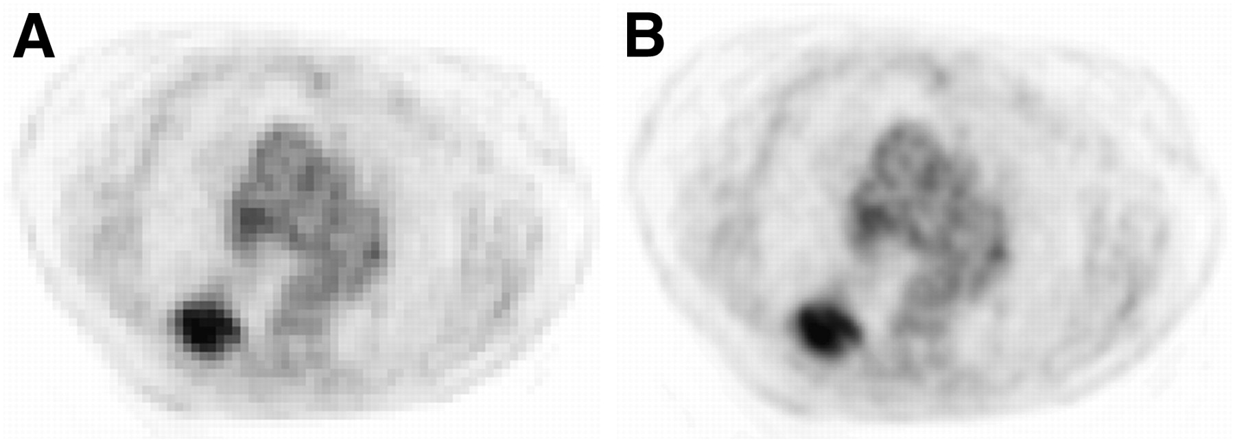
- FIGURE 1.
Illustration of up-sampled PET images (central axial slice). Original PET image with voxel size of 5.31 × 5.31 × 5 mm (A) and PET image up-sampled with voxel size equal to CT (0.98 × 0.98 × 5 mm) (B) using cubic B-spline interpolation.
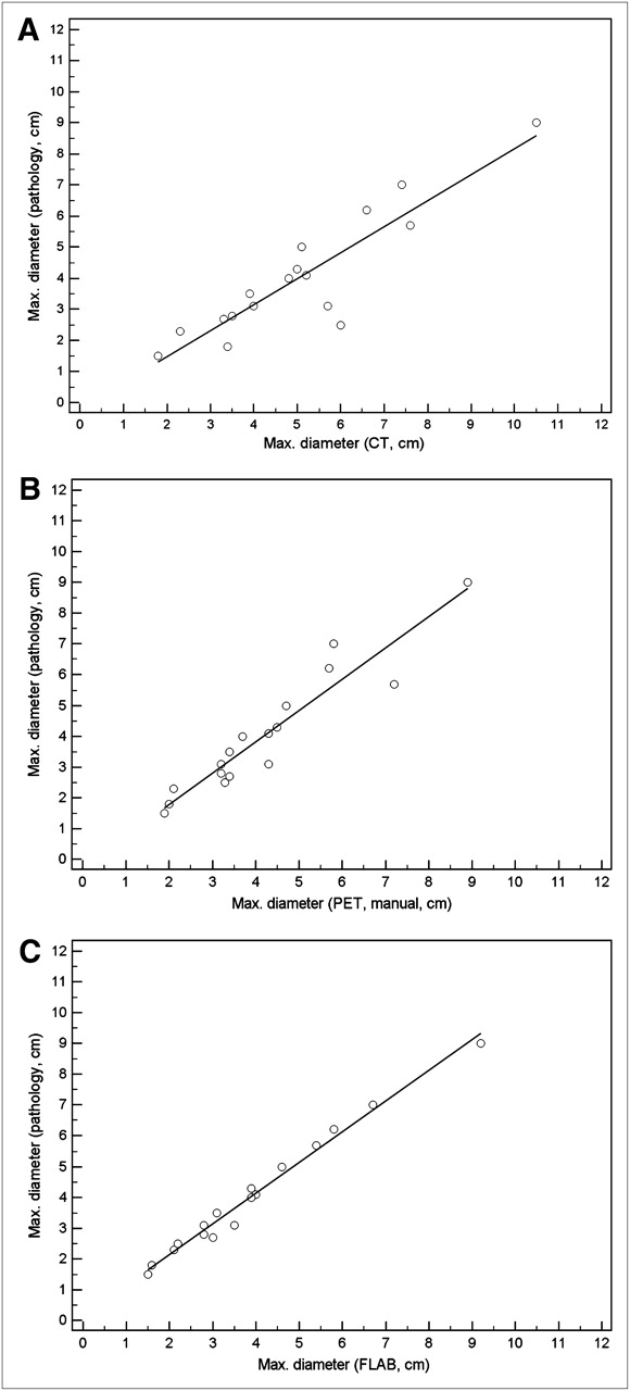
- FIGURE 2.
Correlations with manual delineations on CT (A) and PET (B) and with FLAB delineations on PET (C).
- FIGURE 3.
Absolute (in cm) differences (A) and relative (%) errors (B) between pathology measurements and image-based delineations.
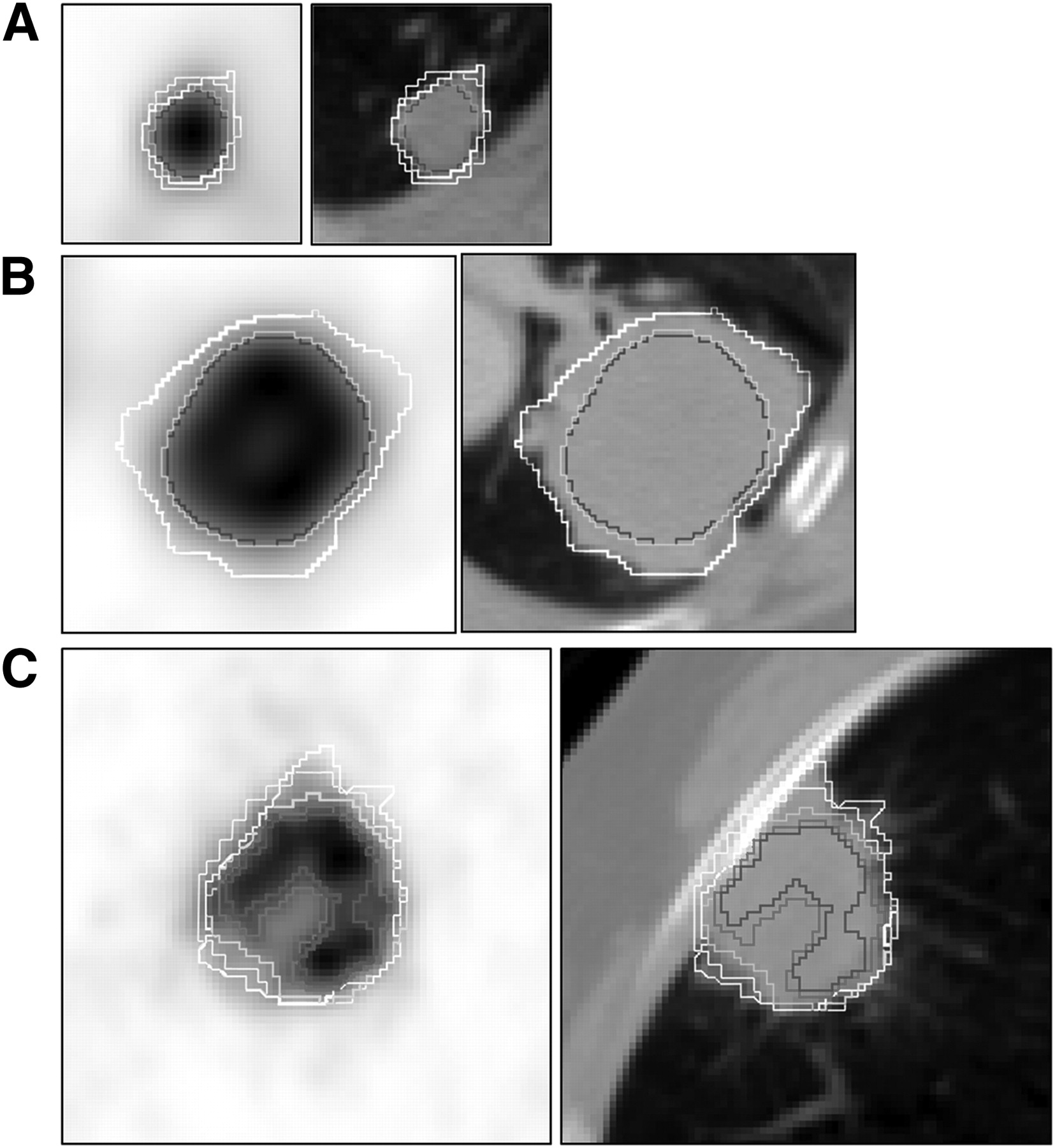
- FIGURE 4.
Small lesions (<2 cm in diameter) (A) and larger lesions with moderate (COVFLAB = 0.23) (B) and higher (COVFLAB = 0.30) (C) heterogeneity. For readability, A1 contours are not shown in B and C and manual PET contours are not shown in B as they were similar to FLAB and T50. White = manual on CT; blue = T50; purple = A1; green = FLAB.
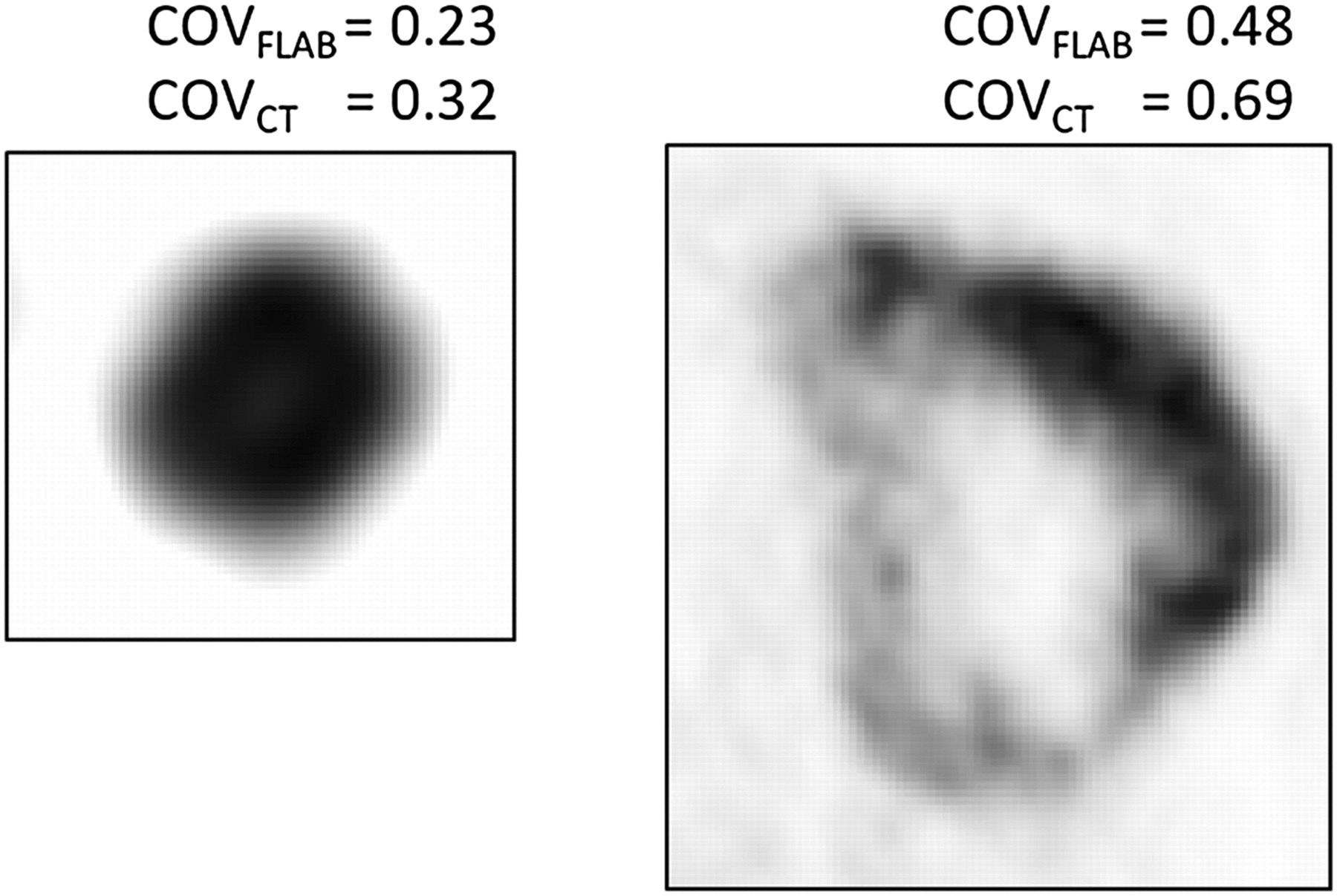
- FIGURE 5.
Heterogeneity estimation for 2 tumors.
- FIGURE 6.
Correlation between anatomic volume (A) or uptake heterogeneity (B) and differences between T50 and FLAB volumes.


Tables
- TABLE 1
Maximum-Diameter Measurements on Pathology and Image Delineations for All 17 Patients
Measurement (cm) Patient no. Pathologic CT1 (manual) CT2 (manual) PET (manual) PET (T50) PET (A1) PET (A2) PET (FLAB) 1 6.2 6.6 6.7 5.7 4.6 5 4.8 5.8 2 2.7 3.3 3.3 3.4 2.8 3.1 2.8 3 3 9 10.5 10.1 8.9 7 7.5 7.7 9.2 4 1.5 1.8 1.9 2.1 1.3 1.6 1.3 1.5 5 1.8 3.4 3.4 2 1.2 1.4 1.3 1.6 6 3.1 4 3.9 3.2 2.4 2.6 2.5 2.8 7 4.3 5 5.1 4.5 3.8 3.9 3.8 3.9 8 3.1 5.7 5.7 5.1 2.8 4 3.7 3.5 9 3.5 3.9 4 3.4 2.7 2.9 3 3.1 10 5.7 7.6 7.7 7.4 7.5 4.7 6.7 5.4 11 5 5.1 5.3 4.7 2.7 3 2.9 4.6 12 2.8 3.5 3.2 3.2 2.4 2.5 2.6 2.8 13 4.1 5.2 5.1 4.3 3.2 3.3 3.3 4 14 4 4.8 4.9 3.7 3.2 3.4 3.2 3.9 15 7 7.4 7.4 5.8 6.2 6.5 6.3 6.7 16 2.3 2.3 2.4 2.1 1.8 1.7 1.9 2.1 17 2.5 6 5.9 4.5 2.5 2.7 2.6 2.2 Mean ± SD 4.0 ± 2.0 5.1 ± 2.2 5.1 ± 2.1 4.2 ± 1.9 3.4 ± 1.9 3.5 ± 1.6 3.6 ± 1.8 3.9 ± 2.0 Median 3.5 5.0 5.1 3.7 2.8 3.1 3.0 3.5 Range 1.5–9 1.8–10.5 1.9–10.1 1.9–8.9 1.2–7.5 1.4–7.5 1.3–7.7 1.5–9.2 Pearson r — 0.90 0.91 0.95 0.89 0.95 0.93 0.99 95% CI for r — 0.74–0.96 0.76–0.96 0.86–0.98 0.72–0.96 0.85–0.98 0.81–0.98 0.98–1.00 CI = confidence interval.
Tumor volume (cm3) (n = 25) Mean ± SD Median Range CT1 (manual) 54.5 ± 74.0 28.2 1.9–338.9 CT2 (manual) 55.1 ± 74.8 29.1 1.8–339.4 PET (manual) 47.3 ± 76.4 21.3 2.1–356.2 PET (T50) 17.7 ± 25.1 9.2 8.5–125.8 PET (A1) 22.6 ± 33.2 11.9 1.2–166.9 PET (A2) 21.8 ± 33.9 11.3 0.9–172.4 PET (FLAB) 39.5 ± 70.5 15.8 1.1–345.1


Jump to section
Related Articles
Cited By...
- Surgical and Oncological Factors Affecting the Successful Engraftment of Patient-derived Xenografts in Pancreatic Ductal Adenocarcinoma
- 18F-FDG PET Uptake Characterization Through Texture Analysis: Investigating the Complementary Nature of Heterogeneity and Functional Tumor Volume in a Multi-Cancer Site Patient Cohort
- Visual Versus Quantitative Assessment of Intratumor 18F-FDG PET Uptake Heterogeneity: Prognostic Value in Non-Small Cell Lung Cancer
- Interobserver Agreement of Qualitative Analysis and Tumor Delineation of 18F-Fluoromisonidazole and 3'-Deoxy-3'-18F-Fluorothymidine PET Images in Lung Cancer
- Tumor Heterogeneity and Permeability as Measured on the CT Component of PET/CT Predict Survival in Patients with Non-Small Cell Lung Cancer
- Comparison Between 18F-FDG PET Image-Derived Indices for Early Prediction of Response to Neoadjuvant Chemotherapy in Breast Cancer