


Article Figures & Data
Figures
- FIGURE 1.
(A) Complexation reaction between [89Zr(C2O4)4]4− and DFO. (B) DFT-optimized structure of 8-coordinate complex [89Zr(HDFO)-cis-(H2O)2]2+ (3-cis).
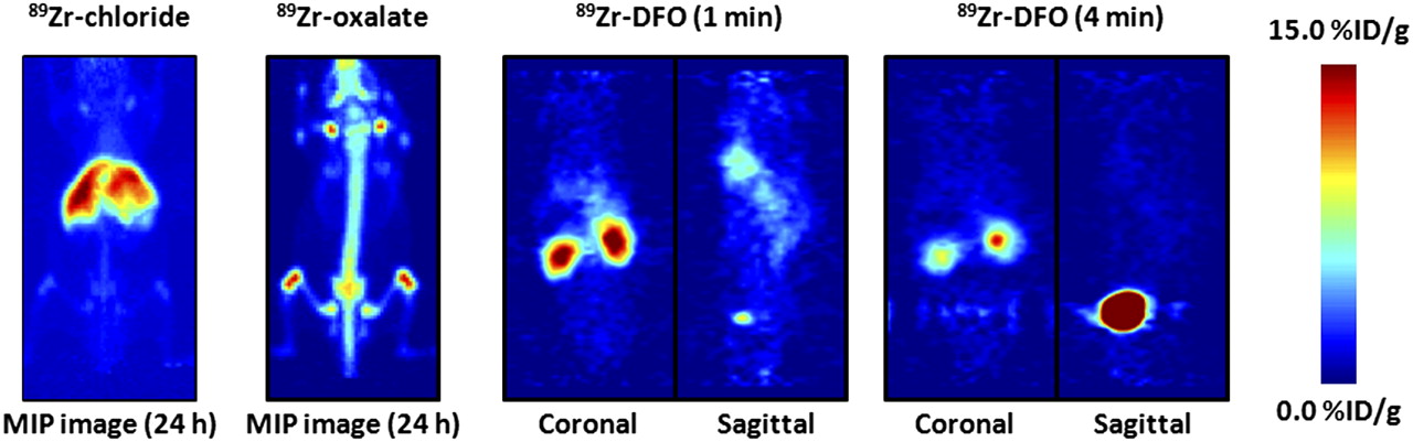
- FIGURE 2.
PET images showing maximum intensity projection of 89Zr-chloride and 89Zr-oxalate at 24 h after intravenous administration and dynamic PET images of 89Zr-DFO at 1 and 4 min after injection. For maximum-intensity-projection images, upper and lower intensity thresholds were set at 100% and 0%, respectively. Further details are presented in Supplemental Figures 3–6. MIP = maximum intensity projection.
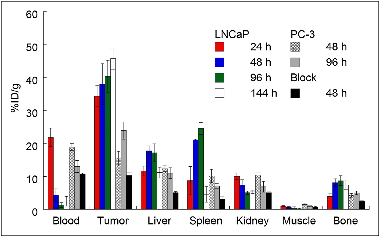
- FIGURE 3.
Bar chart showing selected tissue biodistribution data (%ID/g) for uptake of either high- (181.7 ± 1.1 MBq/mg [4.91 ± 0.03 mCi/mg]; 3–4 μg of mAb per mouse) or low-specific-activity (60-fold decrease, 3.04 MBq/mg [0.082 mCi/mg]; 300 μg of mAb per mouse) formulations of 89Zr-DFO-J591 (0.55–0.74 MBq [15–20 μCi], in 200 μL of sterile saline for injection) in male athymic nu/nu mice bearing subcutaneous LNCaP (PSMA-positive) or PC-3 (PSMA-negative) tumors.
- FIGURE 4.
Temporal immunoPET images of 89Zr-DFO-J591 (10.9–11.3 MBq [295–305 μCi], 60–62 μg of mAb, in 200 μL of sterile saline) recorded in LNCaP tumor–bearing (PSMA-positive, left shoulder) (A) and PC-3 tumor–bearing (PSMA-negative, right shoulder) (B) mice between 3 and 144 h after injection. Transverse and coronal planar images intersect center of tumors, and mean tumor-to-muscle ratios derived from volume-of-interest analysis of immunoPET images are given. Upper thresholds of immunoPET have been adjusted for visual clarity, as indicated by scale bars.
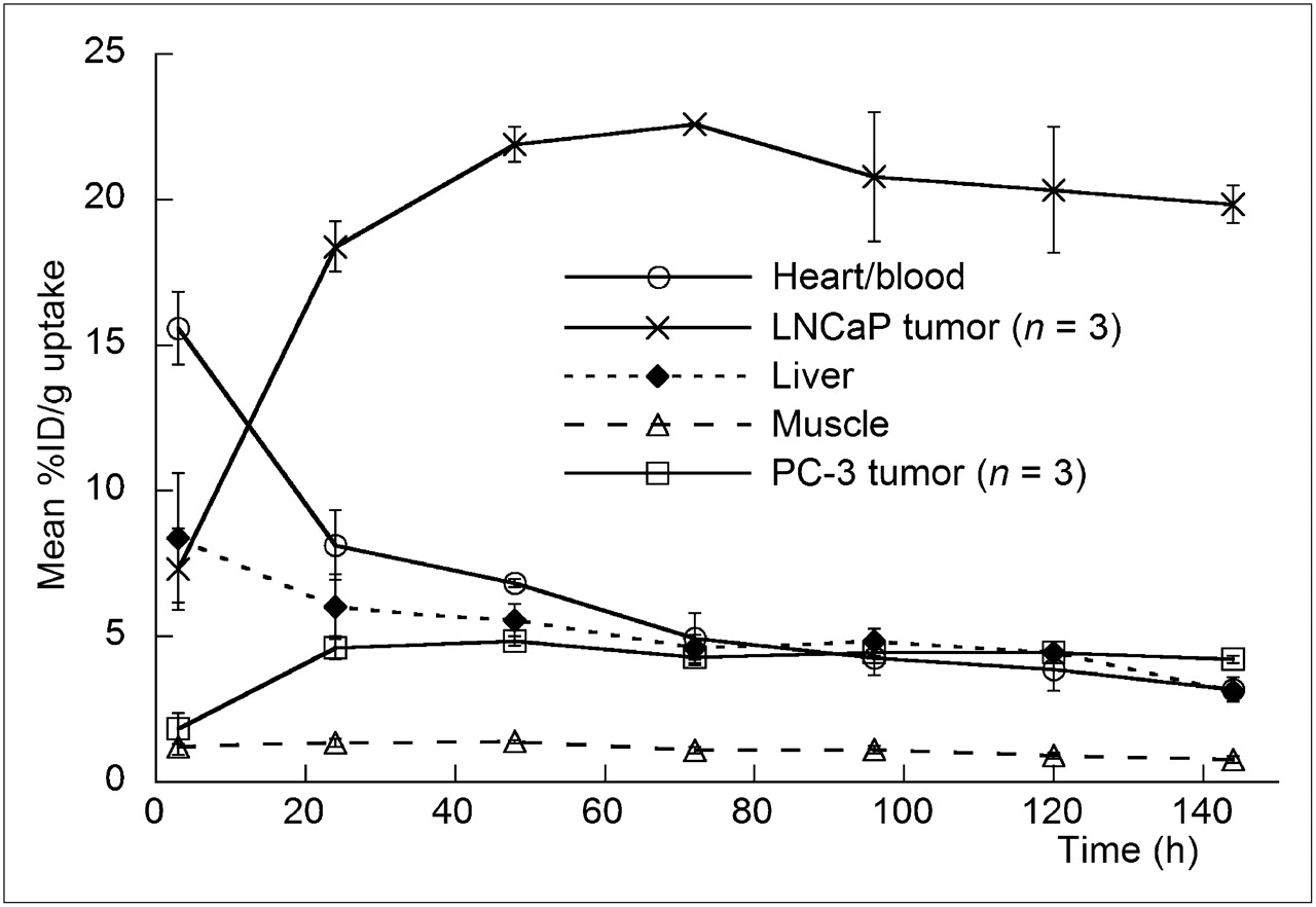
- FIGURE 5.
Time–activity curves derived by volume-of-interest analysis of immunoPET images showing mean %ID/g tissue uptake vs. time/h for 89Zr-DFO-J591 radiotracer accumulation in mice bearing subcutaneous LNCaP (PSMA-positive) or PC-3 (PSMA-negative) tumors. Complete time–activity curve data for 89Zr-DFO-J591 immunoPET imaging is given in supplemental materials (Supplemental Tables 9 and 10; Supplemental Figs. 11–13).


Tables
- TABLE 1.
Biodistribution Data of 89Zr-DFO-J591, Administered Intravenously to Mice Bearing Subcutaneous LNCaP Tumors
Organ 24 h (n = 4) 48 h (n = 5) 96 h (n = 5) 144 h (n = 4) Block (300 μg of mAb) at 48 h (n = 4) Blood 21.8 ± 2.8 4.4 ± 1.9 1.4 ± 0.8 2.6 ± 1.5 10.7 ± 0.4 Tumor 34.4 ± 3.2 38.0 ± 6.2 40.4 ± 4.8 45.8 ± 3.2 10.3 ± 0.8 Heart 7.4 ± 2.2 4.0 ± 1.3 1.7 ± 0.6 1.4 ± 0.5 3.8 ± 0.7 Lung 11.7 ± 1.9 5.7 ± 3.1 2.2 ± 0.9 2.5 ± 0.9 5.7 ± 0.3 Liver 11.7 ± 1.5 17.7 ± 1.6 17.2 ± 2.7 11.2 ± 1.6 5.1 ± 0.4 Spleen 8.8 ± 4.3 21.1 ± 0.3 24.6 ± 1.8 4.6 ± 2.4 3.1 ± 0.7 Kidney 10.1 ± 1.0 7.5 ± 1.5 5.1 ± 0.5 5.3 ± 0.5 5.1 ± 0.2 Muscle 1.1 ± 0.1 0.6 ± 0.3 0.4 ± 0.4 0.2 ± 0.2 0.8 ± 0.2 Bone 4.0 ± 0.8 8.2 ± 1.2 8.7 ± 1.5 7.4 ± 1.3 2.4 ± 0.3 Tumor/blood 1.6 ± 0.2 8.7 ± 4.1 29.7 ± 17.1 18.0 ± 10.5 1.0 ± 0.1 Tumor/heart 4.7 ± 1.5 9.6 ± 3.5 23.4 ± 9.0 31.9 ± 10.7 2.7 ± 0.5 Tumor/lung 2.9 ± 0.5 6.7 ± 3.7 18.4 ± 7.7 18.5 ± 6.8 1.8 ± 0.2 Tumor/liver 2.9 ± 0.5 2.1 ± 0.4 2.3 ± 0.5 4.1 ± 0.7 2.0 ± 0.2 Tumor/spleen 3.9 ± 1.9 1.8 ± 0.3 1.6 ± 0.2 9.9 ± 5.2 3.3 ± 0.8 Tumor/kidney 3.4 ± 0.5 5.1 ± 1.3 7.9 ± 1.2 8.6 ± 1.1 2.0 ± 0.2 Tumor/muscle 32.4 ± 4.6 59.2 ± 28.8 95.9 ± 95.3 306.4 ± 432.2 13.3 ± 3.1 Tumor/bone 8.7 ± 1.9 4.7 ± 1.0 4.6 ± 1.0 6.2 ± 1.2 4.3 ± 0.6 Complete biodistribution data are presented in Supplemental Table 7. Data are expressed as mean %ID/g ± SD. Errors for tumor-to-tissue ratios are calculated as geometric mean of SD. LNCaP tumors: 3–4 μg mAb; PSMA-positive, 50–250 mm3.
- TABLE 2.
Biodistribution Data of 89Zr-DFO-J591, Administered Intravenously to Mice Bearing Subcutaneous PC-3 Tumors (3–4 μg of mAb)
Organ 48 h (n = 4) 96 h (n = 3) Blood 19.0 ± 1.1 13.0 ± 1.8 Tumor 15.6 ± 2.1 24.0 ± 2.6 Heart 6.8 ± 0.1 4.3 ± 0.9 Lung 12.6 ± 1.9 7.0 ± 2.3 Liver 12.4 ± 0.9 11.0 ± 1.6 Spleen 10.2 ± 2.0 7.2 ± 0.7 Kidney 10.5 ± 0.9 6.9 ± 1.6 Muscle 1.5 ± 0.4 0.9 ± 0.2 Bone 4.3 ± 0.6 5.1 ± 0.5 Tumor/blood 0.8 ± 0.1 1.8 ± 0.3 Tumor/heart 2.3 ± 0.3 5.6 ± 1.4 Tumor/lung 1.2 ± 0.3 3.4 ± 1.2 Tumor/liver 1.3 ± 0.2 2.2 ± 0.4 Tumor/spleen 1.5 ± 0.4 3.4 ± 0.5 Tumor/kidney 1.5 ± 0.2 3.5 ± 0.9 Tumor/muscle 10.4 ± 3.3 25.4 ± 5.8 Tumor/bone 3.6 ± 0.7 4.7 ± 0.7 Complete biodistribution data are presented in Supplemental Table 7. Data are expressed as mean %ID/g ± SD. Errors for tumor-to-tissue ratios are calculated as geometric mean of SD. PC-3 tumors: PSMA-negative, 70–90 mm3.
Supplemental Data
Files in this Data Supplement:


Jump to section
Related Articles
Cited By...
- Single Chelator-Minibody Theranostic Agents for 89Zr PET Imaging and 177Lu Radiopharmaceutical Therapy of PSMA-Expressing Prostate Cancer
- DOTA chelation through click chemistry enables favorable biodistribution of 89Zr-radiolabeled antibodies: A comparison with DFO chelation
- CD8-Targeted PET Imaging of Tumor-Infiltrating T Cells in Patients with Cancer: A Phase I First-in-Humans Study of 89Zr-Df-IAB22M2C, a Radiolabeled Anti-CD8 Minibody
- Radiolabeling of PSMA-617 with 89Zr: A Novel Use of DMSO for Radiochemical Yield Enhancement and Preliminary Small-Animal PET Results
- Molecular Imaging of Prostate Cancer Targeting CD46 Using ImmunoPET
- The Effects of Monosodium Glutamate on PSMA Radiotracer Uptake in Men with Recurrent Prostate Cancer: A Prospective, Randomized, Double-Blind, Placebo-Controlled Intraindividual Imaging Study
- Acute Statin Treatment Improves Antibody Accumulation in EGFR- and PSMA-Expressing Tumors
- Detection of incipient pancreatic cancer with novel tumor-specific antibodies in mouse models
- Light-Induced Radiosynthesis of 89Zr-DFO-Azepin-Onartuzumab for Imaging the Hepatocyte Growth Factor Receptor
- First-in-Humans Imaging with 89Zr-Df-IAB22M2C Anti-CD8 Minibody in Patients with Solid Malignancies: Preliminary Pharmacokinetics, Biodistribution, and Lesion Targeting
- Total-Body PET and Highly Stable Chelators Together Enable Meaningful 89Zr-Antibody PET Studies up to 30 Days After Injection
- Exploitation of CD133 for the Targeted Imaging of Lethal Prostate Cancer
- A Novel Fully Human Antibody targeting Extracellular Domain of PSMA Inhibits Tumor Growth in Prostate Cancer
- Immuno-PET of Innate Immune Markers CD11b and IL-1{beta} Detects Inflammation in Murine Colitis
- Monosodium Glutamate Reduces 68Ga-PSMA-11 Uptake in Salivary Glands and Kidneys in a Preclinical Prostate Cancer Model
- Preclinical Evaluation of the Hsp70 Peptide Tracer TPP-PEG24-DFO[89Zr] for Tumor-Specific PET/CT Imaging
- Long-Half-Life 89Zr-Labeled Radiotracers Can Guide Percutaneous Biopsy Within the PET/CT Suite Without Reinjection of Radiotracer
- ImmunoPET of Malignant and Normal B Cells with 89Zr- and 124I-Labeled Obinutuzumab Antibody Fragments Reveals Differential CD20 Internalization In Vivo
- First-in-Human Imaging with 89Zr-Df-IAB2M Anti-PSMA Minibody in Patients with Metastatic Prostate Cancer: Pharmacokinetics, Biodistribution, Dosimetry, and Lesion Uptake
- Internalization of secreted antigen-targeted antibodies by the neonatal Fc receptor for precision imaging of the androgen receptor axis
- Evaluation of Castration-Resistant Prostate Cancer with Androgen Receptor-Axis Imaging
- Molecular Imaging and Quantitation of EphA2 Expression in Xenograft Models with 89Zr-DS-8895a
- Targeted Imaging of the Atypical Chemokine Receptor 3 (ACKR3/CXCR7) in Human Cancer Xenografts
- New Strategies in Prostate Cancer: Prostate-Specific Membrane Antigen (PSMA) Ligands for Diagnosis and Therapy
- Immuno-PET Imaging of CD30-Positive Lymphoma Using 89Zr-Desferrioxamine-Labeled CD30-Specific AC-10 Antibody
- A Phase I/II Study for Analytic Validation of 89Zr-J591 ImmunoPET as a Molecular Imaging Agent for Metastatic Prostate Cancer
- Annotating STEAP1 Regulation in Prostate Cancer with 89Zr Immuno-PET
- CDK9-mediated transcription elongation is required for MYC addiction in hepatocellular carcinoma
- Preclinical Evaluation of Multistep Targeting of Diasialoganglioside GD2 Using an IgG-scFv Bispecific Antibody with High Affinity for GD2 and DOTA Metal Complex
- Glypican-3-Targeted 89Zr PET Imaging of Hepatocellular Carcinoma: Where Antibody Imaging Dares to Tread
- Quantitative ImmunoPET of Prostate Cancer Xenografts with 89Zr- and 124I-Labeled Anti-PSCA A11 Minibody
- Applying PET to Broaden the Diagnostic Utility of the Clinically Validated CA19.9 Serum Biomarker for Oncology
- Interrogating Tumor Metabolism and Tumor Microenvironments Using Molecular Positron Emission Tomography Imaging. Theranostic Approaches to Improve Therapeutics
- Phase II Study of Lutetium-177-Labeled Anti-Prostate-Specific Membrane Antigen Monoclonal Antibody J591 for Metastatic Castration-Resistant Prostate Cancer
- Determination of the In Vivo Selectivity of a New {kappa}-Opioid Receptor Antagonist PET Tracer 11C-LY2795050 in the Rhesus Monkey
- Monitoring Afatinib Treatment in HER2-Positive Gastric Cancer with 18F-FDG and 89Zr-Trastuzumab PET
- Unmet Needs in the Prediction and Detection of Metastases in Prostate Cancer
- Imaging Tumor Burden in the Brain with 89Zr-Transferrin
- 64Cu-p-NH2-Bn-DOTA-hu14.18K322A, a PET Radiotracer Targeting Neuroblastoma and Melanoma
- Advances in Immuno-Positron Emission Tomography: Antibodies for Molecular Imaging in Oncology
- PET of Signal Transduction Pathways in Cancer
- Imaging Androgen Receptor Signaling with a Radiotracer Targeting Free Prostate-Specific Antigen
- 2-(3-{1-Carboxy-5-[(6-[18F]Fluoro-Pyridine-3-Carbonyl)-Amino]-Pentyl}-Ureido)-Pentanedioic Acid, [18F]DCFPyL, a PSMA-Based PET Imaging Agent for Prostate Cancer
- Targeting the Internal Epitope of Prostate-Specific Membrane Antigen with 89Zr-7E11 Immuno-PET
- Noninvasive measurement of androgen receptor signaling with a positron-emitting radiopharmaceutical that targets prostate-specific membrane antigen
- Magnitude of Enhanced Permeability and Retention Effect in Tumors with Different Phenotypes: 89Zr-Albumin as a Model System