Index by author
Cover image

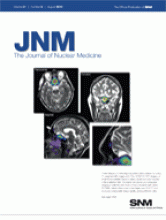
These images of a meningioma patient were created by fusing T2-weighted MR images with 68Ga-DOTATOC PET images. A small frontal satellite lesion is clearly visible and was included in the irradiation field. Structural, functional, and molecular imaging in patients with brain tumors is feasible with hybrid PET/MRI, which offers many advantages over PET/CT and produces comparable image quality and quantitative data.
See page 1198.