


Article Figures & Data
Figures
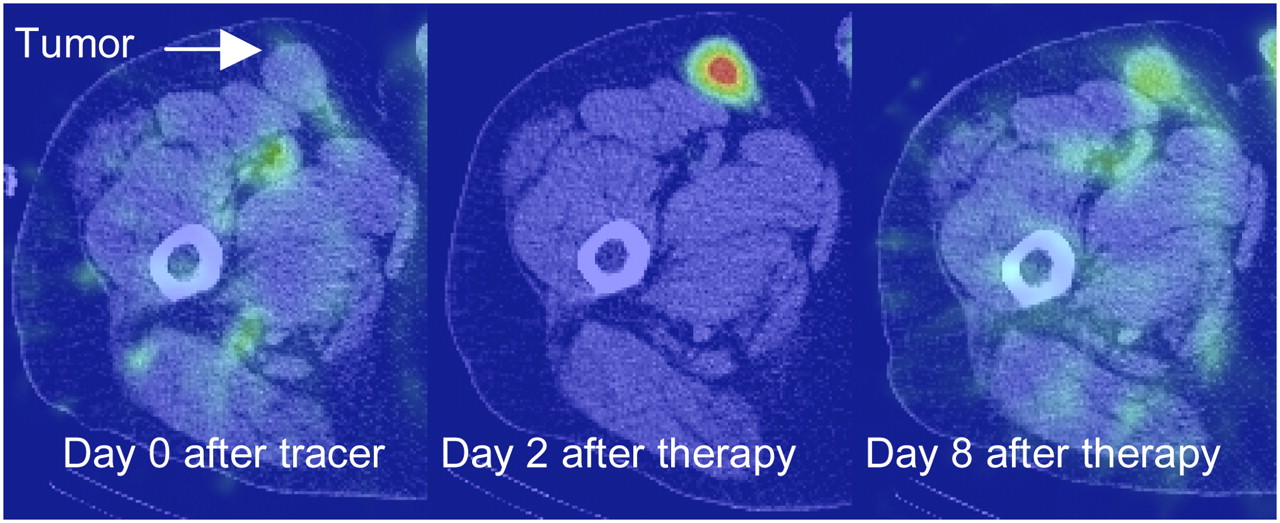
- FIGURE 1.
SPECT/CT images showing uptake in an inguinal tumor at day 0 after tracer, day 2 after therapy, and day 8 after therapy.
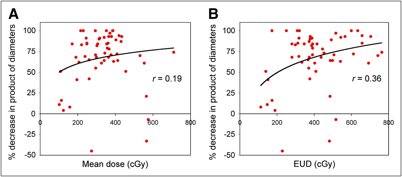
- FIGURE 2.
Response at 2 mo plotted against mean tumor-absorbed dose (A) and EUD (B), at tumor level (n = 57).
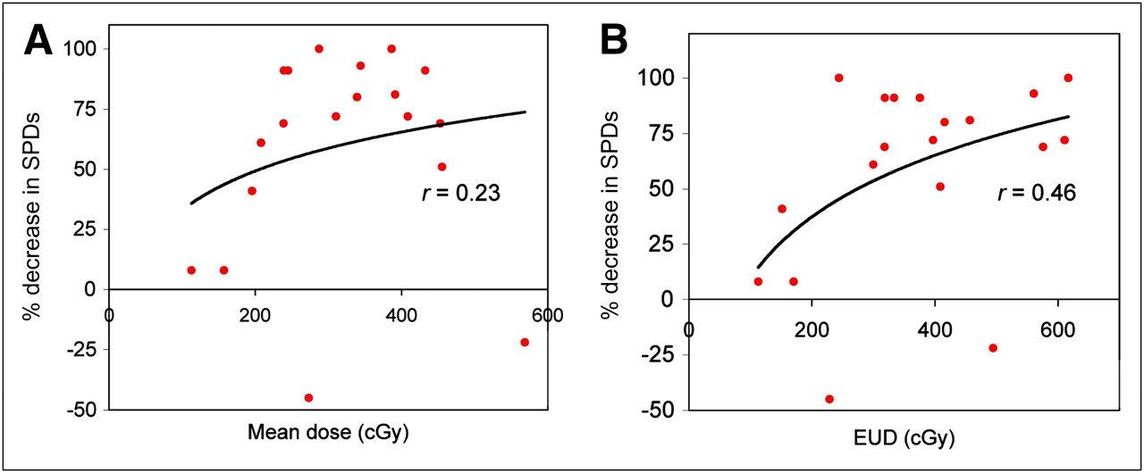
- FIGURE 3.
Response at 2 mo (based on sum of products (SPDs) of perpendicular tumor diameters) plotted against mean tumor-absorbed dose (A) and EUD (B), at patient level (n = 19). Dose values were averaged over multiple tumors of each patient.
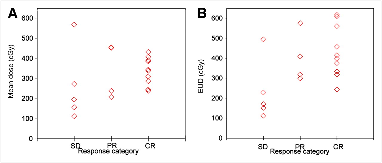
- FIGURE 4.
Mean tumor-absorbed dose (A) and EUD (B) as function of patient response as assessed by Cheson et al. (19).

Tables
Parameter Sample size Median Range Initial tumor volume (mL) 60 34 2 to 423 Volume decrease during tracer SPECT/CT* (%) 60 12 −10 to 49 Volume decrease during therapy SPECT/CT† (%) 60 30 2 to 76 Decrease in product of diameters at 2 mo‡ (%) 60 72 −45 to 100 Average dose (cGy) 57 341 102 to 711 EUD (cGy) 57 391 113 to 764 Maximum dose (cGy) 57 508 162 to 1,404 D99 (cGy) 57 203 35 to 373 D80 (cGy) 57 269 58 to 466 ↵* Difference in volumes defined on first and last posttracer scans (6 d).
↵† Difference in volumes defined on first and last posttherapy scans (∼6 d).
↵‡ Difference in product of largest perpendicular diameters at 2 mo compared with the first posttracer imaging time point.
Results for individual tumors are given in the supplemental material.
- TABLE 2
Pearson Correlations Between Various Dose Measures and Response at 2 Months Determined by Percentage Reduction in Product of Longest Diameters (Tumor Level) or Sum of Product of Diameters (Patient Level)
Tumor level Patient level Parameter rp P rp P Mean tumor dose 0.189 0.159 0.226 0.352 Maximum tumor dose 0.033 0.809 0.086 0.726 D99 0.164 0.222 0.193 0.429 D80 0.200 0.136 0.223 0.359 EUD 0.357 0.006 0.459 0.048
Supplemental Data
Files in this Data Supplement:


Jump to section
Related Articles
Cited By...
- Dosimetry in Radiopharmaceutical Therapy
- Dosimetry for Radiopharmaceutical Therapy: Current Practices and Commercial Resources
- Tumor Response to Radiopharmaceutical Therapies: The Knowns and the Unknowns
- DPD Quantification in Cardiac Amyloidosis: A Novel Imaging Biomarker
- Tumor-Absorbed Dose for Non-Hodgkin Lymphoma Patients Treated with the Anti-CD37 Antibody Radionuclide Conjugate 177Lu-Lilotomab Satetraxetan
- Tumor-Absorbed Dose Predicts Progression-Free Survival Following 131I-Tositumomab Radioimmunotherapy
- MIRD Pamphlet No. 24: Guidelines for Quantitative 131I SPECT in Dosimetry Applications
- Advances in Immuno-Positron Emission Tomography: Antibodies for Molecular Imaging in Oncology
- MIRD Pamphlet No. 23: Quantitative SPECT for Patient-Specific 3-Dimensional Dosimetry in Internal Radionuclide Therapy
- Tumor Dosimetry and Response for 153Sm-Ethylenediamine Tetramethylene Phosphonic Acid Therapy of High-Risk Osteosarcoma
- Combination Radioimmunotherapy and Chemoimmunotherapy Involving Different or the Same Targets Improves Therapy of Human Pancreatic Carcinoma Xenograft Models