


Article Figures & Data
Figures
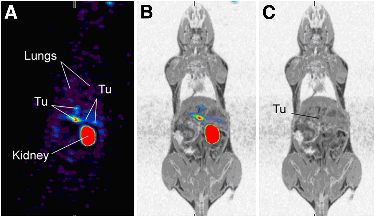
- FIGURE 1.
GLP-1 receptor SPECT/MRI of tumor-bearing Rip1Tag2 mouse at 4 h after injection of 37 MBq of [Lys40(Ahx-HYNIC-99mTc/EDDA)NH2]-exendin-4. Only multipinhole SPECT images (A) and corresponding multipinhole SPECT/MR fused images (B) show 4 tumor lesions (Tu) in pancreas with diameter between 1 and 3.2 mm. Corresponding MR images (C) show only largest tumor. There is intense tracer accumulation in kidneys but only weak uptake of [Lys40(Ahx-HYNIC-99mTc/EDDA)NH2]-exendin-4 in both lungs (A).
- FIGURE 2.
[Lys40(Ahx-DOTA-68Ga)NH2]-exendin-4 PET/CT of 1 Rip1Tag2 mouse after bilateral nephrectomy. Coronal (A) and transverse (B) PET/CT images show intense [Lys40(Ahx-DOTA-68Ga)NH2]-exendin-4 uptake in 2 tumor lesions with maximum diameter of 1.5 mm (d1) and 2.3 mm (d2), respectively. The coregistered CT scan was unremarkable at same location. There is no relevant uptake of [Lys40(Ahx-DOTA-68Ga)NH2]-exendin-4 elsewhere.
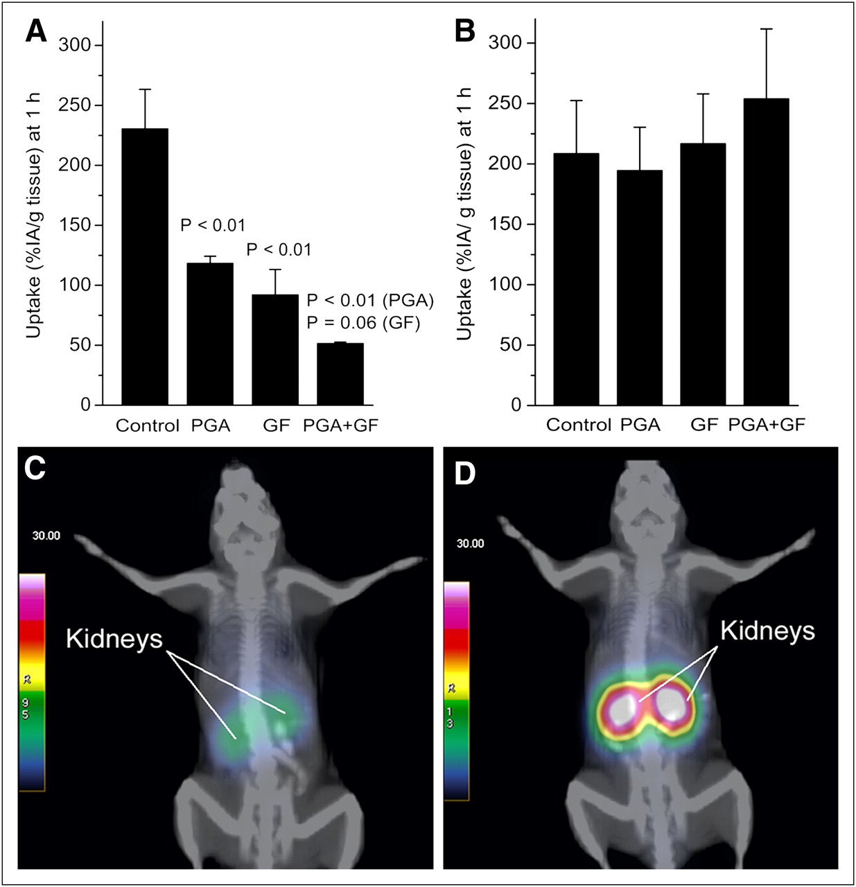
- FIGURE 3.
Biodistribution and PET/CT of Rip1Tag2 mice 1 h after injection of [Lys40(Ahx-DOTA-68Ga)NH2]-exendin-4. PGA, Gelofusine, and combination of the 2 significantly reduce renal accumulation of [Lys40(Ahx-DOTA-68Ga)NH2]-exendin-4 (A). At same time, tumor uptake is not affected by PGA or Gelofusine (B). Combination of PGA and Gelofusine is more efficient than PGA and Gelofusine alone. PGA plus Gelofusine pretreated Rip1Tag2 mouse (C) shows 78% lower kidney uptake than untreated control (D). In both animals, distinct differentiation between tumors and kidneys was not possible.
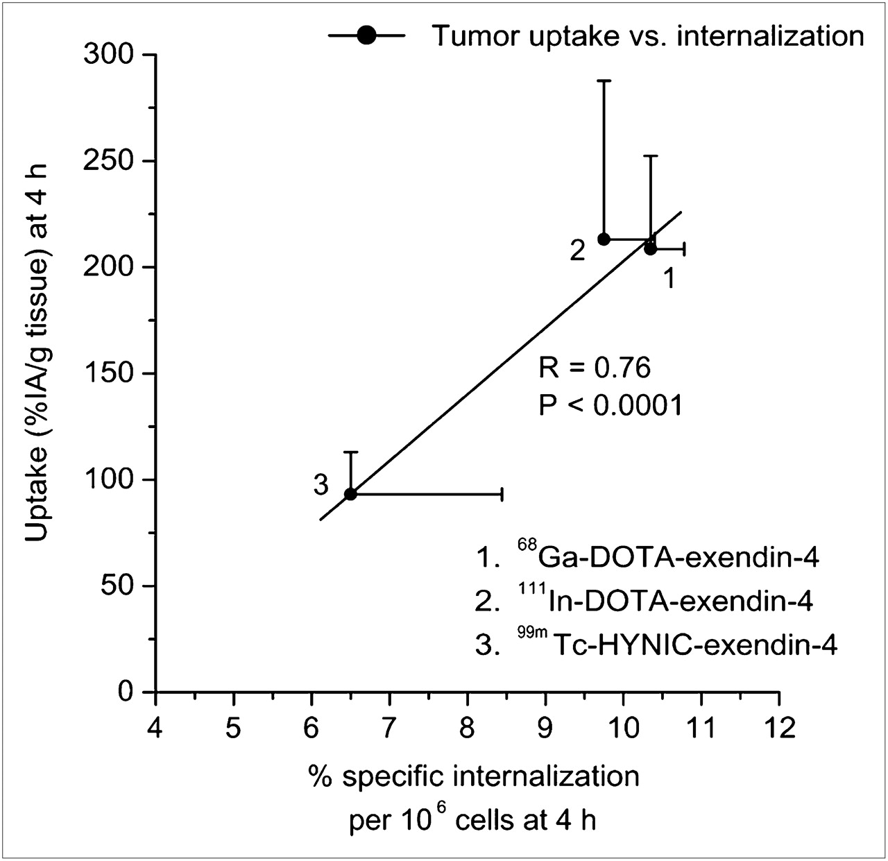
- FIGURE 4.
Correlation of tumor uptake (%IA/g of tissue) and internalization (percentage of specific internalized/106 cells) at 4 h. Each data point shows mean tumor uptake ± SD and mean internalization ± SD.

Tables
- TABLE 1
Comparison of Internalization Kinetics for 111In-, 67Ga-, and 99mTc-Labeled Exendin-4 in β-Tumor Cells
Compound 0.5 h 1 h 2 h 4 h [Lys40(Ahx-DOTA-111In)NH2]-exendin-4 1.03 ± 0.14 2.03 ± 0.18 4.97 ± 0.4 9.75 ± 0.65 [Lys40(Ahx-DOTA-67Ga)NH2]-exendin-4 1.22 ± 0.08 2.48 ± 0.29 5.10 ± 0.26 10.35 ± 0.43 [Lys40(Ahx-HYNIC-99mTc/EDDA)NH2]-exendin-4 0.73 ± 0.29 1.50 ± 0.49 3.37 ± 1.27 6.50 ± 1.94 1-way ANOVA P = 0.002 P = 0.001 P = 0.003 P = 0.0001 Values and SD are result of 2 independent experiments (triplicates in each experiment) and are expressed as specific internalization (% added radioactivity/106 cells ± SD). Significance was analyzed by 1-way ANOVA.
- TABLE 2
Biodistribution in Rip1Tag2 Mice at 0.5, 1, 2, and 4 Hours After Injection of [Lys40(Ahx-DOTA-68Ga)NH2]-Exendin-4
Organ 0.5 h 1 h 2 h 4 h Lungs* 40.8 ± 3.5 42.5 ± 5.1 31.4 ± 2.9 42.5 ± 5.1 Pancreas* 17.0 ± 2.4 13.5 ± 4.4 16.8 ± 6.3 13.5 ± 1.0 Stomach* 4.05 ± 0.33 4.08 ± 0.59 2.56 ± 0.35 2.14 ± 0.77 Tumor* 185 ± 33 209 ± 44 207 ± 60 205 ± 59 Kidneys 255 ± 14 230 ± 33 252 ± 24 202 ± 34 Liver 0.88 ± 0.04 0.61 ± 0.11 0.63 ± 0.12 0.61 ± 0.11 Spleen 2.14 ± 0.12 1.91 ± 0.50 2.10 ± 0.73 2.28 ± 0.59 Muscle 1.30 ± 0.10 1.13 ± 0.51 0.97 ± 0.09 1.00 ± 1.03 Bone 1.03 ± 0.33 1.01 ± 0.91 1.07 ± 0.13 0.89 ± 0.52 Blood 2.08 ± 0.49 1.35 ± 0.17 0.49 ± 0.03 0.29 ± 0.10 Tumor/blood 88.9 155 423 706 Tumor/muscle 142 185 214 205 Tumor/pancreas 10.9 15.5 12.4 15.2 Tumor/lungs 4.52 4.91 6.60 4.82 Tumor/kidneys 0.72 0.91 0.82 1.01 ↵* GLP-1 receptor–positive organs.
Results are expressed as %IA/g (mean ± SD), n ≥ 3.
- TABLE 3
Biodistribution in Rip1Tag2 Mice at 0.5, 2, 4, and 18 Hours After Injection of [Lys40(Ahx-HYNIC-99mTc/EDDA)NH2]-Exendin-4
Organ 0.5 h 2 h 4 h 18 h Lungs* 14.6 ± 4.3 18.5 ± 5.9 15.9 ± 5.6 8.7 ± 1.9 Pancreas* 7.1 ± 2.0 9.6 ± 1.5 7.4 ± 2.2 6.0 ± 1.4 Stomach* 1.18 ± 0.32 1.36 ± 0.32 1.20 ± 0.30 1.05 ± 0.27 Tumor* 67 ± 13 98 ± 19 93 ± 20 50 ± 9 Kidneys 63 ± 10 57 ± 14 60 ± 12 42 ± 12 Liver 0.83 ± 0.20 0.71 ± 0.28 0.72 ± 0.20 0.74 ± 0.19 Spleen 0.59 ± 0.10 0.47 ± 0.17 0.52 ± 0.10 0.58 ± 0.10 Muscle 0.16 ± 0.08 0.13 ± 0.10 0.17 ± 0.08 0.09 ± 0.05 Bone 0.19 ± 0.03 0.16 ± 0.03 0.18 ± 0.03 0.21 ± 0.02 Blood 2.34 ± 0.47 0.55 ± 0.11 0.35 ± 0.13 0.19 ± 0.12 Tumor/blood 29 177 266 262 Tumor/muscle 419 754 547 556 Tumor/pancreas 9.5 10.2 12.6 8.4 Tumor/lungs 4.59 5.29 5.86 10.6 Tumor/kidneys 1.06 1.72 1.55 1.18 ↵* GLP-1 receptor–positive organs.
Results are expressed as %IA/g (mean ± SD), n ≥ 3.
- TABLE 4
Biodistribution Data and Tissue Radioactivity Ratios at 4 Hours After Injection of Respective Radiopeptide
Organs Study parameter* [Lys40(Ahx-DOTA-111In)NH2]-exendin-4 [Lys40(Ahx-DOTA-68Ga)NH2]-exendin-4 [Lys40(Ahx-HYNIC-99mTc/EDDA)NH2]-exendin-4 P (1-way ANOVA) Lungs† Nonblocked 39.7 ± 6.8 42.5 ± 5.1 15.9 ± 5.6 <0.0001 Blocked 0.57 ± 0.01 0.90 ± 0.25 0.65 ± 0.17 Pancreas† Nonblocked 17.8 ± 3.9 13.5 ± 1.0 7.4 ± 2.2 <0.0001 Blocked 0.90 ± 0.26 0.79 ± 0.27 0.34 ± 0.16 Stomach† Nonblocked 3.31 ± 0.86 2.14 ± 0.77 1.20 ± 0.30 <0.0001 Blocked 0.60 ± 0.02 1.22 ± 0.40 0.35 ± 0.07 Tumor† Nonblocked 213 ± 75 205 ± 59 93.1 ± 19.9 <0.0001 Blocked 9.35 ± 4.18 5.62 ± 3.85 5.45 ± 0.43 Kidney Nonblocked 243 ± 17 202 ± 34 60 ± 12 <0.0001 Blocked 257 ± 30 193 ± 81 48 ± 7 Liver Nonblocked 1.03 ± 0.12 0.61 ± 0.11 0.72 ± 0.2 <0.0001 Blocked 0.85 ± 0.18 0.51 ± 0.28 0.54 ± 0.21 Spleen Nonblocked 2.17 ± 0.54 2.28 ± 0.59 0.52 ± 0.1 <0.0001 Blocked 1.77 ± 0.58 1.37 ± 0.47 0.37 ± 0.14 Muscle Nonblocked 1.23 ± 0.76 1.00 ± 1.03 0.17 ± 0.08 0.008 Blocked 0.78 ± 0.32 0.82 ± 0.10 0.08 ± 0.03 Bone Nonblocked 0.36 ± 0.20 0.89 ± 0.52 0.18 ± 0.03 0.01 Blocked 0.13 ± 0.03 0.46 ± 0.26 0.16 ± 0.09 Blood Nonblocked 0.26 ± 0.08 0.29 ± 0.10 0.35 ± 0.13 0.69 Blocked 0.24 ± 0.03 0.39 ± 0.25 0.50 ± 0.29 Tumor/blood 820 706 266 Tumor/muscle 173 205 547 Tumor/pancreas 12.0 15.2 12.6 Tumor/lungs 5.37 4.82 5.86 Tumor/kidneys 0.88 1.01 1.55 - TABLE 5
Radiation Dose Estimation Extrapolated to Humans After Injection of 111In-, 68Ga-, and 99mTc-Labeled Exendin-4
Organ/tissue [Lys40(Ahx-DOTA-111In)NH2]-exendin-4 [Lys40(Ahx-DOTA-68Ga)NH2]-exendin-4 [Lys40(Ahx-HYNIC-99mTc/EDDA)NH2]-exendin-4 Adrenals 0.43 0.050 0.0079 Brain 0.064 0.014 0.0014 Breasts 0.064 0.015 0.0015 Gallbladder wall 0.26 0.029 0.0057 Gastrointestinal 0.14 0.020 0.0032 Lower large intestine wall Small intestine 0.32 0.068 0.0097 Stomach wall 0.20 0.029 0.0046 Upper large intestine wall 0.23 0.026 0.0055 Heart wall 0.10 0.01 0.0025 Kidneys 4.48 1.85 0.083 Liver 0.20 0.020 0.0046 Lungs 0.13 0.044 0.0046 Muscle 0.12 0.019 0.0024 Ovaries 0.16 0.021 0.0039 Pancreas 0.70 0.20 0.020 Red marrow 0.14 0.020 0.0030 Osteogenic cells 0.23 0.028 0.0060 Skin 0.064 0.015 0.0013 Spleen 0.37 0.035 0.068 Thymus 0.086 0.017 0.0020 Thyroid 0.069 0.015 0.0015 Urinary bladder wall 0.11 0.017 0.0026 Uterus 0.15 0.021 0.0036 Total body 0.14 0.029 0.0031 Effective dose (mSv/MBq) 0.155 0.0317 0.00372 Results are expressed as mean absorbed dose (mGy/MBq).
Supplemental Data
Files in this Data Supplement:


Jump to section
Related Articles
Cited By...
- Glucagon-like Peptide-1 Receptor as Emerging Target: Will It Make It to the Clinic?
- Glucose Sensing Mediated by Portal Glucagon-Like Peptide 1 Receptor Is Markedly Impaired in Insulin-Resistant Obese Animals
- PET-Based Human Dosimetry of 68Ga-NODAGA-Exendin-4, a Tracer for {beta}-Cell Imaging
- Molecular imaging in the investigation of hypoglycaemic syndromes and their management
- Glucagon-Like Peptide-1 and Its Class B G Protein-Coupled Receptors: A Long March to Therapeutic Successes
- Glucagon-Like Peptide-1 Receptor PET/CT with 68Ga-NOTA-Exendin-4 for Detecting Localized Insulinoma: A Prospective Cohort Study
- Development of 68Ga- and 89Zr-Labeled Exendin-4 as Potential Radiotracers for the Imaging of Insulinomas by PET
- Localization of Hidden Insulinomas with 68Ga-DOTA-Exendin-4 PET/CT: A Pilot Study
- The Glucose-Dependent Insulinotropic Polypeptide Receptor: A Novel Target for Neuroendocrine Tumor Imaging--First Preclinical Studies
- GLP-1R-Targeting Magnetic Nanoparticles for Pancreatic Islet Imaging
- Development and Evaluation of 18F-TTCO-Cys40-Exendin-4: A PET Probe for Imaging Transplanted Islets
- PET of Glucagonlike Peptide Receptor Upregulation After Myocardial Ischemia or Reperfusion Injury