


Article Figures & Data
Figures
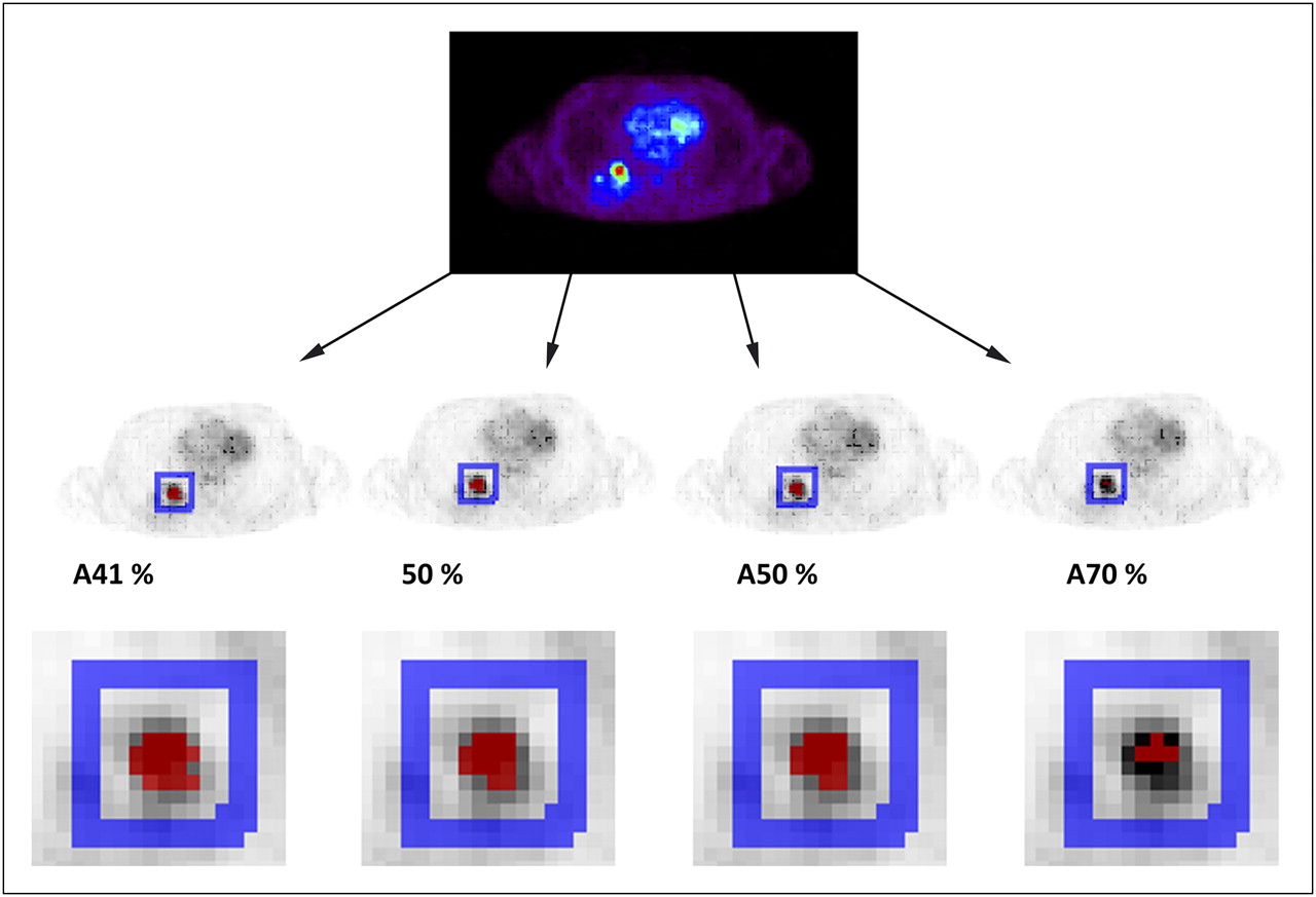
- FIGURE 1.
Typical example of 4 threshold-defined VOIs for 18F-FDG scan, for which red voxels represent resulting VOI and blue voxels local background, used for background correction.
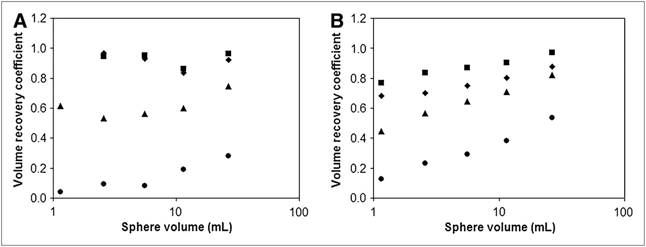
- FIGURE 2.
Plot of volume recovery coefficients per sphere volume in phantom study for different thresholds (same thresholds applied for patient study) with SBRs of 4.5 (A) and 9 (B). In both A and B, VOI A41%, 50%, A50%, and A70% (upper to lower datasets) are represented by ▪, ♦, ▴, and ○, respectively.
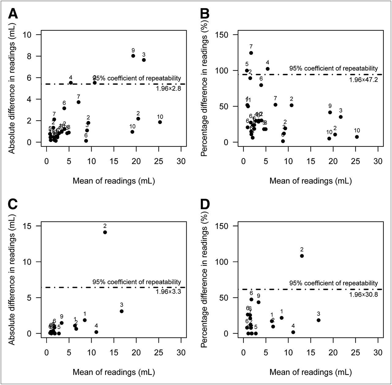
- FIGURE 3.
Plot of absolute difference between 2 scans against their mean for 18F-FDG (A) and 18F-FLT (C), respectively, and of percentage difference between scans against their mean for 18F-FDG (B) and 18F-FLT (D). Difference is proportional to SD of repeated measurements in each individual. The 95% RC is shown. Numbers near dots indicate patient number. One lesion of patient 7, with mean value of 96 for 18F-FDG, is not shown. Absolute difference for this particular lesion was 0.9 mL or 0.93%.
- FIGURE 4.
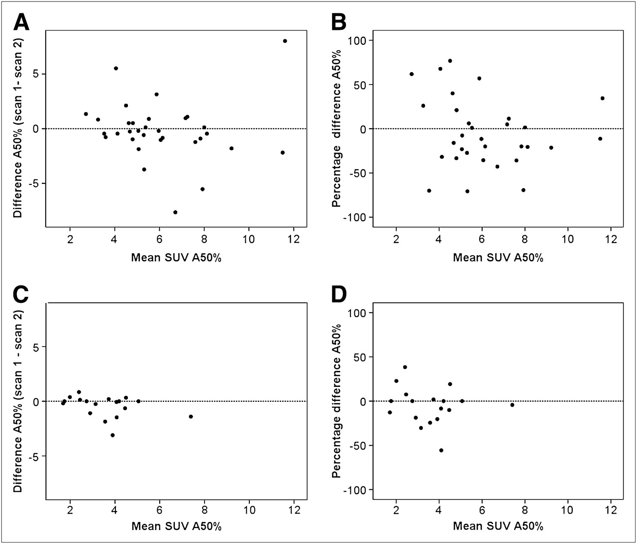
Difference in absolute volume measurement for 18F-FDG (A) and 18F-FLT (C), respectively, and percentage difference for 18F-FDG (B) and 18F-FLT (D) against mean SUV A50%.
- FIGURE 5.
Test and retest image of heterogeneous lesion showing variation in uptake pattern resulting in highly different VOIs, implicating limitation of VOI methodology in (variation in uptake in) heterogeneous lesions. SUVmax = maximum SUV.


Tables
- TABLE 1
Percentage COV of Observed VOIs as Function of Actual Sphere Volume and VOI Method
Sphere volume Percentage COV SBR = 9 SBR = 4.5 A41 50 A50 A70 A41 50 A50 A70 26.52 2.5 2.8 3.5 14.3 6.8 8.0 8.4 40.6 11.49 5.1 7.3 5.2 6.7 6.5 6.9 4.7 40.8 5.57 7.3 4.2 6.6 16.3 6.1 6.5 8.2 30.2 2.57 9.5 8.1 5.9 23.0 19.2 20.7 13.0 54.7 1.15 17.0 11.6 14.1 24.4 16.4 41.6 18.3 53.6 Sphere volume SD SBR = 9 SBR = 4.5 A41 50 A50 A70 A41 50 A50 A70 26.52 0.63 0.65 0.73 1.49 1.71 1.84 1.60 2.03 11.49 0.56 0.71 0.44 0.29 0.69 0.67 0.35 0.81 5.57 0.37 0.18 0.24 0.27 0.31 0.30 0.26 0.29 2.57 0.21 0.14 0.08 0.16 0.52 0.54 0.21 0.20 1.15 0.22 0.13 0.11 0.05 0.28 0.87 0.13 0.06 Tracer VOI Scan No. of lesions Q1 Median Q3 Missing 18F-FDG A41% 1 18 3.29 5.75 8.87 16 2 18 2.86 5.59 11.25 16 All 36 2.96 5.75 9.72 32 50 1 18 3.17 5.69 7.34 16 2 18 2.76 5.11 10.16 16 All 36 2.96 5.69 8.32 32 A50% 1 32 2.12 3.31 8.53 2 2 32 1.86 3.28 9.30 2 All 64 1.91 3.31 8.90 4 A70% 1 34 0.26 0.58 1.41 0 2 34 0.26 0.48 1.78 0 All 68 0.26 0.55 1.43 0 18F-FLT A41% 1 11 2.06 3.54 12.00 9 2 11 2.15 2.64 11.05 9 All 22 2.07 3.44 11.18 18 50% 1 13 2.70 2.83 10.90 7 2 12 2.10 3.28 9.70 8 All 25 2.25 2.83 10.90 15 A50% 1 20 1.67 2.42 6.62 0 2 19 1.35 1.86 8.20 1 All 39 1.54 2.19 7.27 1 A70% 1 20 0.26 0.45 0.64 0 2 20 0.26 0.39 0.93 0 All 40 0.26 0.42 0.66 0 Q1 and Q3 are interquartile ranges.
Radiotracer VOI n (pairs) Mean absolute difference (mL) RC (1.96 × SD) (mL) Mean percentage difference RC (1.96 × SD) (%) 18F-FDG For total lesions A41% 18 1.8 5.9 22.6 44.4 50% 18 2.0 6.0 24.3 52.5 A50% 32 1.8 4.2 35.5 62.4 A70% 34 0.4 1.1 43.7 71.1 For lesions < 4.2 mL A41% 7 0.7 1.3 28.0 47.7 50% 8 0.6 1.4 25.8 57.7 A50% 13 0.7 1.0 39.8 64.5 A70% 31 0.3 0.8 46.2 72.0 For lesions > 4.2 mL A41% 9 2.5 7.8 12.1 21.9 50% 9 2.8 7.9 16.2 28.9 A50% 12 2.9 5.4 21.3 37.2 A70% 3 1.4 2.2 17.2 36.5 18F-FLT For total lesions A41% 10 1.2 2.2 24.1 34.9 50% 12 1.0 2.1 21.0 39.4 A50% 19 1.4 6.3 19.7 50.2 A70% 20 0.5 0.9 56.8 94.0 For lesions < 4.2 mL A41% 6 0.7 0.9 29.5 30.9 50% 7 0.5 0.9 24.4 33.2 A50% 12 0.3 0.9 16.0 33.2 A70% 20 0.5 1.5 56.8 94.0 For lesions > 4.2 mL A41% 4 2.0 2.8 16.1 39.0 50% 5 1.6 2.8 16.2 49.2 A50% 7 3.2 9.6 25.9 72.7 A50%* 6 1.4 2.0 12.2 16.0 A70% 0 — — — — ↵* Data, with exclusion of heterogeneous lesion that highly affected outcome.
None of mean percentage differences were significantly different from 0. Percentage differences were calculated by the following formula: |volume scan 1 – volume scan 2|/(0.5 [volume scan 1 + volume scan 2]) × 100.


Jump to section
Related Articles
Cited By...
- Feasibility of Ultra-Low-Activity 18F-FDG PET/CT Imaging Using a Long-Axial-Field-of-View PET/CT System
- Comparison of Multiple Segmentation Methods for Volumetric Delineation of Primary Prostate Cancer with Prostate-Specific Membrane Antigen-Targeted 18F-DCFPyL PET/CT
- The Impact of Semiautomatic Segmentation Methods on Metabolic Tumor Volume, Intensity, and Dissemination Radiomics in 18F-FDG PET Scans of Patients with Classical Hodgkin Lymphoma
- Interobserver Agreement on Automated Metabolic Tumor Volume Measurements of Deauville Score 4 and 5 Lesions at Interim 18F-FDG PET in Diffuse Large B-Cell Lymphoma
- Quantitative Test-Retest Measurement of 68Ga-PSMA-HBED-CC in Tumor and Normal Tissue
- Variability and Repeatability of Quantitative Uptake Metrics in 18F-FDG PET/CT of Non-Small Cell Lung Cancer: Impact of Segmentation Method, Uptake Interval, and Reconstruction Protocol
- Reproducibility and Repeatability of Semiquantitative 18F-Fluorodihydrotestosterone Uptake Metrics in Castration-Resistant Prostate Cancer Metastases: A Prospective Multicenter Study
- Repeatability of Quantitative Whole-Body 18F-FDG PET/CT Uptake Measures as Function of Uptake Interval and Lesion Selection in Non-Small Cell Lung Cancer Patients
- Repeatability of Quantitative 18F-Fluoromethylcholine PET/CT Studies in Prostate Cancer
- Prognostic Value of Pretherapeutic Tumor-to-Blood Standardized Uptake Ratio in Patients with Esophageal Carcinoma
- 18F-FDG or 3'-Deoxy-3'-18F-Fluorothymidine to Detect Transformation of Follicular Lymphoma
- Value of Metabolic Tumor Volume on Repeated 18F-FDG PET/CT for Early Prediction of Survival in Locally Advanced Non-Small Cell Lung Cancer Treated with Concurrent Chemoradiotherapy
- Using FDG-PET to Measure Early Treatment Response in Head and Neck Squamous Cell Carcinoma: Quantifying Intrinsic Variability in Order to Understand Treatment-Induced Change
- Tumor Microenvironment-Dependent 18F-FDG, 18F-Fluorothymidine, and 18F-Misonidazole Uptake: A Pilot Study in Mouse Models of Human Non-Small Cell Lung Cancer
- Reproducibility of Tumor Uptake Heterogeneity Characterization Through Textural Feature Analysis in 18F-FDG PET
- Effects of Image Characteristics on Performance of Tumor Delineation Methods: A Test-Retest Assessment