


Article Figures & Data
Figures
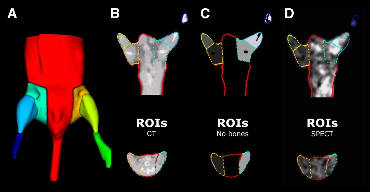
- FIGURE 1.
Method for analysis of micro-SPECT/CT images. (A) Planes were interactively positioned over lower body of mice to segment micro-CT to generate multiple VOIs. (B) Contours of these VOIs are illustrated superimposed on representative micro-CT image. (C) K-means clustering-based segmentation was performed to eliminate bones from VOIs. (D) This complex irregular object map was applied to registered micro-SPECT images to determine mean counts in each VOI.
- FIGURE 2.
Validation of micro-SPECT/CT image-analysis approach. (A) Correlation between I/NI ratios of counts calculated from image analysis and GWC for both proximal and distal regions. Correlation was poor (R2 = 0.05) when all proximal and distal regions were included. (B) Correlation coefficient between imaging analysis and GWC improved significantly (R2 = 0.63) after proximal regions contaminated with scatter from radioactivity in bladder were removed from analysis. Shown are representative 99mTc-NC100692 micro-SPECT/CT images from 3 mice at 7 d after surgical ligation of right femoral artery with superimposed VOIs. (C) Mouse with radioactivity within bladder, which falls within right proximal VOI, resulting in overestimation of counts in ischemic (right) relative to nonischemic (left) proximal leg. (D) Second mouse with bladder located centrally in body and no significant contamination of proximal hindlimb VOIs. (E) Images from third mouse after removal of radioactive urine from bladder by needle aspiration immediately before SPECT acquisition.
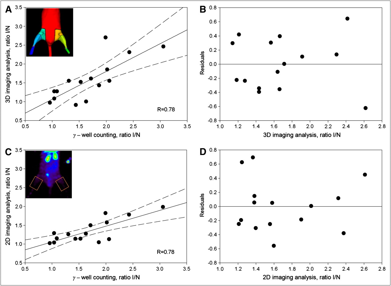
- FIGURE 3.
Validation of analysis of distal hindlimb. There was good correlation (R = 0.78) between ischemic-to-nonischemic counts ratio calculated from 3D volumetric analysis of micro-SPECT/CT images (A) and 2D analysis of maximum-projection images (C) relative to GWC for distal hindlimb. Linear correlation (solid line) and 95% confidence intervals (dashed line) are shown (B and D). Quality of fit was confirmed by residuals plot for micro-SPECT/CT image (B) and 2D maximum-projection image (D) analysis approaches.
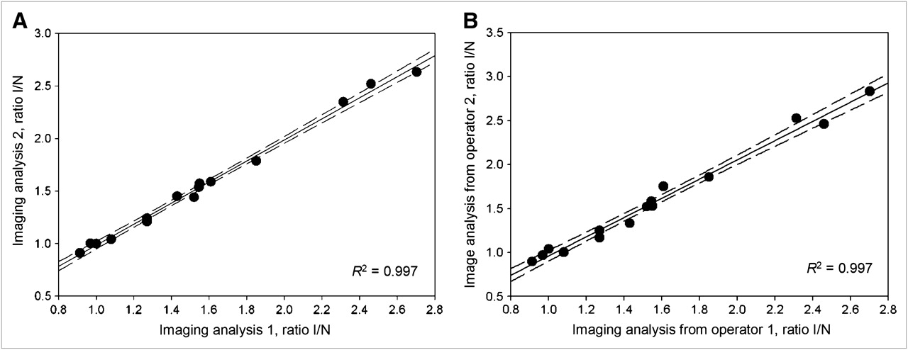
- FIGURE 4.
Reproducibility of image quantification. Intraobserver (A) and interobserver (B) reproducibility of micro-SPECT/CT image analysis were excellent.
- FIGURE 5.
Analysis of wild-type and eNOS-knockout mice. (A) Representative micro-SPECT/CT images of eNOS+/+ and eNOS−/− mice injected with 99mTc-NC100692 at baseline, 7 d, and 4 wk after right femoral artery ligation. Yellow arrows indicate ischemic regions with increased 99mTc-NC100692 retention. Less retention is seen in eNOS−/− mice. (B) Serial micro-SPECT/CT images were analyzed and I/NI 99mTc-NC100692 activity ratios calculated. There was significant (P < 0.05) increase of 99mTc-NC100692 retention in ischemic leg at 7 d after surgery in both groups. However, there was significantly less retention in eNOS−/− mice at 7 d than in wild-type mice. *P < 0.05 vs. wild-type. #P < 0.05 vs. baseline.


Jump to section
Related Articles
Cited By...
- Radiotracer Imaging Allows for Noninvasive Detection and Quantification of Abnormalities in Angiosome Foot Perfusion in Diabetic Patients With Critical Limb Ischemia and Nonhealing Wounds
- Radiotracer Imaging of Peripheral Vascular Disease
- State-of-the-Art Methods for Evaluation of Angiogenesis and Tissue Vascularization: A Scientific Statement From the American Heart Association
- Radiotracer Imaging of Peripheral Vascular Disease
- Report of the National Heart, Lung, and Blood Institute Working Group on the Translation of Cardiovascular Molecular Imaging
- Approaches to Multimodality Imaging of Angiogenesis