


Abstract
An extensive series of radioligands has been developed for imaging central nicotinic acetylcholine receptors (nAChRs) with PET. Two halogeno-derivatives of A-85380 are being used in humans. Nevertheless, these derivatives still display too-slow brain kinetics and low signal-to-noise ratio. Methods: A novel nAChR radioligand, 5-(6-fluorohexyn-1-yl)-3-[2(S)-2-azetidinylmethoxy]pyridine (ZW-104), was characterized in vitro using competition binding assays (nAChR subtypes heterologously expressed in HEK 293 cells and in native α4β2 nAChRs from rat brain). 18F-ZW-104 was prepared as follows: no-carrier-added nucleophilic aliphatic radiofluorination of the corresponding N-Boc-protected tosyloxy derivative 5-(6-tosyloxyhexyn-1-yl)-3-[2(S)-(N-(tert-butoxycarbonyl))-2-azetidinylmethoxy] pyridine) with the activated 4,7,13,16,21,24-hexaoxa-1,10-diazabicyclo-[8,8,8]hexacosane (K-18F-F-Kryptofix 222 [K222] complex), followed by quantitative trifluoroacetic acid–induced removal of the N-Boc protective group. 18F-ZW-104 was then studied in baboons using PET. Results: ZW-104 showed high binding affinities for rat α4β2 nAChRs (Ki, 0.2 nM) and other subtypes containing the β2 subunit but much lower affinities for rat α3β4 nAChRs (Ki, 5,500 nM) and other subtypes containing the β4 subunit. The regional radioactivity distribution in the baboon brain matched that of the α4β2 nAChR, which was similar to that of 2-18F-fluoro-3-(2(S)-azetidinylmethoxy)pyridine (2-18F-A-85380), a radioligand used in humans. Comparison between 18F-ZW-104 and 2-18F-A-85380 demonstrated better in vivo binding properties of the new radioligand: a substantially greater amount of radioactivity accumulated in the brain, and the occurrence of peak uptake in the thalamus was earlier than that of 2-18F-A-85380 and was followed by washout. Distribution volume values in different brain regions were 2-fold higher for 18F-ZW-104 than for 2-18F-A-85380. Displacement by nicotine or unlabeled ZW-104 demonstrated a lower nonspecific binding than that of 2-F-A-85380. Conclusion: These results suggest that 18F-ZW-104 is a promising PET radioligand for studying nAChRs containing the β2 subunits in humans.
A loss of cholinergic neurons has been associated with several pathologic disorders (1), such as dementia of the Alzheimer type and Parkinson disease. PET offers the opportunity to monitor changes in human nicotinic acetylcholine receptors (nAChRs) in vivo. However, there are certain difficulties in the development of suitable radioligands for the in vivo imaging of nAChRs (2), one of them being the low density of nAChRs in extrathalamic areas. Therefore, there is a need for radioligands with high affinities (probably less than 1 nM) and radiosyntheses yielding high specific radioactivities. The role of lipophilicity of the radioligand remains a subject of debate, but a relationship exists between the lipophilicity and nondisplaceable binding. Research efforts from radiochemists have focused on the development of highly specific radioligands with improved brain kinetics (2). A new emerging, promising field is the use of antagonists at nAChRs (3,4), but these compounds need more experiments to confirm their preliminary attractive characteristics.
The agonists 2-fluoro- and 5-iodo-A-85380 (Fig. 1, compounds 1 and 2) are the only PET/SPECT radioligands used in humans today. However, these agonists have 3 drawbacks: slow kinetics, low signal-to-noise ratio, and a large amount of nondisplaceable binding. The presence of a bulky halogen (an iodine) at the 5-position of the pyridinyl ring in 5-iodo-A-85380 does not affect the affinity for nAChRs but leads to an increase of selectivity for α4β2 subtype (5–8). Therefore, recently derivatives of azetidine A-85380 (and also pyrrolidine A-84543, structure not shown) bearing a 5-alkynyl substituent were reported (9), following the idea that a more bulky substituent such as an 5-alkynyl would not decrease but perhaps increase the affinity and selectivity of the compounds at α4β2 nAChRs. Within this series, we have been interested in the pharmacologic properties of 5-(6-hydroxyhexyn-1-yl)-3-[2(S)-2-azetidinylmethoxy]pyridine (Fig. 1, compound 3), which is a selective nAChR agonist (10). This compound is a derivative of A-85380 (11), which itself has high affinity and selectivity for nAChRs containing β2 subunits (12). 5-(6-fluorohexyn-1-yl)-3-[2(S)-2-azetidinylmethoxy]pyridine (ZW-104; Fig. 1, compound 4) is structurally closely related to 5-(6-hydroxyhexyn-1-yl)-3-[2(S)-2-azetidinylmethoxy]pyridine, with the terminal hydroxyl group replaced by a fluorine atom.

Chemical structures of 2-18F-A-85380, 5-123I-I-A-85380, sazetidine, and 18F-ZW-104.
In this study, we characterized the in vitro binding affinity and selectivity of ZW-104. We also labeled ZW-104 with 18F and investigated in rats and baboons the in vivo properties of this novel radioligand to selectively image central α4β2 nAChRs.
MATERIALS AND METHODS
Radiochemistry
Chemicals.
Chemicals were purchased from Aldrich, Fluka, or Sigma France and were used without further purification. ZW-104 (as reference compound) and the corresponding N-Boc-protected tosyloxy derivative 5-(6-tosyloxyhexyn-1-yl)-3-[2(S)-(N-(tert-butoxycarbonyl))-2-azetidinylmethoxy]pyridine (as precursor for labeling with 18F) were synthesized according to literature procedures (9).
High-Performance Liquid Chromatography (HPLC) Analyses.
For HPLC method A, the system was equipped with a Waters 600 pump and Waters 600 controller, a Shimadzu SPD10-AVP ultraviolet (UV)-multiwavelength detector, and a miniature ionisation chamber probe. The column was a semipreparative Zorbax C18 (Hewlett-Packard; 250 × 9.4 mm), porosity was 5 μm, the eluent was 0.9% aqueous NaCl/EtOH/AcOH:800/200/1 (v/v/v), flow rate was 6 mL/min, and absorbance detection was at λ = 254 nm. Room temperature was used. For HPLC method B, the system was equipped with a Waters Alliance 2690 (or a Waters binary HPLC pump 1525) with a UV spectrophotometer (Photodiode Array Detector; Waters 996) and a Berthold LB509 radioactivity detector. The column was an analytic Symmetry-M C18 (Waters; 50 × 4.6 mm); porosity was 5.0 μm; the conditions were isocratic elution with solvent A/solvent B:63/37 (v/v) (solvent A, H2O containing low-UV PIC B7 reagent [Waters; 20 mL for 1,000 mL] and solvent B, H2O/CH3CN:30/70 (v/v) containing low-UV PIC B7 reagent [Waters; 20 mL for 1,000 mL]). The flow rate was 2.0 mL/min, and absorbance detection was at λ = 254 nm. Room temperature was used.
Miscellaneous.
Radiosyntheses using 18F, including the HPLC purifications, were performed in a 7.5-cm lead-shielded cell using a computer-assisted Zymate-XP robot system (Zymark Corp.).
Radioisotope Production.
No-carrier-added aqueous 18F-fluoride ion was produced via the 18O(p, n)18F nuclear reaction by irradiation of 2 mL of 18O-water (>97%-enriched; CortecNet) target on an IBA Cyclone-18/9 cyclotron (18-MeV proton beam) and was transferred to the appropriate hot cell.
Preparation of 4,7,13,16,21,24-hexaoxa-1,10-diazabicyclo[8.8.8]hexacosane (K-18F-F-Kryptofix 222 [K222], Complex.
18F (half-life, 109.8 min) as 18F-fluoride ion was isolated by passing the irradiated 18O-water target, using helium pressure (1.5–2.0 bar), through an anion-exchange resin (Sep-pak Light Accell Plus QMA; Waters) cartridge (chloride form, washed beforehand with aqueous 1 M NaHCO3 (2 mL) and rinsed with water (20 mL) and CH3CN (10 mL)). Helium was blown through the column to maximally extract 18O-water. The 18F-fluoride ion was then eluted from the resin, using an aqueous K2CO3 solution (1.0 mL of a 4.5-mg solution per milliliter), into a Vacutainer tube (Becton, Dickson) containing K-18F-F-K222 (12.0–15.0 mg; Fluka). The resulting solution was then gently concentrated to dryness at 145°C−150°C under a nitrogen stream for 10 min to give no-carrier-added K-18F-F-K222 complex as a white semisolid residue.
Preparation of 18F-ZW-104.
For radiosynthesis, acetonitrile (600 μL) containing 5-(6-tosyloxyhexyn-1-yl)-3-[2(S)-(N-(tert-butoxycarbonyl))-2-azetidinylmethoxy]pyridine (6–10 mg) was added into the Vacutainer tube containing the dried K-18F-F-K222 complex. The nonsealed tube was thoroughly stirred in a vortex mixer (30 s) and then placed in a heating block (at 120°C, for 8 min) without stirring the contents. The reaction vessel was then cooled using an ice-water bath; the reaction mixture was diluted with water (1 mL) and transferred onto a C18 cartridge (PrepSep R-C18 Extraction Column; Fisher Scientific), prefilled with water (2 mL). The tube was rinsed twice with water (1 mL), which was also transferred and added to the diluted reaction mixture on top of the cartridge. An additional portion of water (2 mL) was further added to the diluted reaction mixture on top of the cartridge. The entire contents were then passed through the cartridge, which was then washed with water (3 mL) and partially dried for 0.5 min by applying a nitrogen stream. N-Boc-protected 18F-ZW-104 was eluted from the cartridge with CH2Cl2 (3 mL) into a 5-mL reaction vial containing trifluoroacetic acid (TFA) (0.1 mL). Elution was repeated twice with 1 mL of CH2Cl2 for maximal transfer of the 18F-labeled intermediate. The resulting CH2Cl2/TFA solution (50/1, v/v) was then concentrated to dryness at 65°C−75°C under a gentle nitrogen stream for 3–5 min. The residue was redissolved in CH2Cl2 (2 mL) and concentrated again to dryness to minimize TFA presence (at 65°C−75°C under a gentle nitrogen stream for 2–3 min). Finally, the residue was redissolved in the HPLC solvent used for purification (1.0 mL), and the crude was injected onto HPLC (HPLC method A). Isocratic elution gave pure 18F-ZW-104 (retention time [tR], 17.0–18.0 min). For quality control, final chemical identification of 18F-ZW-104 was performed on an aliquot of the HPLC-collected fraction by analytic HPLC (HPLC method B), with a sample of authentic ZW-104 (tR, 2.34 min). Chemical and radiochemical purities were also assessed on this aliquot by HPLC (HPLC method B). Specific radioactivity of the radioligand was calculated from 3 consecutive HPLC analyses and determined as follows: the area of the UV absorbance peak corresponding to the radiolabeled product was measured (integrated) on the HPLC chromatogram and compared with a standard curve relating mass to UV absorbance. The specific radioactivity follows from the found mass and the associated collected radioactivity.
In Vitro Pharmacology
Stably Transfected Cell Lines and Cell Culture.
Stably transfected cell lines expressing defined rat nAChR subtypes were described previously (6,13). Tissue culture medium and antibiotics were obtained from Invitrogen Corp. Fetal bovine serum was provided by Gemini Bio-Products. Cells were grown at 37°C with 5% CO2 in a humidified incubator.
Radioligand Binding Assay.
Competition binding assays using 3H-epibatidine have been described previously (13). In brief, cultured cells at greater than 80% confluence were removed from their flasks and placed in 10 mL of 50 mM Tris-HCl buffer (pH 7.4, 4°C). The cell suspension was centrifuged at 10,000g for 5 min, and the pellet was collected. The cell pellet was then homogenized in 10 mL of buffer with a Polytron homogenizer (12-mm aggregate, 26,000 rpm, 20 s; model PT2100; Kinematica) and centrifuged at 36,000g for 10 min at 4°C. The membrane pellet was resuspended in fresh buffer, and aliquots of the membrane preparation equivalent to 30–200 μg of protein were used for binding assays. The concentration of 3H-epibatidine used was 100 pM for competition binding. The concentrations of ZW-104 ranged from 0.1 nM to 10 μM. The nonspecific binding was assessed in parallel incubations in the presence of 300 μM nicotine. Bound and free ligands were separated by vacuum filtration through Whatman GF/C filters (Brandel Inc.) treated with 0.5% polyethylenimine. The filter-retained radioactivity was measured by liquid scintillation counting. The Ki (inhibition constant) values for 3H-epibatidine used for calculating Ki values (nanomoles) were 0.02 for α4β2, 0.08 for α2β4, 0.03 for α3β2, 0.3 for α3β4, 0.04 for α4β2, 0.09 for α4β4, and 0.05 for rat forebrain. Data from saturation and competition binding assays were analyzed using Prism 4 (GraphPad Software).
Determination of Plasma and Brain Metabolites in Rats
Before testing the radioligand in baboons, plasma and brain metabolites were assessed in rats. Four male Sprague–Dawley rats (weight, 250 g) were infused through a tail vein with 18F-ZW-104 (32.5 ± 12.2 MBq) and sacrificed 30 or 70 min later, with blood (1 mL) collected at each time point. The brain was excised and homogenized in 4 mL of CH3CN. Plasma (500 μL) was deproteinized (with 700 μL of CH3CN) and injected onto the HPLC column for the determination of percentage of unchanged 18F-ZW-104. The activity of brain homogenates was counted with the γ-counter, and the samples were centrifuged at 3,500g for 2 min at 4°C. The supernatants then were injected onto the HPLC column. The HPLC system included a P680A gradient quaternary pump, an ASI100T auto sampler, a UVD170U UV-VIS detector (Summit Performance), and a LB507 radioisotope detector (MXZ 500-4 cell, Berthold). The eluents consisted of 0.1% TFA in water (solution A) and 0.1% TFA in acetonitrile (solution B). 18F-ZW-104 and its radiolabeled metabolites were separated at room temperature on a C18 μBondapak semipreparative column (300 × 7.8 mm, 10 μm; Waters). The column was equilibrated with 80% of solution A and 20% of solution B, and a linear gradient from 20% to 30% solution B was applied for 10 min. The flow rate was 4 mL/min (UV detection, 220 nm). Data acquisition and processing were performed using Chromeleon software (version 5.0; Dionex). The radioactivity due to unchanged 18F-ZW-104 was expressed as a fraction of the total radioactive peak areas.
In Vivo PET in Baboons
Animals.
All animal-use procedures were in strict accordance with the recommendations of the European Community (86/609/CEE) and the French National Committee (décret 87/848) for the care and use of laboratory animals.
MRI.
For each baboon, MR images were obtained in a separate experiment (1.5-T Signa; GE Healthcare). A T1-weighted inversion-recovery sequence in 3-dimensional mode and a 256 × 192 matrix over 124 slices (1.5-mm thick) were used to generate the MR images compatible with the PET images.
PET.
PET studies of the brain distribution of radiolabeled compound were performed in adult Papio anubis baboons. Two hours before the PET acquisition, the animals received ketamine (10 mg/kg intramuscularly). After being intubated, animals were artificially ventilated and anesthetized with 66% N2O and 1% isoflurane (OAV 7710; Ohmeda). PET experiments were performed with an HR+ Exact positron tomograph (CTI PET Systems). This scanner allowed the simultaneous acquisition of 63 slices every 2.2 mm, with spatial and axial resolutions of 4.5 mm. Transmission scans were acquired for 15 min using 3 retractable 68Ge rod sources. The baboon's head was positioned in the tomograph using a custom-designed stereotactic head holder. Five baboons (mean weight ± SD, 12.6 ± 4.3 kg) underwent a total of 8 PET experiments. They were injected intravenously with 18F-ZW-104 (152 ± 37 MBq; 6.3 ± 1.2 nmol) and imaged for at least 180 min. During PET acquisition, arterial blood samples were withdrawn from the femoral artery at designated times. To better define the kinetics of the radioligand, the duration of 2 PET experiments was extended to 6 h. In 1 baboon after brain imaging, a whole-body scan (180 min after injection of the radioligand) of 18F-ZW-104 distribution was obtained (5 steps; duration of each step, 7 min), followed by a segmented transmission. We examined whether the cerebral uptake of the radioligand could be displaced (n = 2) by injecting, 120 min after the beginning of the PET experiment, either nicotine (2,500 nmol/kg intravenously, n = 1) or unlabeled ZW-104 (500 nmol/kg intravenously, n = 1). PET was continued for an additional 120 min. In 1 experiment, displacement by nicotine (bolus, 1,850 nmol/kg, followed by an infusion of 1,850 nmol/kg over 3 h) was scanned for 4 h. Heart rate, end tidal pCO2, and rectal temperature were continuously monitored during all the PET experiments. Two presaturation experiments were performed using either unlabeled ZW-104 (500 nmol/kg) injected as a slow bolus (10 min) 50 min before injection of the radioligand or nicotine (2,500 nmol/kg) administered 30 min before the radioligand. These PET experiments lasted for 180 min. For comparison of the uptake and brain kinetics of the 2 radioligands, 1 baboon was intravenously injected with 74 MBq (2 mCi (1 nmol)) of 2-18 F-A-85380 and imaged for 3 h.
PET Data Analysis.
For PET data analysis, regions of interest were delineated on images on which anatomic structures (frontal cortex, thalamus, striata, and cerebellum) can be clearly identified. The position of the volumes of interest (VOI) was controlled on the MR images. On the basis of clearly identified anatomic structures, 9 VOIs were delineated in 3 dimensions on T1-weighted MR images: frontal, parietal, and temporal and occipital cortices; caudate nucleus; putamen; thalamus; cerebellum; and hippocampus. Coregistration of PET images to the corresponding MR images was used to ensure the consistency of the anatomic localization of 18F-ZW-104 cerebral binding. To generate each regional time–activity curve, the mean radioactivity in the VOI was calculated for each frame, corrected for 18F decay, plotted versus time, and expressed as standardized uptake value (SUV) (i.e., [MBq/mL of tissue]/injected dose [MBq]/body weight [g]). After a displacement experiment, the percentage changes in thalamic and cerebellar radioactivities were calculated at the end of the PET experiment (240 min) by dividing the difference in radioactivity (control experiment − challenge experiment) by the value of the radioactivity in the control experiment at 240 min.
The Logan graphical (14) analysis of reversible radioligand kinetics was applied to the present data. This method allows for the measurement of the total distribution volume (VT) of the ligand without any assumption on the actual configuration of the tissue compartments. VT was computed by linear regression of the final part of the plot using PMOD software (PMOD Group; http://www.pmod.com).
Determination of Plasma Metabolites in Baboons.
Arterial blood samples (3 mL) were collected at 5, 10, 20, 30, 60, 90, 120, and 160 min after the injection of the tracer and immediately centrifuged (5 min, 2,000g, at 4°C) to obtain cell-free plasma. For deproteinization, 0.5 mL of plasma was mixed with 0.7 mL of acetonitrile. After centrifugation at 3,500g for 5 min, the supernatant (about 1.1 mL) was directly injected onto the HPLC column.
RESULTS
Data are presented as mean ± SD.
Chemistry
ZW-104 was labeled with 18F using the following nonoptimized 2-step radiochemical process: no-carrier-added nucleophilic aliphatic radiofluorination of the corresponding N-Boc-protected tosyloxy derivative (6 mg) with the activated K-18F-F-K222 complex in acetonitrile at 120°C for 8 min, followed by quantitative TFA-induced removal of the N-Boc protective group and finally semipreparative HPLC purification on a Zorbax C18 column (Hewlett-Packard) using a mixture of solvents directly compatible with an intravenous injection (0.9% aqueous NaCl/EtOH/AcOH:800/200/1 [v/v/v]). Typically, 1.11–1.85 GBq of radiochemically pure (>99%) 18F-ZW-104 (37–74 GBq/μmol) could be obtained within 100 min, starting from 37.0 GBq of 18F-fluoride.
Binding Affinities of ZW-104 for nAChR Subtypes
ZW-104 bound with high affinities to rat α4β2, α3β2, and α2β2 nAChR subtypes heterologously expressed in HEK 293 cells and to native α4β2 nAChRs from rat brain (Table 1). The binding affinity of ZW-104 for α4β2 receptors is 28,000 times higher than that for α3β4 receptors. The selectivity of ZW-104 for α4β2 receptors over α3β4 receptors was much greater than that of 5-iodo-A-85380 (Ki ratio, 4,700) or 2-F-A-85380 (Ki ratio, 2,700), both of which are considered as relatively selective ligands for α4β2 nAChRs. ZW-104 also binds to α2β2 and α3β2 subtypes with high affinities, though they are slightly lower than that to α4β2 subtype. Therefore, ZW-104 appears to be selective for the β2 containing nAChRs.
Inhibition Constants (Ki, nM) at nAChR Subtypes of Nicotinic Reference Ligands and of PET SPECT Radioligands
Metabolites in Rat Plasma and Brain
Plasma HPLC analysis showed that unchanged 18F-ZW-104 represented 38% ± 9% and 17.5% ± 2.3% at 30 and 70 min after injection, respectively. In the brain, only authentic 18F-ZW-104 could be detected until 70 min after injection.
PET Studies in Baboons
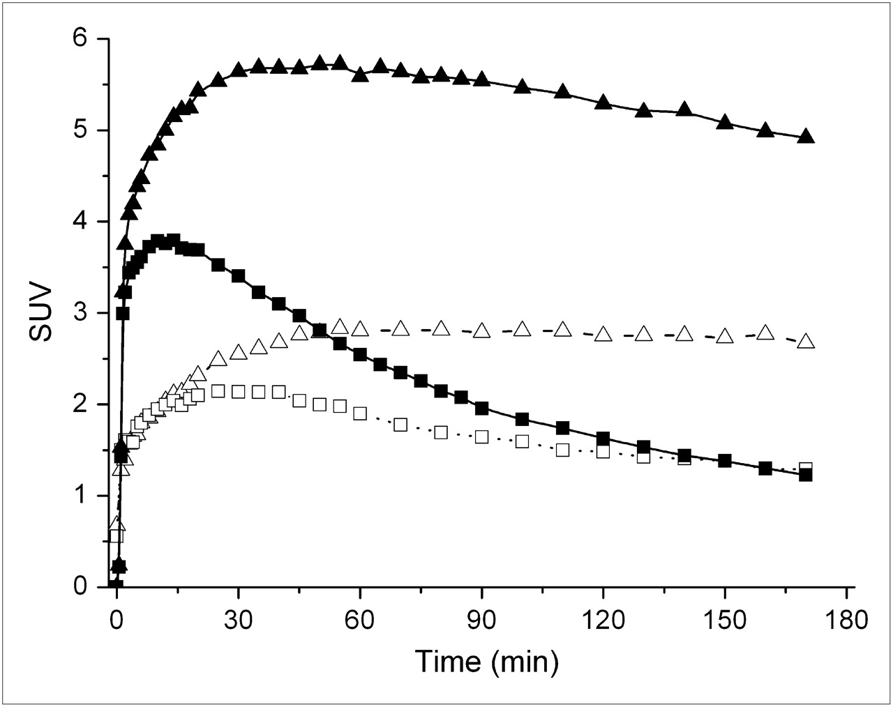
Brain kinetics in control animals are presented in Figure 2. As expected, the thalamus had the higher uptake (maximum standardized uptake value [SUVmax] 5.72 at 35–50 min after injection), followed by a slow washout (3.49 at 350 min after injection). Caudate nucleus, putamen, and cortices had an intermediate uptake, whereas the cerebellum showed the lowest uptake (2.80 SUVmax at 35 min after injection) and the fastest washout (0.71 SUV at 350 min after injection). The radioactivity ratio for thalamus to cerebellum increased with time until 230 min and remained stable at a value of 4.6–4.8 until 360 min. Logan analysis demonstrated that 30, 45, and 55 min are required for VT to become time-invariant in the cerebellum, frontal cortex, and thalamus, respectively (Fig. 3). Ratio of thalamic VT to cerebellar VT (Logan plot) was 3.5 at 180 min and remained unchanged at 360 min after injection. For comparison, this ratio was 2.38 at 180 min after injection with 2-F-A-85380. Pretreatment with nicotine reduced uptake in the thalamus to 2.80 SUV at 35 min (Fig. 4). Pretreatment with ZW-104 reduced uptake in the thalamus to 3.05 SUVmax at 35 min, and it was followed by a steep washout (Fig. 4). At 180 min after injection, uptake in the thalamus was drastically reduced 1.23 and 117 SUV after nicotine or ZW-104 before treatment, respectively; Fig. 4). These reductions represented a saturation of the radioligand uptake by 80% and 75%, respectively.

Baboon brain time–activity curves (SUV) after intravenous injection of 18F-ZW-104. Peak radioactivity in thalamus (▴) occurred at 50–60 min and was followed by clear washout. In regions with low nAChR densities, frontal cortex (•) and cerebellum (▪) washout was faster. Plasma (continuous line) and plasma-unchanged ZW-104 (dotted line) radioactivities are also displayed.

Logan plots in thalamus, frontal cortex, and cerebellum. For VT to be time-invariant in cerebellum, frontal cortex, and thalamus, 30, 45, and 55 min, respectively, are required.

Thalamic time–activity curves in control animal (▴) and after pretreatment with intravenous nicotine (•), 2,500 nmol/kg administered 30 min before the radiotracer, or unlabeled ZW-104 (♦), 500 nmol/kg administered 50 min before the radiotracer.
Injection of unlabeled ZW-104 produced a steep washout (t1/2, 65 min). Radioactivity in the thalamus decreased from 4.18 SUV (just before the displacement) to 1.23 in 2 h. Radioactivity in the cerebellum decreased from SUV 1.53 to 0.74. Injection of nicotine produced a washout (t1/2 = 67 min). Radioactivity in the thalamus decreased from 5.63 SUV (just before the displacement) to 1.76 SUV in 4 h. Radioactivity in the cerebellum decreased from 2.19 to 1.06 SUV. Therefore, when these results are compared with the 6-h control experiment, the percentage of displaceable binding could be estimated at 83% with nicotine and at 80% with ZW-104.
The whole-body images (obtained 185 min after injection of 18F-ZW-104; Fig. 5) showed a high accumulation of radioactivity in the kidney and bladder and in the gallbladder and intestine, suggesting 2 main routes of elimination through the renal and the hepatobiliary systems. Uptake in the bone was low.

Whole-body distribution of 18F-ZW-104. Projection obtained 3 h after injection. Elimination of radioligand through biliary system and kidneys.
Plasma Metabolites in Baboons
Unchanged fraction of 18F-ZW-104 in baboon (n = 3) plasma at 30, 50, and 120 min represented 64% ± 5%, 50% ± 8%, and 32% ± 12%, respectively, of total radioactivity. The tR of ZW-104, in the present experimental conditions, was 8.2 min. At 90 min after injection, 5 labeled metabolites were detected: 4 had shorter tR (1.3, 3.33, 4.67, and 6.83 min), and 1 tR was longer (9.5 min). This latter accounted for 7% of the radioactivity at 160 min after injection, and unchanged 18F-ZW-104 represented 27% of plasma radioactivity. The HPLC profile of ZW-104 was obtained in acidic conditions, and therefore no conclusions about the lipophilicity of the radioactive metabolites at physiologic pH can be made from these data.
DISCUSSION
The present study shows that ZW-104 has a high binding affinity for the α4β2 nAChR subtype. Although it has slightly lower affinities for the heterologously expressed α4β2 subtype and native α4β2 receptors from rat forebrain than those of 5-iodo-A-85380, binding affinities of ZW-104 for this receptor subtype are higher than those of 2-F-A-85380 (Table 1). ZW-104 displays better selectivity for α4β2 receptors over the α3β4 receptors than do 5-iodo-A-85380 and 2-F-A-85380, which indicates that ZW-104 is less likely to cause side effects mediated by α3β4 receptors of the autonomic nervous system.
The present PET results demonstrate that, compared with 2-F-A-85380, ZW-104 exhibits some superior properties for in vivo imaging. The regional distributions of radioactivity in Papio anubis brain after a bolus administration of both radioligands are notably similar and correspond to the distribution and regional densities of nAChRs in this species (Fig. 6). The contrast between nAChR-rich and -poor regions is higher with 18F-ZW-104. ZW-104 SUVmax was higher than 2-F-A-85380 SUVmax (5.72 vs. 2.8). This value of SUVmax is in the same range of that observed with the nAChR antagonist (−)-7-methyl-2-exo-[3′-(6-[18F]fluoropyridin-2-yl)-5′-pyridinyl]-7-azabicyclo[2.2.1]heptane (4). But, with this latter the maximal accumulated radioactivity was reached earlier (7–28 min, n = 2, but the anesthesia was performed using propofol). VT values for 18F-ZW-104 were roughly 2-fold higher than for 2-18F-A-85380 (thalamus, +132%; frontal cortex, +91%; cerebellum, +63%); brain and dissociation kinetics for 18F-ZW-104 were also faster than those of 2-18F-A-85380 (Fig. 7). The rate of 18F-ZW-1004 metabolism in plasma after intravenous injection is similar to that of 2-18F-A-85380, and no metabolites were detected in the rat brain.

Sagittal sum images (from 45 to 90 min) after injection of 18F-ZW-104 and 2-18F-A-85380.

SUV curves of 18F-ZW-104 (filled symbols) and 2-18F-A-85380 (open symbols) in thalamus (▴) and in cerebellum (▪).
However, 18F-ZW-104 may not be the best choice for imaging areas with high concentrations of nAChRs such as the thalamus because the PET emission scan would be rather long (at least 40–50 min are needed to reach the SUVmax). Although the toxicity of ZW-104 was not addressed in the present article, the low affinity for α3β4 nAChRs and the high dose administered during the displacement experiment suggest a rather good safety. The following are drawbacks of 18F-ZW-104: it has a radiosynthesis with a lower specific radioactivity than that of 2-18F-A-85380. Average specific radioactivity values were 675 ± 226 mCi/μmol (25.0 ± 8.4 Bq/μmol) for 18F-ZW-104, and for 2-18F-A-85380 the corresponding values were 3,128 ± 1,314 mCi/μmol (115.7 ± 48.6 GBq/μmol) (n = 34; H. Valette, unpublished data, 2007–2008). Therefore, the radiosynthesis has to be optimized. We did not directly study the dose effect of 18F-ZW-104 on the VT values, but, for example, a baboon injected with 0.3 nmol/kg had the same VT value (31.5 vs. 32) as a baboon injected with 0.71 nmol/kg. 18F-ZW-104 has nonnegligible affinity for α3β2 and for α2β2 nAChR subtypes, albeit the density of α3β2 and of α2β2 nAChRs is low in the baboon brain when compared with that of α4β2: they represent only 20% of the nAChRs containing the β2 subunit in the striata and frontal cortex (15).
CONCLUSION
These properties suggest that ZW-104 has potential as a radioligand for the in vivo imaging of nAChRs containing the β2 subunit because it demonstrated faster and higher brain uptake and higher specific binding than 2-fluoro-A-85380. This work should be further confirmed by in vivo studies using a multiinjection protocol and a compartmental analysis to determine a clear superiority over 2-fluoro-A-85380, in particular the importance of the nonspecific binding.
Acknowledgments
We thank Françoise Hinnen, Sandrine Bourgeois, and Sébastien Goutal for their technical assistance.
Footnotes
-
COPYRIGHT © 2009 by the Society of Nuclear Medicine, Inc.
References
- Received for publication December 16, 2008.
- Accepted for publication April 8, 2009.


