


Article Figures & Data
Figures
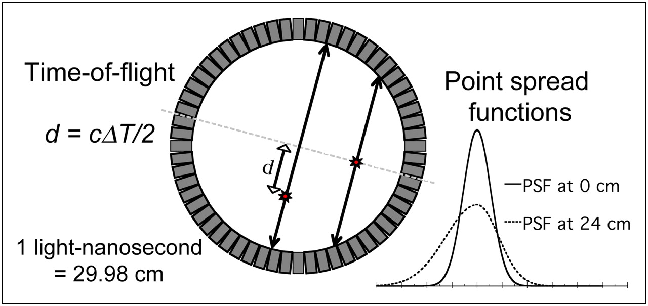
- FIGURE 1.
Recent advances in PET technology include tomographs with time-of-flight capability and point spread function modeling. TOF PET utilizes very fast detectors and electronics to measure the time difference between detection of each photon of annihilation pair, providing an estimate of depth d along the line-of-response where event originated. When events originate away from central axis of the scanner, the point response function becomes broad and asymmetric due to depth-of-interaction and other effects. Consideration of both of these effects during reconstruction can provide lower noise images with improved spatial resolution.
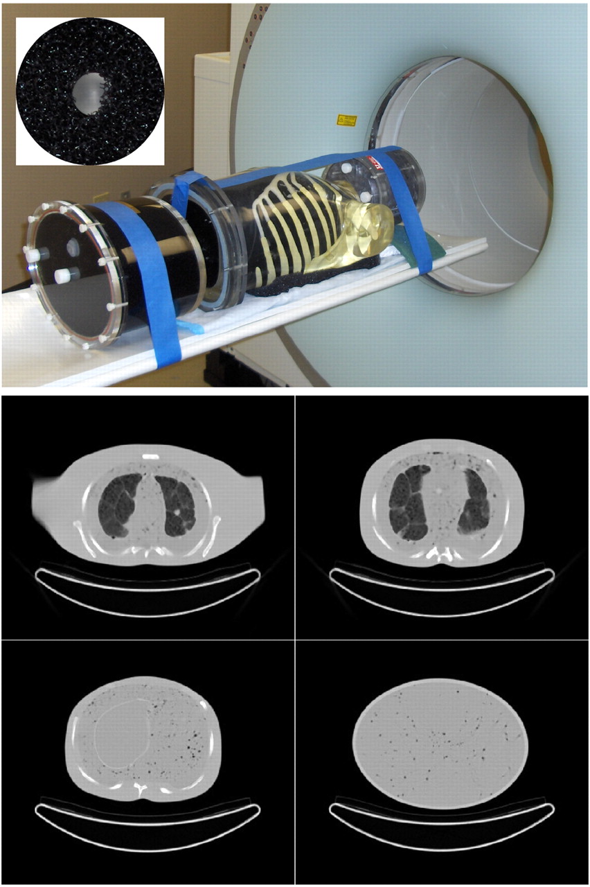
- FIGURE 2.
Whole-body phantom (top) consisted of anthropomorphic thorax phantom with lungs and liver, 3D brain phantom, and pelvis compartment with bladder. Inset (top) shows shell-less 68Ge lesion embedded in black open cell foam. Custom modifications to the soft-tissue and lung compartments with open-cell foam and nylon bead bags provided inhomogeneous density and structures as shown by CT (bottom). Such heterogeneities more accurately mimic patient structures, providing a more realistic and challenging lesion-detection task than encountered for phantoms with uniform background compartments.
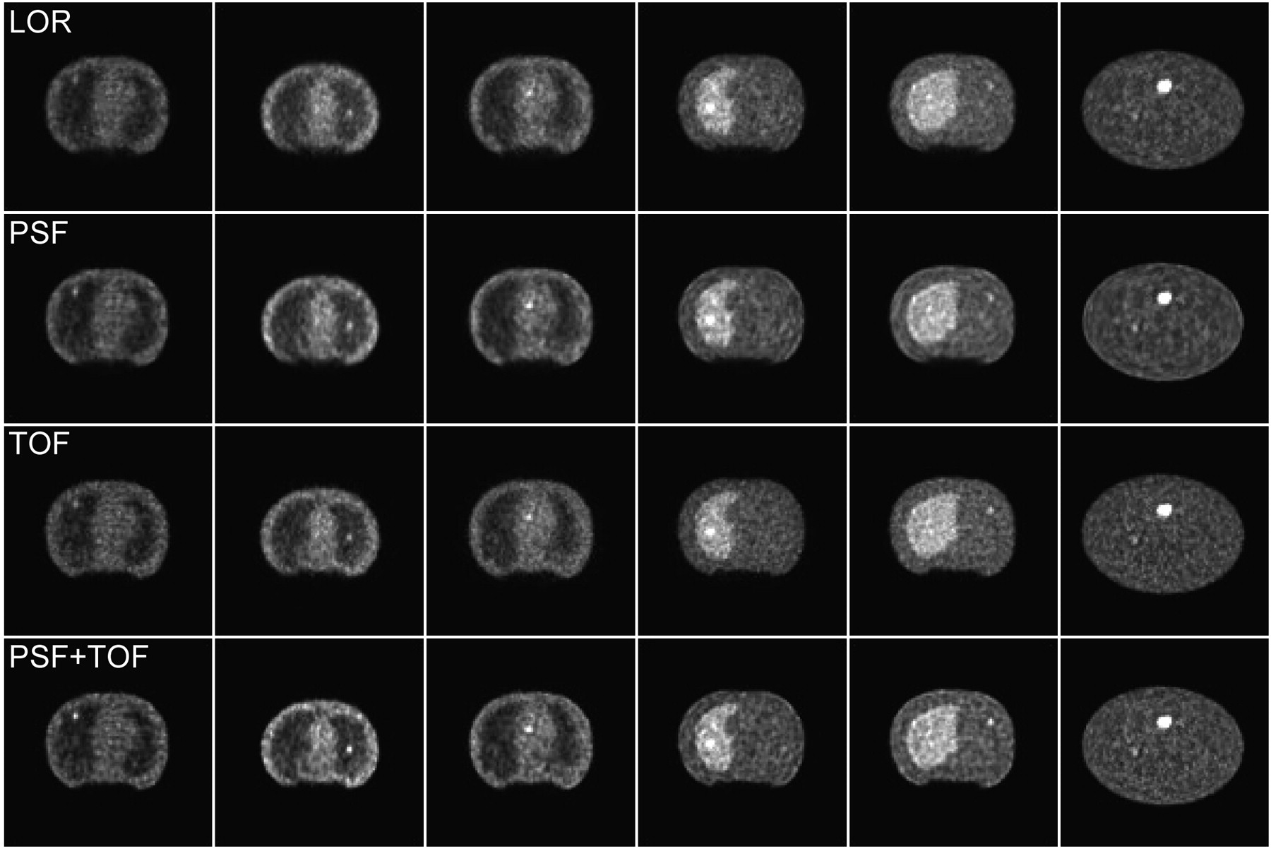
- FIGURE 3.
Example reconstructed images for (top-to-bottom) LOR, PSF, TOF, and PSF+TOF. Each image slice shown contains exactly 1 lesion, where relatively obvious cases are shown for purposes of this example. Number of iterations and post-reconstruction filter strength used for each algorithm are given in Table 1. Note that while images show broad similarities, differences in lesion depiction and noise texture exist between algorithms. The effect of these differences upon the task of identifying and localizing focal lesions has been evaluated in this study.
- FIGURE 4.
Results of CNPW model observer analysis, plotting area under the LROC curve versus filter strength for each algorithm. Separate lines are shown on each plot for iterations 1–10. These data were used to select optimal reconstruction parameters for images read by human observers. The algorithms including PSF model (B, D) required little or no filter for maximal performance, whereas other algorithms (A, C) required moderate filtering for peak performance.
- FIGURE 5.
Final results of model observer study (A) and human observer study (B), providing comparison and ranking of 4 algorithms studied. Model observer results plot area under LROC curve versus number of iterations, where optimal filter for each iteration and algorithm was applied (as determined from results shown in Fig. 4). Note that reconstructions with TOF provided markedly higher performance at earliest iterations, showing that the most important image features for lesion detection are recovered more quickly when using TOF. Human observer LROC curves demonstrate distinctions in performance for 4 algorithms, and confirm model observer results. Quantitative results for these curves are provided in Table 2.
- FIGURE 6.
Coronal (top) and sagittal (bottom) sections through 18F-FDG PET/CT scan of a patient with history of colon cancer. The 71-y-old male patient had BMI of 33.5 and was injected with 396 MBq (10.7 mCi) of 18F-FDG. After 90-min uptake period, patient underwent CT scan (130 kV; 180 mAs) with both intravenous and oral contrast, followed by PET scan acquired at 6 bed positions for 3 min per bed. PET data were reconstructed into 168 × 168 matrix with (A) baseline algorithm and PSF (4 iterations, 14 subsets, no smoothing), and (B) baseline algorithm with PSF and TOF (2 iterations, 14 subsets, no smoothing) and time resolution kernel of 590 ps.
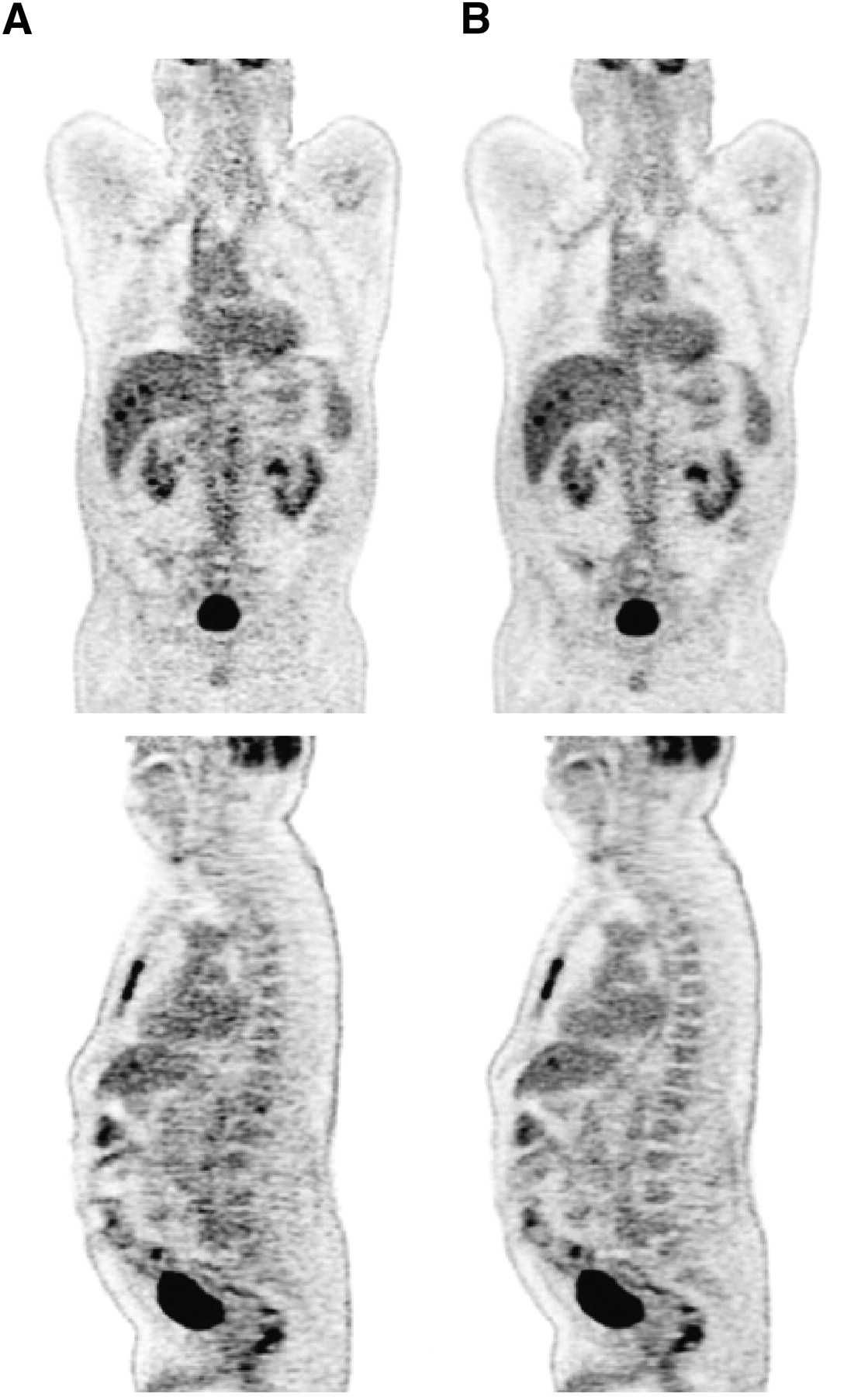
- FIGURE 7.
Coronal (top) and sagittal (bottom) sections through 18F-FDG PET/CT scan of a patient with esophageal cancer. The 61-y-old male patient had a BMI of 27 and was injected with 366.3 MBq (9.9 mCi) of 18F-FDG. After 90-min uptake period, patient underwent CT scan (130 kV; 180 mAs), followed by PET scan that was acquired at 5 bed positions for 3 min per bed. PET data were reconstructed into 168 × 168 matrix by (A) baseline algorithm with PSF (4 iterations, 14 subsets, no smoothing), and (B) baseline algorithm with PSF and TOF (2 iterations, 14 subsets, no smoothing) and time resolution kernel of 590 ps.


Tables
Optimal parameters CNPW observer results Algorithm No. of iterations† Filter SD (voxels) PLOC ± SD ALROC ± SD LOR 6 0.5 0.588 ± 0.068 0.418 ± 0.051 PSF 8 0.1 0.706 ± 0.063 0.516 ± 0.052 TOF 6 0.5 0.804 ± 0.056 0.673 ± 0.054 PSF+TOF 8 0.1 0.882 ± 0.045 0.813 ± 0.046 Human observer results Tukey all-pairs comparison (P)* Algorithm PLOC ± SD ALROC ± SD LOR PSF TOF PSF+TOF LOR 0.549 ± 0.039 0.486 ± 0.045 N/A 0.002 0.001 <0.001 PSF 0.690 ± 0.064 0.662 ± 0.087 0.002 N/A 0.882 0.001 TOF 0.741 ± 0.043 0.691 ± 0.046 0.001 0.882 N/A 0.002 PSF+TOF 0.886 ± 0.056 0.873 ± 0.062 <0.001 0.001 0.002 N/A ↵* Tukey all-pairs comparison performed on ALROC figure-of-merit.
N/A = not applicable.
Supplemental Data
Files in this Data Supplement:


Jump to section
Related Articles
Cited By...
- Impact of PET Reconstruction on Amyloid-{beta} Quantitation in Cross-Sectional and Longitudinal Analyses
- Performance Evaluation of the uMI Panorama PET/CT System in Accordance with the National Electrical Manufacturers Association NU 2-2018 Standard
- Impact of PET reconstruction on A{beta}-amyloid quantitation in cross-sectional and longitudinal analyses
- Protocols for Harmonized Quantification and Noise Reduction in Low-Dose Oncologic 18F-FDG PET/CT Imaging
- Time-of-Flight Information Improved the Detectability of Subcentimeter Spheres Using a Clinical PET/CT Scanner
- Clinical Impact of Respiratory Motion Correction in Simultaneous PET/MR, Using a Joint PET/MR Predictive Motion Model
- The Effect of Misregistration Between CT-Attenuation and PET-Emission Images in 13N-Ammonia Myocardial PET/CT
- Dual-Gated Motion-Frozen Cardiac PET with Flurpiridaz F 18
- Impact of Image Reconstruction Settings on Texture Features in 18F-FDG PET
- Improving the Detection of Small Lesions Using a State-of-the-Art Time-of-Flight PET/CT System and Small-Voxel Reconstructions
- Update on Time-of-Flight PET Imaging
- Effect of Time-of-Flight Technique on the Diagnostic Performance of 18F-FDG PET/CT for Assessment of Lymph Node Metastases in Head and Neck Squamous Cell Carcinoma
- Determination of Accuracy and Precision of Lesion Uptake Measurements in Human Subjects with Time-of-Flight PET
- Improvement in PET/CT Image Quality with a Combination of Point-Spread Function and Time-of-Flight in Relation to Reconstruction Parameters
- Impact of Time-of-Flight PET on Whole-Body Oncologic Studies: A Human Observer Lesion Detection and Localization Study
- An Assessment of the Impact of Incorporating Time-of-Flight Information into Clinical PET/CT Imaging