
Main menu

  • Home
  • Content
    • Current
    • Ahead of print
    • Past Issues
    • JNM Supplement
    • SNMMI Annual Meeting Abstracts
    • Continuing Education
    • JNM Podcasts
  • Subscriptions
    • Subscribers
    • Institutional and Non-member
    • Rates
    • Journal Claims
    • Corporate & Special Sales
  • Authors
    • Submit to JNM
    • Information for Authors
    • Assignment of Copyright
    • AQARA requirements
  • Info
    • Reviewers
    • Permissions
    • Advertisers
  • About
    • About Us
    • Editorial Board
    • Contact Information
  • More
    • Alerts
    • Feedback
    • Help
    • SNMMI Journals
  • SNMMI
    • JNM
    • JNMT
    • SNMMI Journals
    • SNMMI

User menu

  • Subscribe
  • My alerts
  • Log in
  • My Cart

Search

  • Advanced search
Journal of Nuclear Medicine
  • SNMMI
    • JNM
    • JNMT
    • SNMMI Journals
    • SNMMI
  • Subscribe
  • My alerts
  • Log in
  • My Cart
Journal of Nuclear Medicine

Advanced Search

  • Home
  • Content
    • Current
    • Ahead of print
    • Past Issues
    • JNM Supplement
    • SNMMI Annual Meeting Abstracts
    • Continuing Education
    • JNM Podcasts
  • Subscriptions
    • Subscribers
    • Institutional and Non-member
    • Rates
    • Journal Claims
    • Corporate & Special Sales
  • Authors
    • Submit to JNM
    • Information for Authors
    • Assignment of Copyright
    • AQARA requirements
  • Info
    • Reviewers
    • Permissions
    • Advertisers
  • About
    • About Us
    • Editorial Board
    • Contact Information
  • More
    • Alerts
    • Feedback
    • Help
    • SNMMI Journals
  • View or Listen to JNM Podcast
  • Visit JNM on Facebook
  • Join JNM on LinkedIn
  • Follow JNM on Twitter
  • Subscribe to our RSS feeds
OtherBASIC SCIENCE INVESTIGATIONS

Uptake of 18F-Labeled 6-Fluoro-6-Deoxy-d-Glucose by Skeletal Muscle Is Responsive to Insulin Stimulation

Chandra Spring-Robinson, Visvanathan Chandramouli, William C. Schumann, Peter F. Faulhaber, Yanming Wang, Chunying Wu, Faramarz Ismail-Beigi and Raymond F. Muzic
Journal of Nuclear Medicine June 2009, 50 (6) 912-919; DOI: https://doi.org/10.2967/jnumed.109.062687
Chandra Spring-Robinson
  • Find this author on Google Scholar
  • Find this author on PubMed
  • Search for this author on this site
Visvanathan Chandramouli
  • Find this author on Google Scholar
  • Find this author on PubMed
  • Search for this author on this site
William C. Schumann
  • Find this author on Google Scholar
  • Find this author on PubMed
  • Search for this author on this site
Peter F. Faulhaber
  • Find this author on Google Scholar
  • Find this author on PubMed
  • Search for this author on this site
Yanming Wang
  • Find this author on Google Scholar
  • Find this author on PubMed
  • Search for this author on this site
Chunying Wu
  • Find this author on Google Scholar
  • Find this author on PubMed
  • Search for this author on this site
Faramarz Ismail-Beigi
  • Find this author on Google Scholar
  • Find this author on PubMed
  • Search for this author on this site
Raymond F. Muzic Jr.
  • Find this author on Google Scholar
  • Find this author on PubMed
  • Search for this author on this site
  • Article
  • Figures & Data
  • Info & Metrics
  • PDF
Loading

Article Figures & Data

Figures

  • Tables
  • FIGURE 1. 
    • Download figure
    • Open in new tab
    • Download powerpoint
    FIGURE 1. 

    (Top) Glucose infusion rate over duration of typical experiment. Insulin infusion was begun at 35 min, and glucose infusion rate was increased to maintain plasma glucose concentration at approximately 6 mM. (Middle) Observed glucose concentrations. Data showed that glucose concentrations were similar under basal and insulin-stimulated conditions. (Bottom) Times of injection of 18F-6FDG (arrows) and resultant time courses of activity in gastrocnemius muscle (solid curves) under basal and insulin-stimulated conditions. Contribution of activity from that remaining from first scan was removed from second scan by extrapolation (dashed curve) and subtraction.
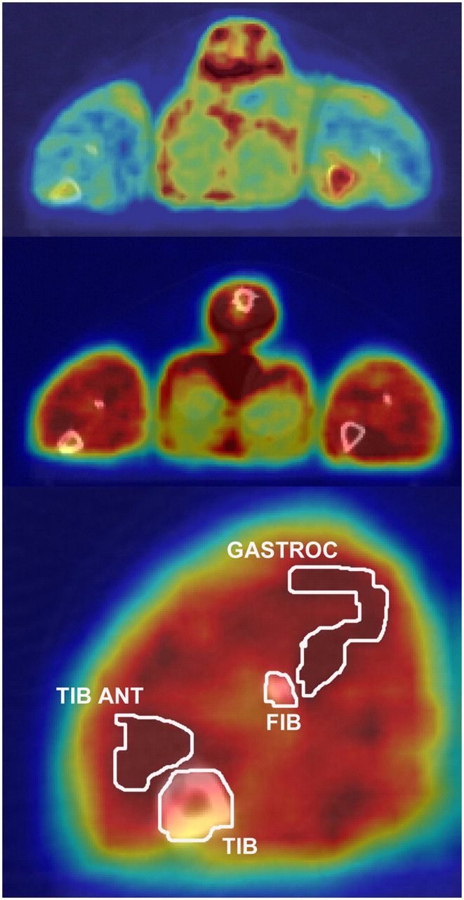
  • FIGURE 2. 
    • Download figure
    • Open in new tab
    • Download powerpoint
    FIGURE 2. 

    CT images (gray scale) fused with PET images (color scale) of same rat in basal state (top) and high-insulin state (middle and bottom), illustrating data analysis procedure and typical imaging results. Contours illustrate boundaries of VOIs. Rat was lying prone with its hind legs extended caudally. These images reflected tissue activity between 0 and 30 min after 18F-6FDG injection. FIB = fibula; GASTROC = gastrocnemius; TIB = tibia; TIB ANT = tibialis anterior.
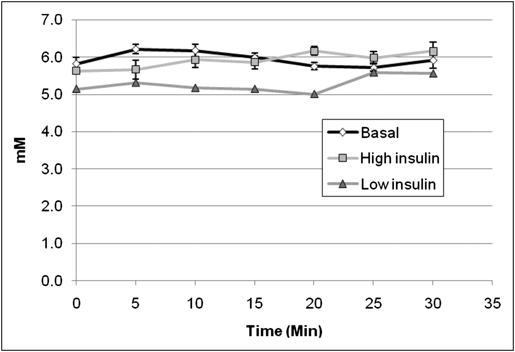
  • FIGURE 3. 
    • Download figure
    • Open in new tab
    • Download powerpoint
    FIGURE 3. 

    Plasma glucose concentrations measured in arterial blood samples collected every 5 min. With perfect glucose clamp, concentrations should be same in all states. Concentration of 5.3 ± 0.3 mM measured during low-insulin state was slightly lower than concentration of 6.2 ± 0.4 mM measured during basal state in these animals, although this small difference was statistically significant. Concentrations in high-insulin state and basal state were both 5.9 ± 0.5 mM. Error bars denote SE for basal state (n = 9) and high-insulin state (n = 7) and were omitted for low-insulin state (n = 2).
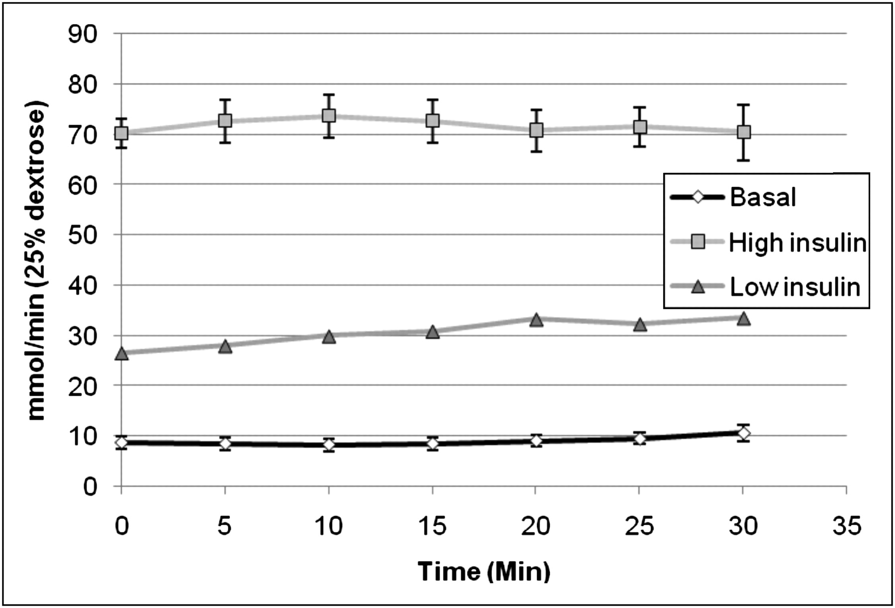
  • FIGURE 4. 
    • Download figure
    • Open in new tab
    • Download powerpoint
    FIGURE 4. 

    Average GIR needed to maintain 6 mM plasma glucose concentration. This value was higher during low-insulin (n = 2) and high-insulin (n = 7) infusions than at baseline (n = 9). Error bars denote SE.
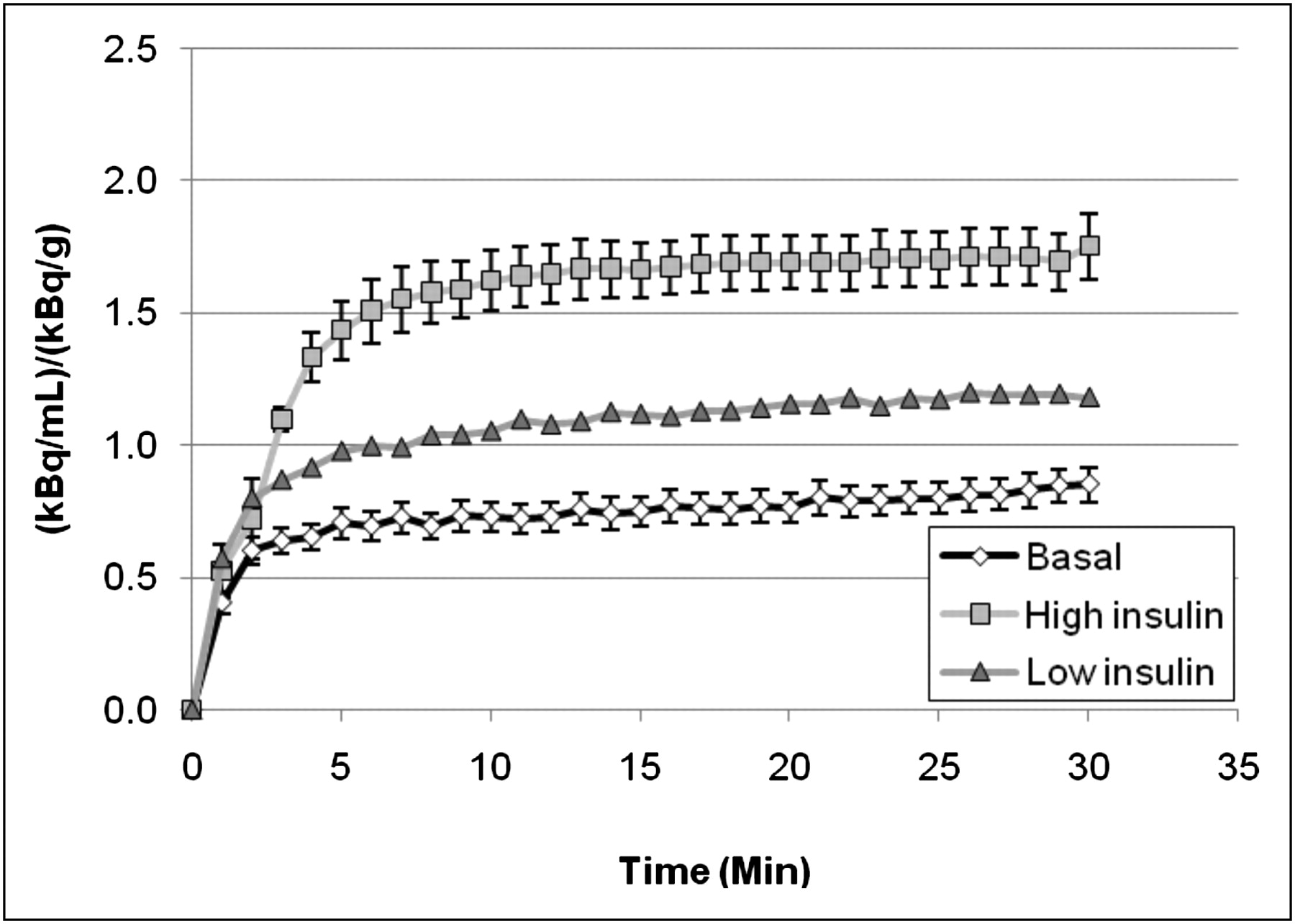
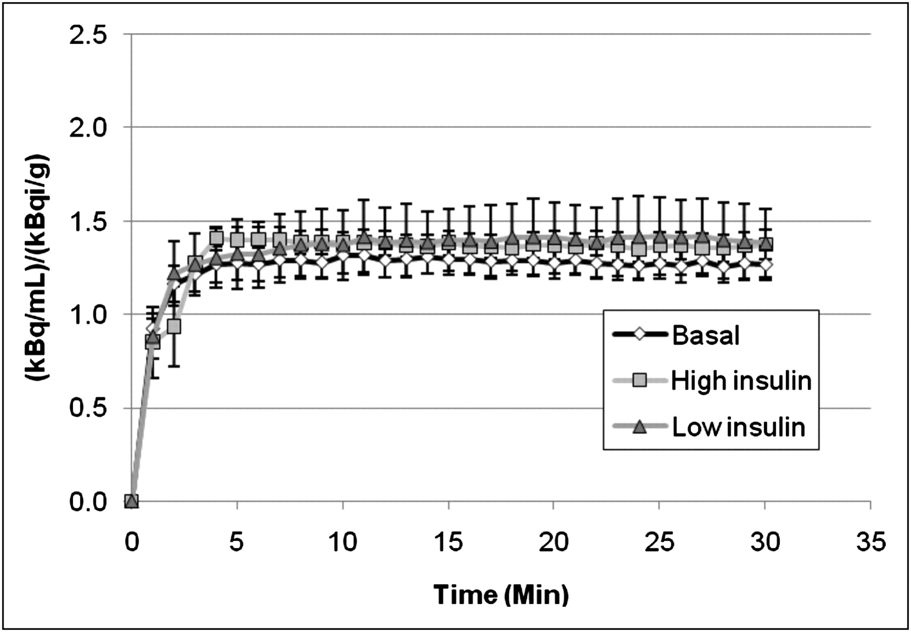
  • FIGURE 5. 
    • Download figure
    • Open in new tab
    • Download powerpoint
    FIGURE 5. 

    Tissue time–activity curves for gastrocnemius muscle during scans acquired under high-insulin conditions (n = 7), low-insulin conditions (n = 2), and basal conditions (n = 9), demonstrating increased 18F-6FDG uptake caused by insulin administration. Error bars are not shown for low insulin because of small number of subjects, although maximum difference between individual curves from 2 rats was 25%.

  • FIGURE 6. 
    • Download figure
    • Open in new tab
    • Download powerpoint
    FIGURE 6. 

    Bone marrow time–activity curves in basal, low-insulin, and high-insulin states. These curves were comparable, indicating lack of effect of insulin stimulation on glucose transport in marrow. This result was in contrast to effect observed in skeletal muscle in Figure 5.

Tables

  • Figures
    • View popup
    TABLE 1

    Metabolic Parameters Evaluated in Glucose Clamp Study

    Value in following state:
    ParameterBasalLow insulinHigh insulin
    No. of rats927
    Arterial glucose concentration (mM)*5.9 ± 0.45.3 ± 0.35.9 ± 0.5
    Arterial insulin concentration (pmol/mL)*0.5 ± 0.4Not available12.2 ± 1.1
    GIR (μmol/min)*9.0 ± 3.830.6 ± 2.871.7 ± 10.3
    • ↵* Reported as mean ± SD.

PreviousNext
Back to top

In this issue

Journal of Nuclear Medicine: 50 (6)
Journal of Nuclear Medicine
Vol. 50, Issue 6
June 2009
  • Table of Contents
  • Table of Contents (PDF)
  • About the Cover
  • Index by author
Print
Download PDF
Article Alerts
Sign In to Email Alerts with your Email Address
Email Article

Thank you for your interest in spreading the word on Journal of Nuclear Medicine.

NOTE: We only request your email address so that the person you are recommending the page to knows that you wanted them to see it, and that it is not junk mail. We do not capture any email address.

Enter multiple addresses on separate lines or separate them with commas.
Uptake of 18F-Labeled 6-Fluoro-6-Deoxy-d-Glucose by Skeletal Muscle Is Responsive to Insulin Stimulation
(Your Name) has sent you a message from Journal of Nuclear Medicine
(Your Name) thought you would like to see the Journal of Nuclear Medicine web site.
Citation Tools
Uptake of 18F-Labeled 6-Fluoro-6-Deoxy-d-Glucose by Skeletal Muscle Is Responsive to Insulin Stimulation
Chandra Spring-Robinson, Visvanathan Chandramouli, William C. Schumann, Peter F. Faulhaber, Yanming Wang, Chunying Wu, Faramarz Ismail-Beigi, Raymond F. Muzic
Journal of Nuclear Medicine Jun 2009, 50 (6) 912-919; DOI: 10.2967/jnumed.109.062687

Citation Manager Formats

  • BibTeX
  • Bookends
  • EasyBib
  • EndNote (tagged)
  • EndNote 8 (xml)
  • Medlars
  • Mendeley
  • Papers
  • RefWorks Tagged
  • Ref Manager
  • RIS
  • Zotero
Share
Uptake of 18F-Labeled 6-Fluoro-6-Deoxy-d-Glucose by Skeletal Muscle Is Responsive to Insulin Stimulation
Chandra Spring-Robinson, Visvanathan Chandramouli, William C. Schumann, Peter F. Faulhaber, Yanming Wang, Chunying Wu, Faramarz Ismail-Beigi, Raymond F. Muzic
Journal of Nuclear Medicine Jun 2009, 50 (6) 912-919; DOI: 10.2967/jnumed.109.062687
Twitter logo Facebook logo LinkedIn logo Mendeley logo
  • Tweet Widget
  • Facebook Like
  • Google Plus One
Bookmark this article

Jump to section

  • Article
    • Abstract
    • MATERIALS AND METHODS
    • RESULTS
    • DISCUSSION
    • CONCLUSION
    • Acknowledgments
    • Footnotes
    • References
  • Figures & Data
  • Info & Metrics
  • PDF

Related Articles

  • This Month in JNM
  • PubMed
  • Google Scholar

Cited By...

  • No citing articles found.
  • Google Scholar

More in this TOC Section

  • PET/MRI of Hypoxic Atherosclerosis Using 64Cu-ATSM in a Rabbit Model
  • Tumor Uptake of Anti-CD20 Fabs Depends on Tumor Perfusion
  • How Sensitive Is the Upper Gastrointestinal Tract to 90Y Radioembolization? A Histologic and Dosimetric Analysis in a Porcine Model
Show more Basic Science Investigations

Similar Articles

SNMMI

© 2025 SNMMI

Powered by HighWire