


Article Figures & Data
Figures
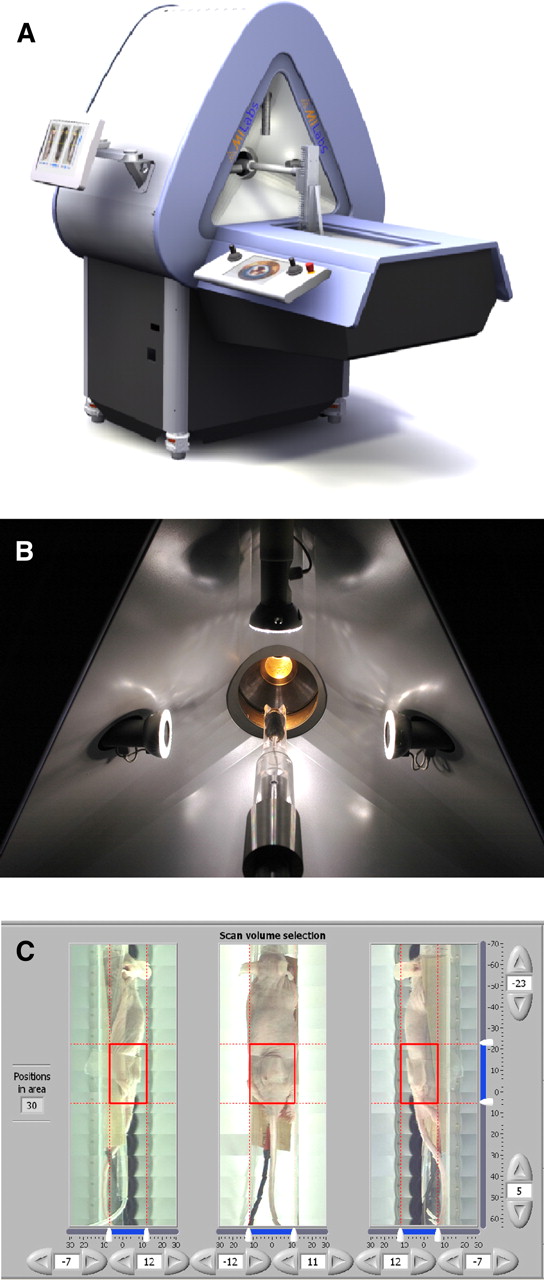
- FIGURE 1.
(A) U-SPECT-II system. (B) Mouse observed with 3 optical cameras for ROI selection. (C) User interface for selection of ROI, for example, a tumor. ROI = region of interest.
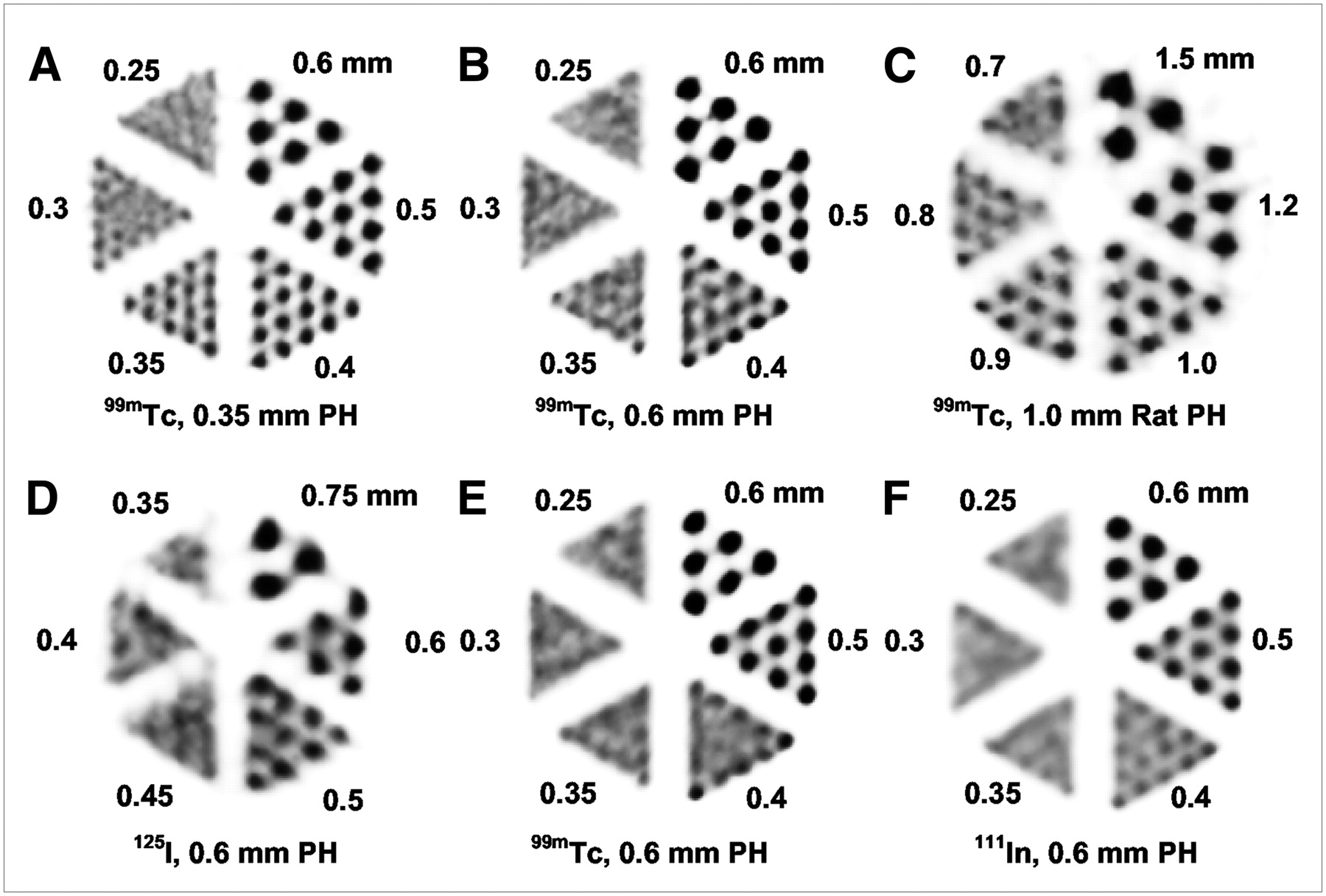
- FIGURE 2.
Capillary phantom reconstructions, with slice thickness of 3 mm. Top row shows reconstructions with different collimators with 99mTc (600 MBq/mL, 2-h scan time): 0.35-mm-pinhole mouse collimator (A), 0.6-mm-pinhole mouse collimator (B), and 1.0-mm-pinhole rat collimator (C). Smallest discernable rod sizes were 0.35, 0.4, and 0.8 mm, respectively. Bottom row shows reconstructions with different isotopes—125I (D), 99mTc (E), and 111In (F)—and 0.6-mm-pinhole mouse collimator (74 MBq/mL, 2-h scan time).
- FIGURE 3.
Maximum-intensity-projection images of rat 99mTc-HDP bone scan. Reconstruction used 100% down to 1% of available counts from list-mode data. Movie of gated reconstruction is shown in Supplemental Video 1.
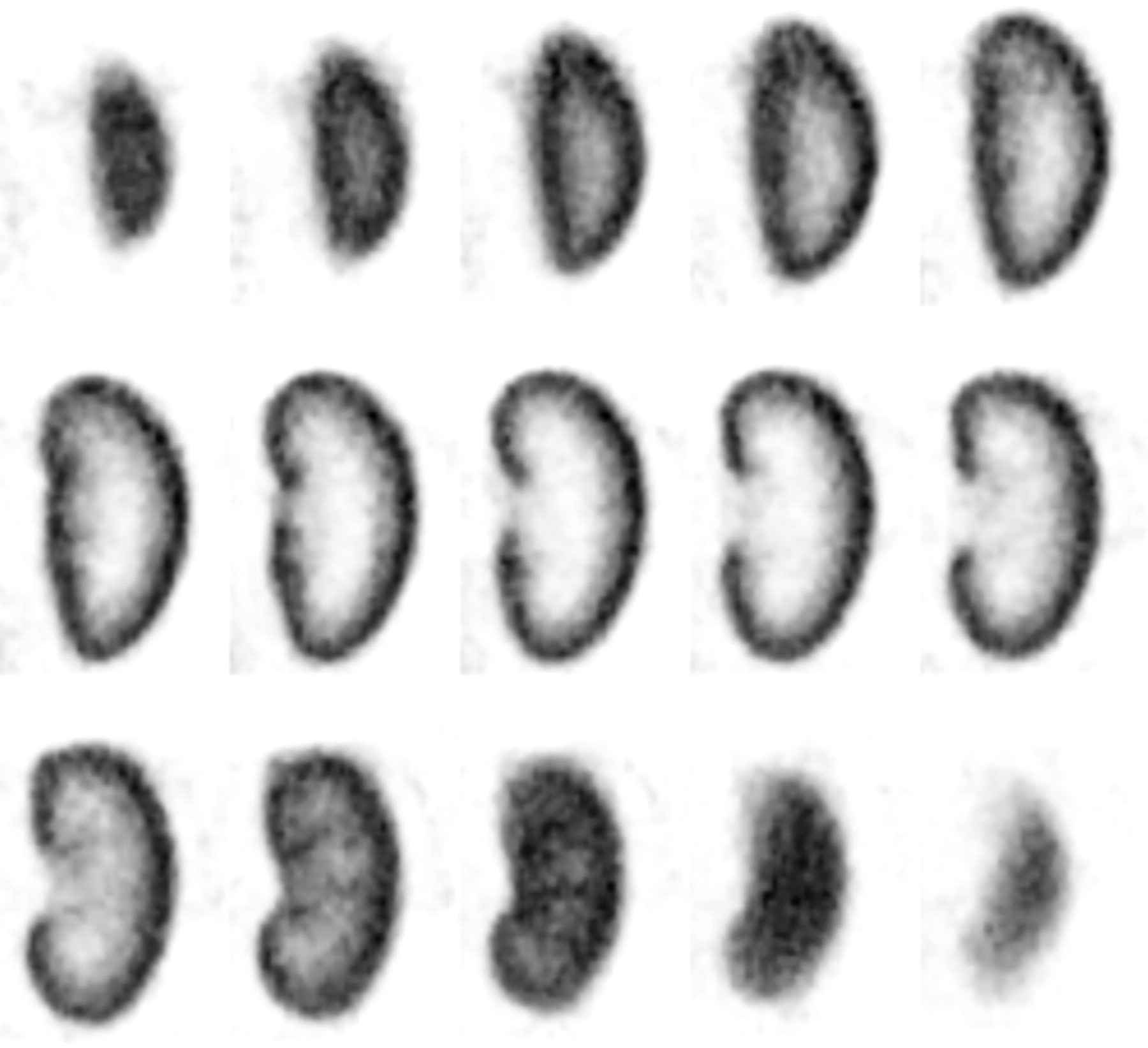
- FIGURE 4.
99mTc-DMSA scan of mouse kidney (slice thickness, 0.375 mm). Reconstruction was performed using 100% (shown) and 10% (Supplemental Fig. 1) of acquired counts. Image shows distribution of functioning kidney tissue. Such scans can be used to localize defects in parenchyma, to assess relative contribution of subcompartments of kidney.
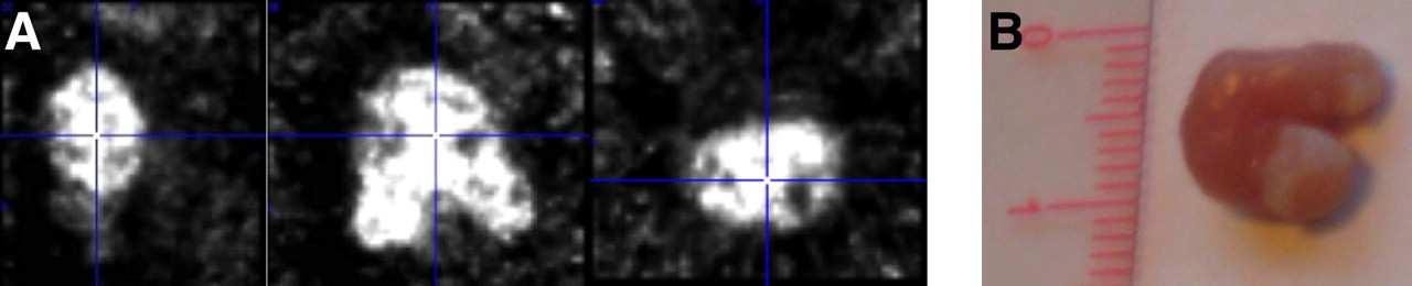
- FIGURE 5.
111In-DTPA-14C5 mouse study. (A) Three orthogonal views of reconstructed data. (B) Photograph of tumor.


Tables
Parameter U-SPECT-I U-SPECT-II Detectors Crystal length × width × thickness (mm) 410 × 250 × 9.5 508 × 381 × 9.5 No. of detectors 3 3 No. of PMTs per detector 49 55 PMT readout method Analog, resistor network Each PMT signal digitized Data collection Planar pixel image List-mode data Pinhole collimator configuration No. of rings and pinholes per ring 5 × 15 5 × 15 Mouse single-bed position FOV diameter (mm) 10.5 12 Mouse single-bed position FOV axial (mm) 5 7 Rat single-bed position FOV diameter (mm) N/A 27 Rat single-bed position FOV axial (mm) N/A 11 Mouse collimator bore diameter (mm) 39 44 Mouse radial position of pinholes (mm) 22 24 Rat collimator bore diameter (mm) N/A 98 Rat radial position of pinholes (mm) N/A 53 Mouse pinhole material and diameters (mm) Gold, 0.3, 0.6 Gold, 0.35, 0.6 Rat pinhole material and diameters (mm) N/A Tungsten, 0.7–1.5, gold optional Mouse pinhole tube material Tungsten Tungsten Mouse shielding tube material Lead Tungsten Rat collimator tube material N/A Tungsten General features Mouse collimator, 0.6-mm-pinhole peak efficiency (%) 0.22 0.18 Mouse collimator, 0.35-mm-pinhole peak efficiency (%) 0.07 0.07 Rat collimator, 1.0-mm-pinhole peak efficiency (%) N/A 0.09 Gating possible No Yes, multiple Dead time between measurements (s) 12 <2 Dynamic sequence imaging Manual start Automatic Coordination of scan sequence with acquisition Manual Automatic GUI-based navigator available No Yes PMTs = photomultiplier tubes; N/A = not applicable; GUI = graphical user interface.
Collimator FOV sensitivity (cps/MBq 99mTc) FOV geometric sensitivity (%) Mouse, 0.6-mm pinhole 1,500 0.18 Mouse, 0.35-mm pinhole 525 0.07 Rat, 1.0-mm pinhole 700 0.09
Supplemental Data
Files in this Data Supplement:


Jump to section
Related Articles
Cited By...
- A pair of congenic mice for imaging of transplants by positron emission tomography using anti-transferrin receptor nanobodies
- PD-L1 microSPECT/CT Imaging for Longitudinal Monitoring of PD-L1 Expression in Syngeneic and Humanized Mouse Models for Cancer
- In Vivo Imaging of Antileukemic Drug Asparaginase Reveals a Rapid Macrophage-Mediated Clearance from the Bone Marrow
- Liposomal Treatment of Experimental Arthritis Can Be Monitored Noninvasively with a Radiolabeled Anti-Fibroblast Activation Protein Antibody
- Noninvasive Imaging of Tumor PD-L1 Expression Using Radiolabeled Anti-PD-L1 Antibodies
- Immuno-PET and Immuno-SPECT of Rheumatoid Arthritis with Radiolabeled Anti-Fibroblast Activation Protein Antibody Correlates with Severity of Arthritis
- Ultra-High-Sensitivity Submillimeter Mouse SPECT
- Can 111In-RGD2 Monitor Response to Therapy in Head and Neck Tumor Xenografts?
- Performance Assessment of a Preclinical PET Scanner with Pinhole Collimation by Comparison to a Coincidence-Based Small-Animal PET Scanner
- Optical Imaging of Renal Cell Carcinoma with Anti-Carbonic Anhydrase IX Monoclonal Antibody Girentuximab
- Three-Dimensional Histologic Validation of High-Resolution SPECT of Antibody Distributions Within Xenografts
- Imaging Integrin {alpha}v{beta}3 on Blood Vessels with 111In-RGD2 in Head and Neck Tumor Xenografts
- Imaging of Epidermal Growth Factor Receptor Expression in Head and Neck Cancer with SPECT/CT and 111In-Labeled Cetuximab-F(ab')2
- Imaging Capabilities of the Inveon SPECT System Using Single-and Multipinhole Collimators
- VECTor: A Preclinical Imaging System for Simultaneous Submillimeter SPECT and PET
- Fast Spiral SPECT with Stationary {gamma}-Cameras and Focusing Pinholes
- Imaging of Human Epidermal Growth Factor Receptor Type 2 Expression with 18F-Labeled Affibody Molecule ZHER2:2395 in a Mouse Model for Ovarian Cancer
- Serial Semiquantitative Imaging of Brain Damage Using Micro-SPECT and Micro-CT After Endothelin-1-Induced Transient Focal Cerebral Ischemia in Rats
- 99mTc-(CO)3 His-Annexin A5 Micro-SPECT Demonstrates Increased Cell Death by Irinotecan During the Vascular Normalization Window Caused by Bevacizumab
- Using the NEMA NU 4 PET Image Quality Phantom in Multipinhole Small-Animal SPECT
- Dynamic and Static Small-Animal SPECT in Rats for Monitoring Renal Function After 177Lu-Labeled Tyr3-Octreotate Radionuclide Therapy
- ImmunoSPECT and ImmunoPET of IGF-1R Expression with the Radiolabeled Antibody R1507 in a Triple-Negative Breast Cancer Model