


Article Figures & Data
Figures
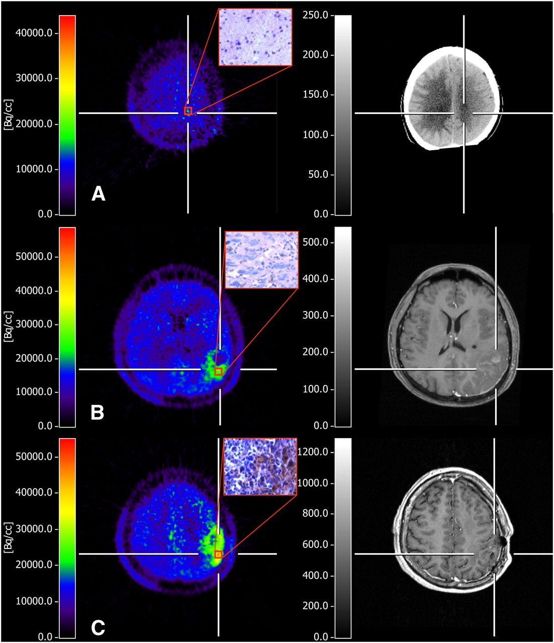
- FIGURE 1.
11C-MET PET of 39-y-old man with malignant progression of recurrent glioma. (A) Newly diagnosed grade II astrocytoma with average 11C-MET uptake of 1.3 to contralateral gray matter, with no enhancement on contrast-enhanced CT and no immunohistochemical VEGF expression. (B) One year later, patient presented with malignant progression to grade III astrocytoma associated with significant increase in 11C-MET uptake (to 2.1-fold) and only slight contrast enhancement outside metabolically active tumor. Histologic analysis from resection showed increase in cellularity, numerous pleomorphic nuclei, and low VEGF expression. (C) In following year, resection of tumor again confirmed malignant progression to glioblastoma multiforme, showing markedly increased uptake of 11C-MET (to 2.8-fold), marginal contrast enhancement on MRI, and ∼35% of tumor cells expressing VEGF (original magnification, ×400). PET-guided biopsies were taken from region with highest 11C-MET uptake, which was in different locations within same tumor over time.
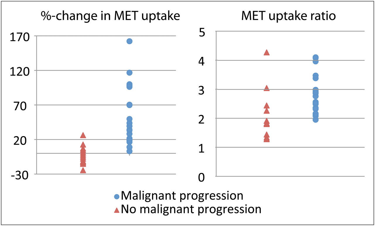
- FIGURE 2.
Comparison of value of 11C-MET uptake ratio at time of biopsy vs. percentage change in 11C-MET uptake in distinguishing malignant from nonmalignant progression.
- FIGURE 3.
Receiver operating characteristic analysis to identify change in 11C-MET uptake for differentiation between malignant progression of tumor grade and no malignant progression. Percentage increase that best distinguished malignant progression from no malignant progression was at threshold of 14.6%, with sensitivity of 90% and specificity of 92.3%.
- FIGURE 4.
Correlation between changes in expression of VEGF and changes in 11C-MET uptake during first follow-up investigation.


Tables
Patient no. Age (y) Sex Initial histology and WHO grade Resection Radiotherapy before PET Chemotherapy before PET 1 32 F Astrocytoma II T/P Yes (9)/yes (3) Yes (9)/no 2 46 F Oligoastrocytoma II S/P No/no No/no 3 10 M Astrocytoma II T/T No/yes (6) No/yes (6) 4 40 F Astrocytoma II P/P/P No/no/yes (36) No/no/yes (36) 5 28 M Astrocytoma II S/S No/yes (150) No/no 6 49 F Oligoastrocytoma II T/T No/no No/no 7 26 M Oligoastrocytoma II T/P/P No/no/yes (159) No/no/no 8 35 M Astrocytoma III S/S No/yes (24) No/yes (24) 9 32 F Oligoastrocytoma II T/T/T/T No/yes (376)/yes (53)/no No/no/no/no 10 35 F Oligoastrocytoma II P/P Yes (156)/no No/no 11 53 M Oligodendroglioma II T/P/P No/yes (126)/no No/no/no 12 29 M Astrocytoma II T/S No/no No/no 13 40 M Oligoastrocytoma II T/T/P No/no/yes (12) No/no/yes (12) 14 30 M Oligoastrocytoma III P/P No/no No/no 15 23 M Astrocytoma III P/T No/yes (50) No/yes (50) 16 27 F Oligodendroglioma III S/P No/yes (9) No/yes (9) 17 39 M Oligoastrocytoma II T/T No/no No/no 18 38 F Astrocytoma II T/T/S No/no/yes (102) No/no/no 19 57 F Astrocytoma II S/T No/no No/no 20 37 M Astrocytoma II S/T/T No/no/yes (25) No/no/yes (25) 21 34 M Astrocytoma II P/S No/no No/no 22 59 F Astrocytoma II P/P/P No/no/no No/no/no 23 47 M Oligoastrocytoma III O/P No/yes (25) No/yes (25) 24 58 M Astrocytoma III S/P No/no No/no T = total resection; P = partial resection; S = stereotactic biopsy; O = open biopsy.
Data in parentheses are interval (in weeks) between PET scan and chemo- or radiotherapy.
- TABLE 2
Changes in 11C-MET Uptake in Patients Without Tumor Progression or with Various Degrees of Progression
Malignant progression Parameter None (n = 13) From grade II to III (n = 11) From grade III to IV (n = 6) From grade II to IV (n = 3) Mean 3.9% 72.9% 33.5% 38.1% SD 13.7% 46.2% 35.4% 33.6% - TABLE 3
Changes in Contrast Enhancement in Relation to Histologically Proven Malignant Progression
Newly appearing contrast enhancement on MRI/CT Malignant progression Present Absent Present 8 9 Absent 3 8


Jump to section
Related Articles
Cited By...
- Definition of Primary and Secondary Glioblastoma--Letter
- Role of O-(2-18F-Fluoroethyl)-L-Tyrosine PET as a Diagnostic Tool for Detection of Malignant Progression in Patients with Low-Grade Glioma
- Multimodality Assessment of Brain Tumors and Tumor Recurrence
- Novel Positron Emission Tomography Tracer Distinguishes Normal from Cancerous Cells