


Article Figures & Data
Figures
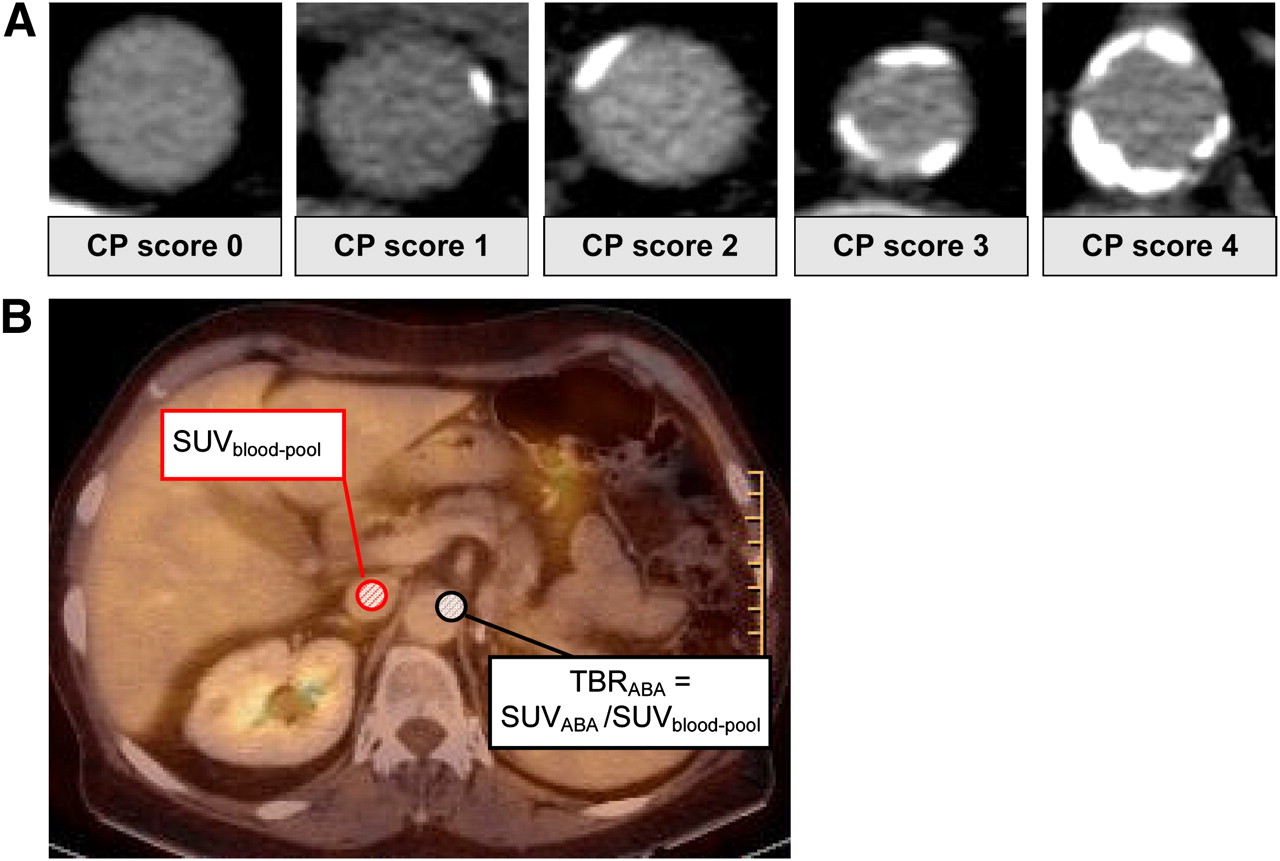
- FIGURE 1.
PET/CT image analysis. (A) Example of modified calcified plaque scoring system (16). Images from left to right show scores of 0 (no calcified plaque), 1 (calcified plaque involving <10% of vessel circumference), 2 (calcified plaque involving 10%−25%), 3 (calcified plaque involving 25%−50%), and 4 (calcified plaque involving >50%). (B) Example of TBR calculation method. ROIs were drawn in center of vena cava inferior (red) and in arterial wall of abdominal aorta (black). ABA = abdominal aorta; CP = calcified plaque.
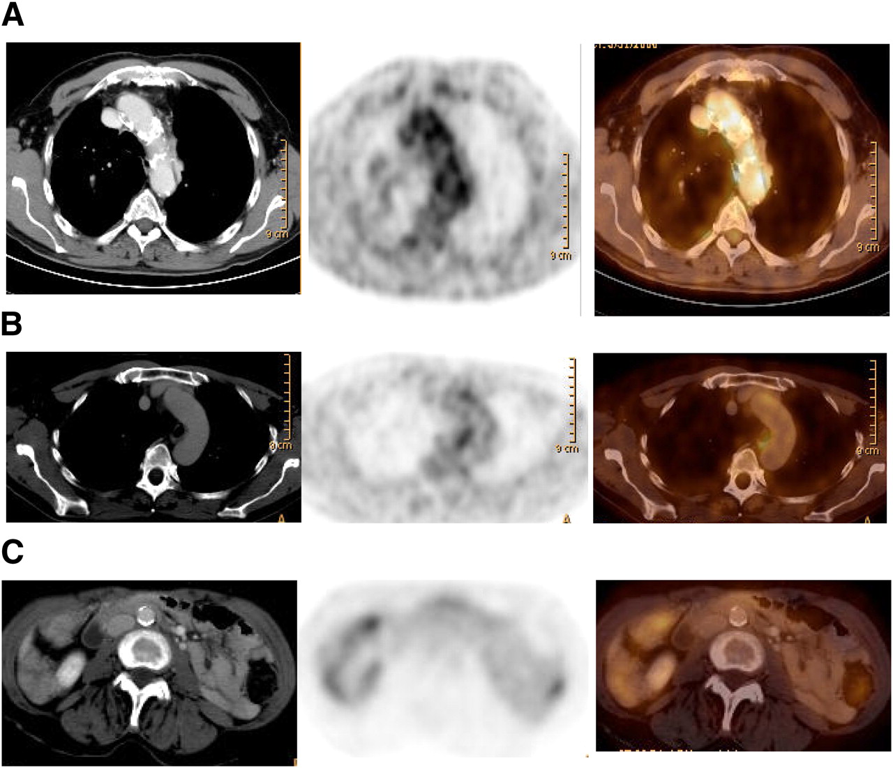
- FIGURE 2.
Arterial 18F-FDG uptake and calcifications in relation to patient outcome. From left to right are representative transaxial CT, 18F-FDG PET, and fused PET/CT images. (A) 81-y-old man with significantly increased uptake in region of aortic arch (TBR, 1.78; mean TBR, 1.62). Calcified plaque in aortic arch was 3, and calcified plaque sum was 21. Patient experienced myocardial infarction 14 mo later. (B) 45-y-old man with calcified plaque score of 0 in aortic arch (calcified plaque sum, 2) but aortic arch TBR of 1.90 (mean TBR, 1.74), who experienced vascular event 14 mo after PET/CT. (C) 64-y-old woman with calcified plaque score of 4 in abdominal aorta (calcified plaque sum, 14) and TBR of 1.49 in abdominal aorta (mean TBR, 1.56), who experienced no vascular event during follow-up.
- FIGURE 3.
Kaplan–Meier cumulative event-free survival curves: mean TBR using mean TBR cutoff of ≥1.7 (A), calcified plaque sum using calcified plaque sum cutoff of ≥15 (B), and combined subgroups of mean TBR and calcified plaque sum (C). *P < 0.01. **P < 0.001.
- FIGURE 4.
Distribution of arterial segments with different patterns of 18F-FDG uptake and calcifications in patients with and without vascular event during follow-up: proportional and disproportional distribution patterns in event group vs. control group (A); arterial segments with high TBR and low calcified plaque values vs. segments with low TBR and high calcified plaque values (B).


Tables
Parameter Control group (n = 319) Event group (n = 15) P Median age (y) 59.0 (48.0, 66.0) 66.0 (55.0, 71.5) <0.05 Men (n) 123 (38.6%) 10 (66.7%) <0.05 Body mass index 25.2 ± 4.6 (15.0, 48.0) 25.8 ± 5.6 (17.0, 35.0) NS Median follow-up time (mo) 30.0 (20.0, 36.0) 27.0 (21.0, 33.0) NS Events (n) 15 Cardiac 10 Cerebral 5 Cardiovascular risk factors (n) Hypercholesterolemia 57 (17.9%) 3 (20%) NS Hypertension 60 (18.8%) 11 (73.3%) <0.05 Smoker 30 (9.4%) 4 (26.7%) NS Diabetes mellitus 15 (4.7%) 1 (6.7%) NS Family history of cardiovascular disease 76 (25.2%) 5 (33.3%) NS Mean calcified plaque sum 4.6 ± 5.6 (0, 30) 10.9 ± 8.3 (0, 23) <0.05 Mean TBR 1.3 ± 0.2 (0.8, 2.0) 1.9 ± 0.6 (1.4, 3.9) <0.05 Therapy (n) Current statin medication 14 (4.4%) 1 (6.7%) NS Ongoing chemotherapy 43 (13.5%) 2 (13.3%) NS NS = not statistically significant.
Values are mean ± SD followed by range, median followed by (Q1, Q3), or number followed by percentage.
Future cerebrovascular or cardiovascular event Univariate analysis Multivariate analysis Parameter HR 95% CI P HR 95% CI P Age (≥65 y) 2.181 0.790–6.023 0.132 NS Sex (M) 3.355 1.144–9.804 0.027 NS Cardiovascular risk factor (+)* 12.130 1.595–92.238 0.016 8.711 1.126–67.386 0.038 Calcified plaque sum (≥15) 9.200 3.232–26.192 <0.0001 3.560 1.171–10.821 0.025 Mean TBR (≥1.7) 20.291 7.214–57.071 <0.0001 14.144 4.823–41.482 <0.0001 ↵* Minimum of 1 cardiovascular risk factor present, such as family history, hypercholesterolemia, diabetes, hypertension, or smoking.
HR = hazard ratio; CI = confidence interval; NS = not statistically significant.


Jump to section
Related Articles
Cited By...
- Continuous positive airway pressure but not Liraglutide-mediated weight loss improves early cardiovascular disease in obstructive sleep apnea: Data from a randomized proof-of-concept study
- Vascular Inflammation in Subclinical Atherosclerosis Detected by Hybrid PET/MRI
- Unraveling Vascular Inflammation: From Immunology to Imaging
- Association Between Osteogenesis and Inflammation During the Progression of Calcified Plaque Evaluated by 18F-Fluoride and 18F-FDG
- Head-to-Head Comparison of Inflammation and Neovascularization in Human Carotid Plaques: Implications for the Imaging of Vulnerable Plaques
- Arterial and Cellular Inflammation in Patients with CKD
- Positron Emission Tomography Assessment of Left Main Coronary Arterial Inflammation With Coronary Computed Tomographic Angiography Validation Before and After Statin Therapy: More Promise for Fluorodeoxyglucose Vascular Uptake?
- Coronary Plaque Morphology and the Anti-Inflammatory Impact of Atorvastatin: A Multicenter 18F-Fluorodeoxyglucose Positron Emission Tomographic/Computed Tomographic Study
- GlycA Is a Novel Biomarker of Inflammation and Subclinical Cardiovascular Disease in Psoriasis
- Evaluation of 68Ga-Glutamate Carboxypeptidase II Ligand Positron Emission Tomography for Clinical Molecular Imaging of Atherosclerotic Plaque Neovascularization
- Thresholds for Arterial Wall Inflammation Quantified by 18F-FDG PET Imaging: Implications for Vascular Interventional Studies
- GM-CSF Enhances Macrophage Glycolytic Activity In Vitro and Improves Detection of Inflammation In Vivo
- Arterial Stiffness Is Positively Associated With 18F-fluorodeoxyglucose Positron Emission Tomography-Assessed Subclinical Vascular Inflammation in People With Early Type 2 Diabetes
- Relationship Between Measures of Adiposity, Arterial Inflammation, and Subsequent Cardiovascular Events
- Radiotracer Imaging of Peripheral Vascular Disease
- Systemic Atherosclerotic Inflammation Following Acute Myocardial Infarction: Myocardial Infarction Begets Myocardial Infarction
- Carotid FDG Uptake Improves Prediction of Future Cardiovascular Events in Asymptomatic Individuals
- Nonpharmacological Lipoprotein Apheresis Reduces Arterial Inflammation in Familial Hypercholesterolemia
- A Randomized Trial on the Optimization of 18F-FDG Myocardial Uptake Suppression: Implications for Vulnerable Coronary Plaque Imaging
- Oxidized Low-Density Lipoprotein Stimulates Macrophage 18F-FDG Uptake via Hypoxia-Inducible Factor-1{alpha} Activation Through Nox2-Dependent Reactive Oxygen Species Generation
- Molecular imaging of atherosclerosis: clinical state-of-the-art
- Is Carotid Intima-Media Thickness as Predictive as Other Noninvasive Techniques for the Detection of Coronary Artery Disease?
- Imaging and Nanomedicine in Inflammatory Atherosclerosis
- Measurement of Arterial Activity on Routine FDG PET/CT Images Improves Prediction of Risk of Future CV Events
- 18F-FDG Imaging of Carotid Arteries for Identifying the Vulnerable Patient: Are We at the Beginning of the End?
- Radiotracer Imaging of Peripheral Vascular Disease
- Imaging of Arterial Inflammation: Keeping Our Cool on a Hot Topic
- Relation of Carotid Artery 18F-FDG Uptake to C-Reactive Protein and Framingham Risk Score in a Large Cohort of Asymptomatic Adults
- Bioenergetic Profiles Diverge During Macrophage Polarization: Implications for the Interpretation of 18F-FDG PET Imaging of Atherosclerosis
- Focal Arterial Inflammation Precedes Subsequent Calcification in the Same Location: A Longitudinal FDG-PET/CT Study
- Subclinical Vasculitis as a Potential Mechanism to Explain the Heightened Cardiovascular Risk in Rheumatoid Arthritis
- Impact of Noninsulin-Dependent Type 2 Diabetes on Carotid Wall 18F-Fluorodeoxyglucose Positron Emission Tomography Uptake
- Feasibility of 11C-Acetate PET/CT for Imaging of Fatty Acid Synthesis in the Atherosclerotic Vessel Wall
- Prevalence and Risk Factors of Carotid Vessel Wall Inflammation in Coronary Artery Disease Patients: FDG-PET and CT Imaging Study
- Imaging Inflammatory Changes in Atherosclerosis: Multimodal Imaging Hitting Stride
- Incidental Diagnosis of Thrombus Within an Aneurysm on 18F-FDG PET/CT: Frequency in 926 Patients
- Imaging Atherosclerosis With F18-Fluorodeoxyglucose Positron Emission Tomography: What Are We Actually Seeing?
- Correlation of Inflammation Assessed by 18F-FDG PET, Active Mineral Deposition Assessed by 18F-Fluoride PET, and Vascular Calcification in Atherosclerotic Plaque: A Dual-Tracer PET/CT Study
- Vessel Wall Inflammation in Spontaneous Cervical Artery Dissection: A Prospective, Observational Positron Emission Tomography, Computed Tomography, and Magnetic Resonance Imaging Study
- Imaging the Vulnerable Plaque
- Association of Adiponectin, Resistin, and Vascular Inflammation: Analysis With 18F-Fluorodeoxyglucose Positron Emission Tomography
- In Vivo Imaging of Mineral Deposition in Carotid Plaque Using 18F-Sodium Fluoride PET/CT: Correlation with Atherogenic Risk Factors
- The Year in Molecular Imaging
- Imaging Atherosclerotic Plaque Inflammation by Fluorodeoxyglucose With Positron Emission Tomography: Ready for Prime Time?
- The Role of 18F-FDG PET in Aortic Dissection
- In Vivo Imaging of Macrophage Activity in the Coronary Arteries Using 68Ga-DOTATATE PET/CT: Correlation with Coronary Calcium Burden and Risk Factors