


Article Figures & Data
Figures
- FIGURE 1.
Coronal slices of an 18F-FDG PET scan of a patient with mesothelioma demonstrating contiguous involvement of the right pleural surface, including infiltration of the oblique fissure. There is additional subcarinal, precarinal, right paratracheal, and right hilar lymph node involvement.
- FIGURE 2.
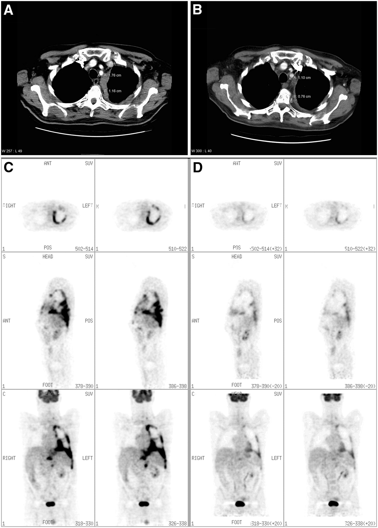
Representative CT transaxial slices of a patient with mesothelioma (A) before chemotherapy and (B) after chemotherapy. Measurements according to modified RECIST criteria have been applied. The patient had a radiological partial response after 1 cycle of chemotherapy. The challenge of defining a measurement site to determine response is demonstrated. Representative 18F-FDG PET transverse, sagittal, and coronal slices in the same patient (C) before chemotherapy and (D) after 1 cycle of chemotherapy. A significant reduction in intensity and extent of 18F-FDG uptake in the left pleural cavity is demonstrated. The response is more clearly visualized on the 18F-FDG PET imaging, and the degree of change compared with baseline in the patient was greater (TGV fell to 11% of baseline on the postchemotherapy scan, compared with a fall to 63% of baseline on CT measurements).
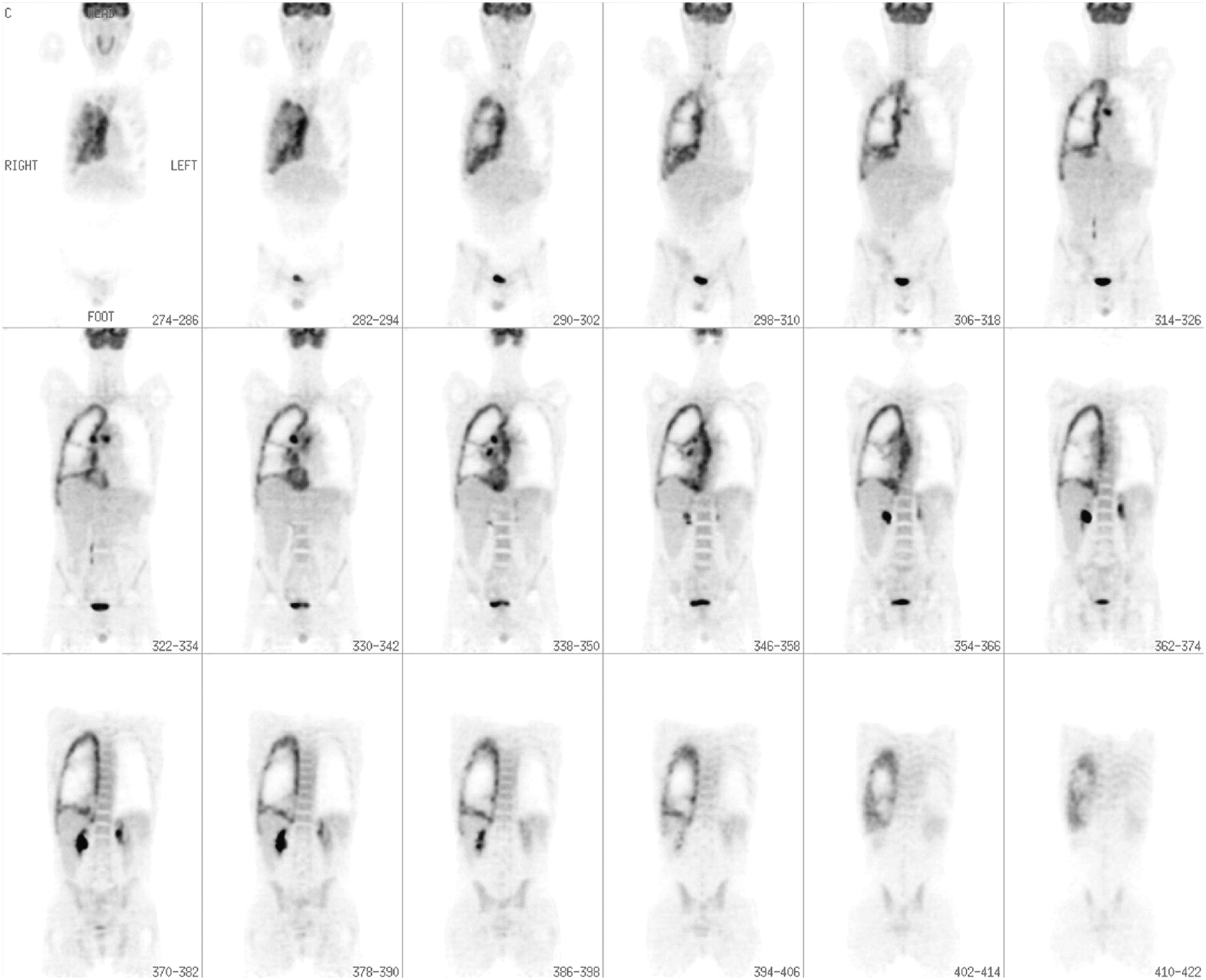
- FIGURE 3.
Representative 18F-FDG PET coronal slices in a patient with left pleural mesothelioma (A) before chemotherapy and (B) after 1 cycle of chemotherapy, demonstrating reduction in the extent and intensity of 18F-FDG activity. The region generated by the semiautomated region-growing algorithm is shown on the coronal slice (C) before chemotherapy and (D) after chemotherapy. (A–D) illustrate one representative coronal slice both before and after chemotherapy; however, in practice the region is grown in 3 dimensions to define an overall volume of interest (VOI). (E) Histogram of the SUV voxel values of the VOI generated by the region-growing algorithm in this patient before chemotherapy (red line) and after chemotherapy (green line). The histogram demonstrates both a reduction in the numerical SUV values and in the overall volume of metabolically active tumor. The TGV fell to 30% of the prechemotherapy value.
- FIGURE 4.
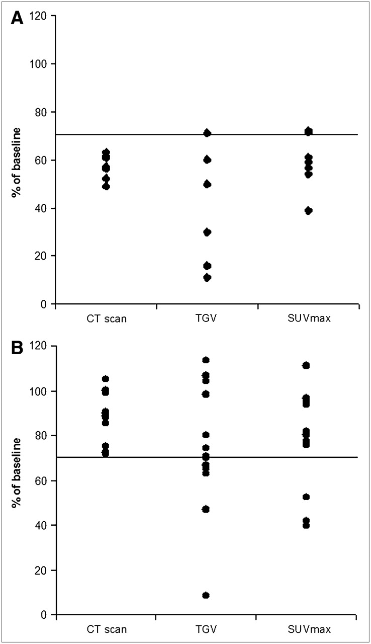
18F-FDG PET TGV and SUVmax percentage response values compared with CT response values in the 7 patients with CT-defined PR (A) and 13 patients with CT-defined SD after 1 cycle of chemotherapy (B). All values are expressed as a percentage of the baseline value. The solid line represents the 70% value used in CT to define a PR.
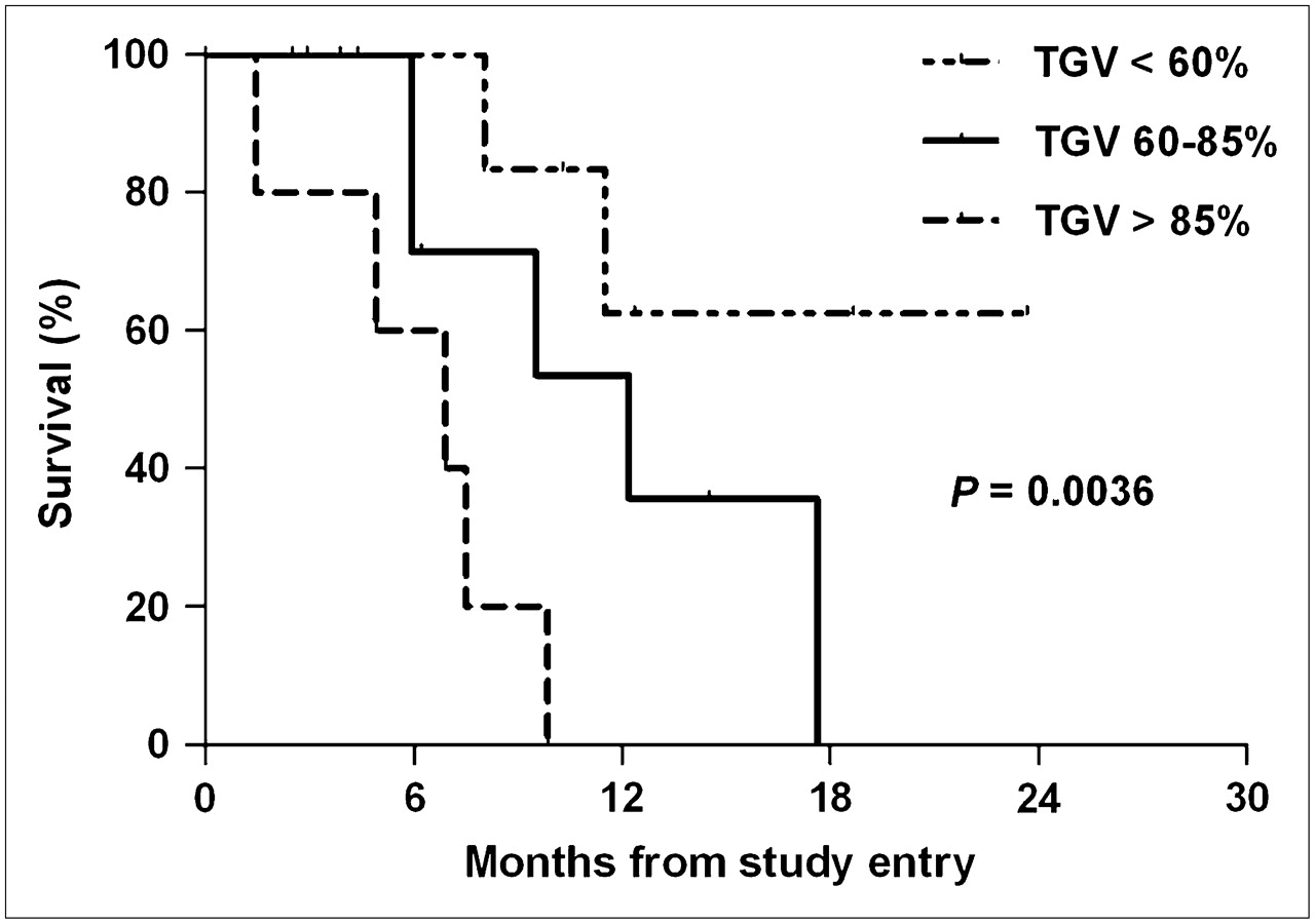
- FIGURE 5.
Kaplan–Meier survival curves illustrate the relationship between the degrees of reduction in TGV compared with baseline and survival. TGV <60% represents a reduction in TGV after 1 cycle of chemotherapy to less than 60% of the baseline value. TGV 60%–85% represents a reduction to 60%–85% of the baseline value. TGV >85% includes patients whose TGV after chemotherapy was 85% or greater than the baseline value and patients whose TGV increased after chemotherapy.


Tables
- TABLE 1
Patient Demographics and 18F-FDG PET Response of the 23 Patients in the Study, with Patients Grouped According to CT Response Attained After 1 Cycle of Chemotherapy
CT response Patient demographics PR SD nm No. of patients (%) 7 (30) 13 (57) 3 (13) Age (y) Median 67 64 67 Range 61–71 54–75 52–73 Male sex (%) 5 (71) 12 (92) 3 (100) Baseline TGV (SUV·mL) Median 3,372 5,056 844* Range 278–16,101 897–15,810 834–853 Baseline SUVmax (SUV) Median 12.2 12.8 6.4* Range 5.4–13.9 5.5–18.1 5.3–7.5 TGV1/TGV0 (%)† Median 30 71 92* Range 11–71 8–113 83–101 SUVmax1/SUVmax0 (%)‡ Median 59 80 98* Range 39–72 40–111 92–103 ↵* A TGV or SUVmax measurement could be obtained in only 2 of 3 patients. Therefore, median and range are derived from only 2 data points.
↵† TGV after 1 cycle of chemotherapy divided by baseline TGV value and expressed as a percentage.
↵‡ SUVmax after 1 cycle of chemotherapy divided by baseline SUVmax value and expressed as a percentage.
PR = partial response; SD = stable disease; nm = not measurable.
- TABLE 2
Baseline and After 1 Cycle of Chemotherapy, TGV and SUVmax Values for All Patients, Grouped According to CT Response
Status Sex Age (y) TGV0 TGV1 TGV1/TGV0 (%) SUV0 SUV1 SUV1/SUV0 (%) Survival (mo) CT PR 1 M 69 278 138 50 5.6 4.0 71 23.7† 2 M* 67 1,100 177 16 13.9 7.5 54 8.0 3 F 61 16,101 4,814 30 13.2 7.5 57 18.7† 4 M 69 7,205 1,130 16 11.5 8.3 72 11.5 5 M 71 9,840 1,089 11 13.1 5.1 39 10.3† 6 M 66 3,372 2,033 60 12.2 7.4 61 3.9† 7 F 65 549 390 71 5.4 3.2 59 4.4† CT SD 8 M 68 9,177 10,412 113 12.8 12.2 95 4.9 9 M 55 4,701 5,012 107 5.5 4.5 82 1.4 10 M 63 5,139 5,057 98 9.0 8.7 97 6.9 11 M 68 3,048 2,268 74 18.1 14.0 77 9.5 12 M 54 5,631 3,762 67 9.0 6.8 76 17.6 13 M 64 4,398 2,862 65 13.0 6.8 52 5.9 14 M* 60 15,809 11,104 70 13.6 11.1 82 14.5† 15 M 55 3,104 265 8 9.3 3.7 40 12.4† 16 F 71 1,413 1,470 104 13.2 10.6 80 9.9 17 M 74 5,484 3,440 63 10.9 10.2 94 6.2† 18 M 65 9,635 7,754 80 13.0 10.4 80 5.9 19 M 75 897 423 47 17.8 7.5 42 2.9† 20 M 54 5,056 3,574 71 10.0 11.1 111 2.5† CT nonmeasurable 21 M 52 853 861 101 5.3 4.9 92 7.5 22 M 73 834 689 83 7.5 7.7 103 12.2 23 M 67 n/e n/e n/e n/e n/e n/e 8.4† ↵* Previous talc pleurodesis.
↵† Survival at time of censorship (alive).
TGV0 = TGV at baseline (before chemotherapy); TGV1 = TGV after 1 cycle of chemotherapy; TGV1/TGV0 (%) = TGV after 1 cycle of chemotherapy divided by baseline TGV value and expressed as a percentage; SUV0 = SUVmax at baseline (before chemotherapy); SUV1 =SUVmax after 1 cycle of chemotherapy; SUV1/SUV0 (%) = SUVmax after 1 cycle of chemotherapy divided by baseline SUVmax value and expressed as a percentage; n/e =not evaluable, as a VOI could not be generated on PET images.
- TABLE 3
Cox Proportional Regression Analysis of Relationship Between Change in CT, SUVmax, and TGV after 1 Cycle of Chemotherapy and Survival
10% change Hazard ratio 95% CI P value CT 0.71 0.21–1.07 0.131 SUVmax 0.65 0.12 –1.05 0.097 TGV 0.64 0.26–0.94 0.015 The boldface P value 0.015 indicates the value is statistically significant.


Jump to section
Related Articles
Cited By...
- PET/CT Imaging in Lung Cancer
- Volumetric PET Response Assessment Outperforms Conventional Criteria in Patients Receiving High-Dose Pembrolizumab for Malignant Mesothelioma
- 18F-FDG PET Assessment of Malignant Pleural Mesothelioma: Total Lesion Volume and Total Lesion Glycolysis--The Central Role of Volume
- Early PET/CT Scan Is More Effective Than RECIST in Predicting Outcome of Patients with Liver Metastases from Colorectal Cancer Treated with Preoperative Chemotherapy Plus Bevacizumab
- Prognostic Ability of 18F-FDG PET/CT in the Assessment of Colorectal Liver Metastases
- Volume-of-Interest Assessment of Oncologic Response Using 18F-FDG PET/CT: A Phantom Study
- Serum Soluble Mesothelin Concentrations in Malignant Pleural Mesothelioma: Relationship to Tumor Volume, Clinical Stage and Changes in Tumor Burden
- Reproducibility of 18F-FDG and 3'-Deoxy-3'-18F-Fluorothymidine PET Tumor Volume Measurements
- Investigation of a unilateral pleural effusion in adults: British Thoracic Society pleural disease guideline 2010
- A Novel Prognostic Model for Malignant Mesothelioma Incorporating Quantitative FDG-PET Imaging with Clinical Parameters
- Guidelines of the European Respiratory Society and the European Society of Thoracic Surgeons for the management of malignant pleural mesothelioma
- From RECIST to PERCIST: Evolving Considerations for PET Response Criteria in Solid Tumors
- Role of 18F-FDG PET in Assessment of Response in Non-Small Cell Lung Cancer
- Tumor Volume Assessment by 18F-FDG PET/CT in Patients with Oral Cavity Cancer with Dental Artifacts on CT or MR Images