


Abstract
Integrin αvβ3 plays a critical role in tumor angiogenesis and metastasis. Suitably radiolabeled cyclic arginine-glycine-aspartic (RGD) peptides can be used for noninvasive imaging of αvβ3 expression and targeted radionuclide therapy. In this study, we developed 64Cu-labeled multimeric RGD peptides, E{E[c(RGDyK)]2}2 (RGD tetramer) and E(E{E[c(RGDyK)]2}2)2 (RGD octamer), for PET imaging of tumor integrin αvβ3 expression. Methods: Both RGD tetramer and RGD octamer were synthesized with glutamate as the linker. After conjugation with 1,4,7,10-tetra-azacyclododecane-N,N′,N″,N″′-tetraacetic acid (DOTA), the peptides were labeled with 64Cu for biodistribution and small-animal PET imaging studies (U87MG human glioblastoma xenograft model and c-neu oncomouse model). A cell adhesion assay, a cell-binding assay, receptor blocking experiments, and immunohistochemistry were also performed to evaluate the αvβ3-binding affinity/specificity of the RGD peptide-based conjugates in vitro and in vivo. Results: RGD octamer had significantly higher integrin αvβ3-binding affinity and specificity than RGD tetramer analog (inhibitory concentration of 50% was 10 nM for octamer vs. 35 nM for tetramer). 64Cu-DOTA-RGD octamer had higher tumor uptake and longer tumor retention than 64Cu-DOTA-RGD tetramer in both tumor models tested. The integrin αvβ3 specificity of both tracers was confirmed by successful receptor-blocking experiments. The high uptake and slow clearance of 64Cu-DOTA-RGD octamer in the kidneys was attributed mainly to the integrin positivity of the kidneys, significantly higher integrin αvβ3-binding affinity, and the larger molecular size of the octamer, as compared with the other RGD analogs. Conclusion: Polyvalency has a profound effect on the receptor-binding affinity and in vivo kinetics of radiolabeled RGD multimers. The information obtained here may guide the future development of RGD peptide-based imaging and internal radiotherapeutic agents targeting integrin αvβ3.
Angiogenesis is an invasive process characterized by endothelial cell proliferation, modulation of the extracellular matrix, and cell adhesion/migration (1). Angiogenesis has been shown to be required for both tumor growth and metastasis (2–4). Among many angiogenic factors, the cell adhesion molecule integrin is an important mediator in many tumor types, including glioma and breast cancer (5,6). Integrin αvβ3, in particular, was found to be necessary for the formation, survival, and maturation of new blood vessels (7,8), and its expression correlates with tumor grade and histologic type in several cancer types (9). Antagonists of αvβ3 integrin have been shown to inhibit tumor angiogenesis and metastasis (10,11). Molecular imaging of αvβ3 integrin expression during tumor angiogenesis will play a pivotal role in visualizing and quantifying αvβ3 integrin expression level, in more appropriately selecting patients considered for anti-integrin αvβ3 treatment, and in monitoring treatment efficacy in αvβ3-positive patients.
In the past decade or so, integrin αvβ3 has been found to serve as a receptor for a variety of proteins and small peptides with the exposed arginine-glycine-aspartic (RGD) sequence (1,7–11). Significant progress has also been made in the development of radiolabeled RGD-containing peptides to target integrin αvβ3 overexpressed in various tumors (12–17). We and others have found that multimeric RGD peptides can significantly enhance the affinity of the receptor–ligand interaction through the polyvalency effect (13,18–23). Recent reports on the use of multimeric RGD peptides for ligand endocytosis, imaging of angiogenesis, and targeting of tumors have demonstrated that polyvalency is an efficient strategy for discovering and developing novel RGD-based compounds with better targeting capability and higher cellular uptake because of the increased integrin recognition ability (24–27). Although we and others have already successfully developed dimeric and tetrameric RGD peptides that are suitable for diagnosis, further improvement on tumor retention and absolute uptake is still needed before they may be applied for effective peptide receptor radiotherapy.
Herein, we report the design, synthesis, and evaluation of the new tetrameric and octameric RGD peptides based on the polyvalency principle. These multimeric RGD peptides were constructed on the c(RGDyK) motif with glutamate as the branching unit. They were conjugated with the macrocylic chelator 1,4,7,10-tetraazacyclododecane-N,N′,N″,N″′-tetraacetic acid (DOTA) and labeled with 64Cu for small-animal PET imaging of integrin αvβ3 expression in both the c-neu oncomouse model (murine mammary carcinoma) and a subcutaneous U87MG xenograft (human glioblastoma) model. The aim of this study was to investigate the αvβ3-targeting characteristics of 64Cu-DOTA-RGD multimers in vitro and in vivo, thereby providing information for the future design of radiolabeled RGD peptide agents based on the polyvalency effect.
MATERIALS AND METHODS
All commercially available reagents were used without further purification. DOTA was purchased from Macrocyclics, Inc. Dicyclcohexylcarbodiimide, 1-ethyl-3-[3-(dimethylamino)propyl]carbodiimide (EDC), N-hydroxysulfonosuccinimide (SNHS), trifluoroacetic acid (TFA), and Chelex 100 resin (50-100 mesh) were purchased from Aldrich. Water and all buffers were passed through a Chelex 100 column (1 × 15 cm) before radiolabeling. Reversed-phase extraction C-18 Sep-Pak cartridges were obtained from Waters. The syringe filter and polyethersulfone membranes (pore size, 0.2 μm; diameter, 13 mm) were obtained from Nalge Nunc International. 125I-Echistatin (specific activity, 74,000 GBq/mmol) was purchased from GE Healthcare. Female athymic nude mice (4–6 wk old) were supplied from Harlan. 64Cu (half-life, 12.7 h; β+, 17.4%; β−, 30%) was obtained by using the 64Ni(p,n)64Cu nuclear reaction from University of Wisconsin–Madison. The dimeric RGD peptide E[c(RGDyK)]2 was synthesized by Peptides International, Inc. Analytic and semipreparative reversed-phase high-performance liquid chromatography (HPLC) was performed on a Dionex 680 chromatography system with a UVD 170U absorbance detector and model 105S single-channel radiation detector (Carroll and Ramsey Associates). DOTA-conjugated peptides and 64Cu-labeled peptides were isolated using a Vydac protein and peptide column (218TP510; 5 μm, 250 × 10 mm). The flow rate was 3 mL/min for semipreparative HPLC, with the mobile phase starting from 95% solvent A (0.1% TFA in water) and 5% solvent B (0.1% TFA in acetonitrile) (0–2 min) to 35% solvent A and 65% solvent B at 32 min. The analytic HPLC was performed with the same gradient system but with a Vydac 218TP54 column (5 μm, 250 × 4.6 mm) at a flow rate of 1 mL/min. The ultraviolet absorbance was monitored at 218 nm.
Preparation of E{E[c(RGDyK)]2}2 (RGD Tetramer) and E(E{E[c(RGDyK)]2}2)2 (RGD Octamer)
The Boc-protected glutamic acid activated ester Boc-E(OSu)2 was prepared as previously reported (20). To a solution of Boc-E(OSu)2 (4.4 mg, 0.01 mmol) in 1 mL of anhydrous N,N-dimethylformamide, 3 equivalents of RGD dimer (E[c(RGDyK)]2, 40 mg, 0.03 mmol) or RGD tetramer were added. The pH of the resulting mixture was adjusted to 8.5–9.0 with diisopropylethyl amine. After stirring at room temperature overnight, the desired product, Boc-RGD tetramer or Boc-RGD octamer, was isolated by preparative HPLC. The Boc group was then removed by anhydrous TFA, and the crude product was again purified by HPLC. Seventeen milligrams of RGD tetramer were obtained as a white powder with 58% overall yield (analytic HPLC retention time Rt, 13.3 min). Matrix-assisted laser desorption/ionization (MALDI) time-of-flight (TOF) mass spectrometry (MS): m/z 2,811.0 for [MH]+ (C123H180N39O38, calculated molecular weight 2,811.3). RGD octamer was obtained in 46% overall yield (analytic HPLC Rt, 14.3 min). MALDI-TOF-MS: m/z 5,735.5 for [MH]+ (C251H364N79O78, calculated molecular weight 5,734.7).
DOTA Conjugation and Radiolabeling
DOTA was activated and conjugated to RGD multimers as reported earlier (20). DOTA-RGD multimers were purified by semipreparative HPLC. Details of the 64Cu-labeling procedure were reported earlier (20). In brief, 20 μL of 64CuCl2 (74 MBq in 0.1N HCl) were diluted in 400 μL of 0.1 mol/L sodium acetate buffer (pH 6.5) and added to the DOTA-RGD multimer (a 1 mg/mL solution of peptide was made and separated into aliquots; 5 μg of DOTA-RGD tetramer and 10 μg of DOTA-RGD octamer per 37 MBq of 64Cu were used for the labeling). The reaction mixture was incubated for 1 h at 50°C. 64Cu-DOTA-RGD tetramer/octamer was then purified by semipreparative HPLC, and the radioactive peak containing the desired product was collected. After removal of the solvent by rotary evaporation, the residue was reconstituted in 800 μL of phosphate-buffered saline and passed through a 0.22-μm syringe filter for in vivo animal experiments.
Cell Adhesion Assay
Ninety-six–well plates were coated with 2 μg of fibronectin or vitronectin (Sigma-Aldrich) per milliliter in phosphate-buffered saline at 4°C overnight and treated with 2% bovine serum albumin for 1 h at 37°C. U87MG cells (human glioblastoma, American Type Culture Collection; 2 × 10⁵ cells/mL) with various concentrations of RGD multimers (50, 200, and 800 nM) in 100 μL of serum-free Dulbecco's modified Eagle's medium containing 0.1% bovine serum albumin were incubated for 20 min at 37°C. The resulting mixture was added to the plates and incubated for 1 h at 37°C. Plates treated with only bovine serum albumin were used as a negative control. After removal of the medium by aspiration, 0.04% crystal violet solution was added and incubated for 10 min at room temperature. The wells were washed 3 times with phosphate-buffered saline, and 20 μL of Triton X-100 (Union Carbide) were added for permeabilization. Distilled water (80 μL) was then added, and the number of adherent cells was assessed with a microplate reader (Tecan; measurement wavelength, 550 nm; reference wavelength, 630 nm).
Cell Integrin Receptor-Binding Assay
The in vitro integrin-binding affinity and specificity of RGD multimers and DOTA-RGD multimers were assessed via competitive cell-binding assays using 125I-echistatin as the integrin αvβ3-specific radioligand (20). The best-fit 50% inhibitory concentration (IC50) values for U87MG cells were calculated by fitting the data with nonlinear regression using GraphPad Prism with a 450- to 600-keV energy window (GraphPad Software, Inc.). Experiments were performed on triplicate samples.
Animal Models
Animal procedures were performed according to a protocol approved by Stanford University Institutional Animal Care and Use Committee. The U87MG xenograft model was generated by subcutaneous injection of 1 × 10⁷ U87MG cells (integrin αvβ3-positive) into the front left flank of female athymic nude mice. Three to 4 wk after inoculation (tumor volume, 100–400 mm3), the mice (about 9–10 wk old, with a body weight of 20–25 g) were used for biodistribution and PET studies. The c-neu oncomouse (integrin αvβ3-positive; Charles River Laboratories) is a spontaneous tumor-bearing model that carries an activated c-neu oncogene driven by a mouse mammary tumor virus promoter. Transgenic mice uniformly expressing the mouse mammary tumor virus/c-neu gene develop mammary adenocarcinomas (4–8 mo postpartum) that involve the entire epithelium in each gland. The animals were scanned at 7 mo old at about a 20-g body weight, and the tumors were on both sides of the body (28).
Biodistribution Studies
Female nude mice received a 0.74- to 1.11-MBq injection of 64Cu-DOTA-RGD tetramer or 64Cu-DOTA-RGD octamer to evaluate the distribution of these tracers in the major organs of mice (20). A blocking experiment was also performed by coinjecting radiotracer with a saturating dose of c(RGDyK) (10 mg/kg of mouse body weight). All mice were sacrificed and dissected at 20 h after injection of the tracer. Blood, U87MG tumor, major organs, and tissues were collected and weighed wet. The radioactivity in the tissue was measured using a γ-counter (Packard). The results were presented as percentage injected dose per gram of tissue (%ID/g). For each mouse, the radioactivity of the tissue samples was calibrated against a known aliquot of the injectate and normalized to a body mass of 20 g. Values were expressed as mean ± SD for a group of 3 animals.
Small-Animal PET Studies
PET scans and image analysis were performed using a rodent scanner (microPET R4; Siemens Medical Solutions) as previously reported (13,20). About 9.3 MBq of 64Cu-DOTA-RGD multimer were intravenously injected into each mouse under isoflurane anesthesia. Five-minute static scans were acquired at 30 min and at 1, 2, 6, and 20 h after injection. The images were reconstructed by a 2-dimensional ordered-subsets expectation maximum algorithm, and no correction was applied for attenuation or scatter. For each PET scan, regions of interest were drawn over the tumor, normal tissue, and major organs on decay-corrected whole-body coronal images. The radioactivity concentration (accumulation) within a tumor was obtained from the maximum value within the multiple regions of interest and then converted to %ID/g (20). For a receptor-blocking experiment, mice bearing U87MG tumors on the front left flank were scanned (5-min static) after coinjection of 9.3 MBq of 64Cu-DOTA-RGD multimer and 10 mg of c(RGDyK) per kilogram.
Statistical Analysis
Quantitative data were expressed as mean ± SD. Means were compared using 1-way ANOVA and the Student t test. P values of less than 0.05 were considered statistically significant.
RESULTS
Chemistry and Radiochemistry
RGD tetramer and RGD octamer were synthesized through an active ester method by coupling Boc-E(OSu)2 with RGD dimer/tetramer followed by TFA deprotection. In aqueous solution, DOTA was activated with EDC/SNHS, and the resulting DOTA-sulfosuccinimide ester was conjugated with RGD tetramer/octamer to yield DOTA-RGD tetramer and DOTA-RGD octamer (Fig. 1). DOTA-RGD tetramer was synthesized in 70% yield (analytic HPLC Rt, 14.5 min). MALDI-TOF-MS: m/z 3,199.0 for [MH]+ (C140H207N42O45, calculated molecular weight 3,198.4). DOTA-RGD octamer was produced in 67% (analytic HPLC Rt, 14.5 min). MALDI-TOF-MS: m/z 6,122.3 for [MH]+ (C267H390N83O85, calculated molecular weight 6,121.9). On the analytic HPLC, no significant difference in retention time was observed between 64Cu-DOTA-RGD multimer and DOTA-RGD multimer. 64Cu was labeled in 80%–90% decay-corrected yield with radiochemical purity of more than 98%. The specific activity of 64Cu-DOTA-RGD tetramer and 64Cu-DOTA-RGD octamer was about 23 MBq/nmol (0.62 Ci/μmol).

Chemical structures of DOTA-RGD tetramer and DOTA-RGD octamer.
Cell Adhesion Assay
The effect of RGD multimers on U87MG cell adhesion ability was investigated. Both fibronectin and vitronectin are ligands for integrin αvβ3. Fibronectin binds to several other integrins besides αvβ3, whereas vitronectin is integrin αvβ3–specific (29,30). For fibronectin-coated plates, no significant difference in U87MG cell adhesion ability was observed in the presence of RGD multimers at the tested concentration range (Fig. 2A). For vitronectin-coated plates, RGD multimers inhibited cell adhesion in a concentration-dependent manner. The ability of different RGD peptides to inhibit cell adhesion at the same concentration followed the order of monomer < dimer < tetramer < octamer (Fig. 2B). The calculated IC50 values for RGD monomer, dimer, tetramer, and octamer were (2.7 ± 0.7) × 10−6, (7.0 ± 1.0) × 10−7, (3.2 ± 0.9) × 10−7, and (1.1 ± 0.2) × 10−7 mol/L, respectively (Supplemental Fig. 1; supplemental figures are available online only at http://jnm.snmjournals.org). RGD octamer was 3 times as effective as RGD tetramer and 27 times as effective as RGD monomer.
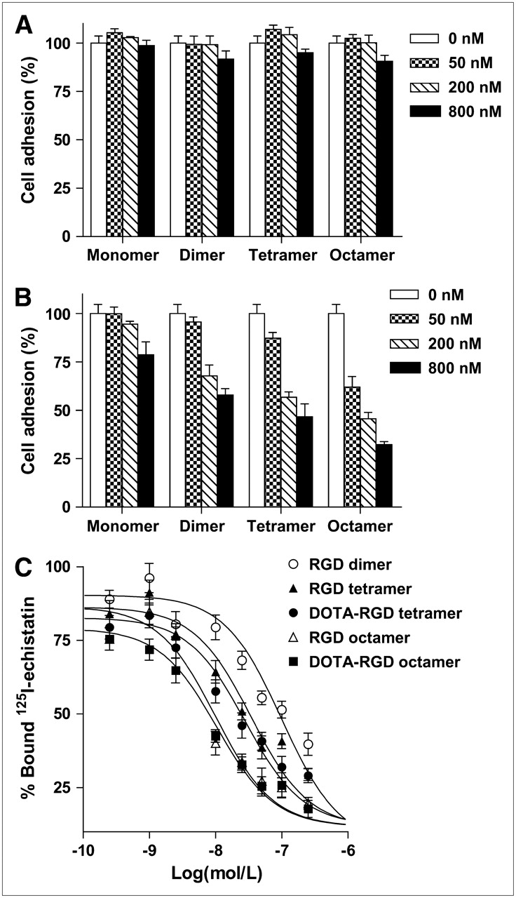
In vitro cell adhesion assay and cell-binding assay using U87MG human glioblastoma cells. (A) Cell adhesion assay of RGD monomer, dimer, tetramer, and octamer on fibronectin-coated plates (n = 4, mean ± SD). (B) Cell adhesion assay of RGD monomer, dimer, tetramer, and octamer on vitronectin-coated plates (n = 4, mean ± SD). (C) Inhibition of 125I-echistatin (integrin αvβ3–specific) binding to αvβ3 integrin on U87MG cells by RGD dimer, tetramer, octamer, DOTA-RGD tetramer, and DOTA-RGD octamer (n = 3, mean ± SD).
Cell-Binding Assay
We compared the receptor-binding affinity of RGD dimer, tetramer, and octamer; DOTA-RGD tetramer; and DOTA-RGD octamer using a competitive cell-binding assay (Fig. 2C). All peptides inhibited the binding of 125I-echistatin to αvβ3 integrin-positive U87MG cells in a dose-dependent manner. The IC50 values for RGD dimer, tetramer, and octamer were (1.0 ± 0.1) × 10−7, (3.5 ± 0.3) × 10−8, and (1.0 ± 0.2) × 10−8 mol/L, respectively (n = 3). DOTA conjugation had a minimal effect on the receptor-binding avidity, and the IC50 values for DOTA-RGD tetramer and DOTA-RGD octamer were (2.8 ± 0.4) × 10−8 and (1.1 ± 0.2) × 10−8 mol/L, respectively. The cell-binding assay demonstrated that RGD tetramer had about 3-fold higher integrin αvβ3 avidity than RGD dimer, and RGD octamer further increased the integrin avidity by another 3-fold (attributed to the polyvalency effect). The IC50 values measured from such a cell-binding assay are always lower than those obtained from purified αvβ3 integrin protein fixed on a solid matrix (e.g., an ELISA and solid-phase receptor-binding assay) (31).
PET Imaging of U87MG Tumor-Bearing Mice and c-Neu Oncomice
The tumor-targeting efficacy of 64Cu-DOTA-RGD tetramer and 64Cu-DOTA-RGD octamer in U87MG tumor-bearing nude mice (n = 3 per tracer) was evaluated by multiple time-point static PET scans. Representative decay-corrected coronal images at different times after injection are shown in Figure 3A. The U87MG tumors were clearly visualized with high tumor-to-background contrast for both tracers. The uptake of 64Cu-DOTA-RGD tetramer in U87MG tumors was rapid and high, reaching 10.3 ± 1.6, 9.6 ± 1.4, 8.6 ± 1.0, 7.7 ± 1.6, and 6.4 ± 0.7 %ID/g at 0.5, 1, 2, 6, and 20 h after injection, respectively (Fig. 4A). The activity accumulation of 64Cu-DOTA-E{E[c(RGDyK)]2}2 (the D-Tyr analog) in U87MG tumor was slightly higher than of 64Cu-DOTA-E{E[c(RGDfK)]2}2 (the D-Phe analog) (20), and no significant difference in liver and kidney uptake was observed between D-Tyr and D-Phe RGD tetramer analogs, similar to previous reports for RGD dimers (32).

Small-animal PET studies of U87MG tumor-bearing mice and c-neu oncomice. (A) Decay-corrected whole-body coronal images of athymic female nude mice bearing U87MG tumor at 30 min and at 1, 2, 6, and 20 h after injection of about 9 MBq of 64Cu-DOTA-RGD tetramer or 64Cu-DOTA-RGD octamer. (B) Coronal images of U87MG tumor-bearing mice at 2 h after injection of 64Cu-DOTA-RGD tetramer or 64Cu-DOTA-RGD octamer without and with (denoted as “blocking”) coinjection of 10 mg of c(RGDyK) per kilogram of mouse body weight. (C) Decay-corrected whole-body coronal images of c-neu oncomice at 1, 5, and 20 h after injection of about 9 MBq of 64Cu-DOTA-RGD tetramer or 64Cu-DOTA-RGD octamer. These mice are 7 mo old, and all have multiple tumors. 64Cu-DOTA-RGD tetramer and 64Cu-DOTA-RGD octamer are denoted as “RGD tetramer” and “RGD octamer,” respectively. All images shown are of 5- or 10-min static scans and representative of 3 mice per group. Tumors are indicated by arrows.
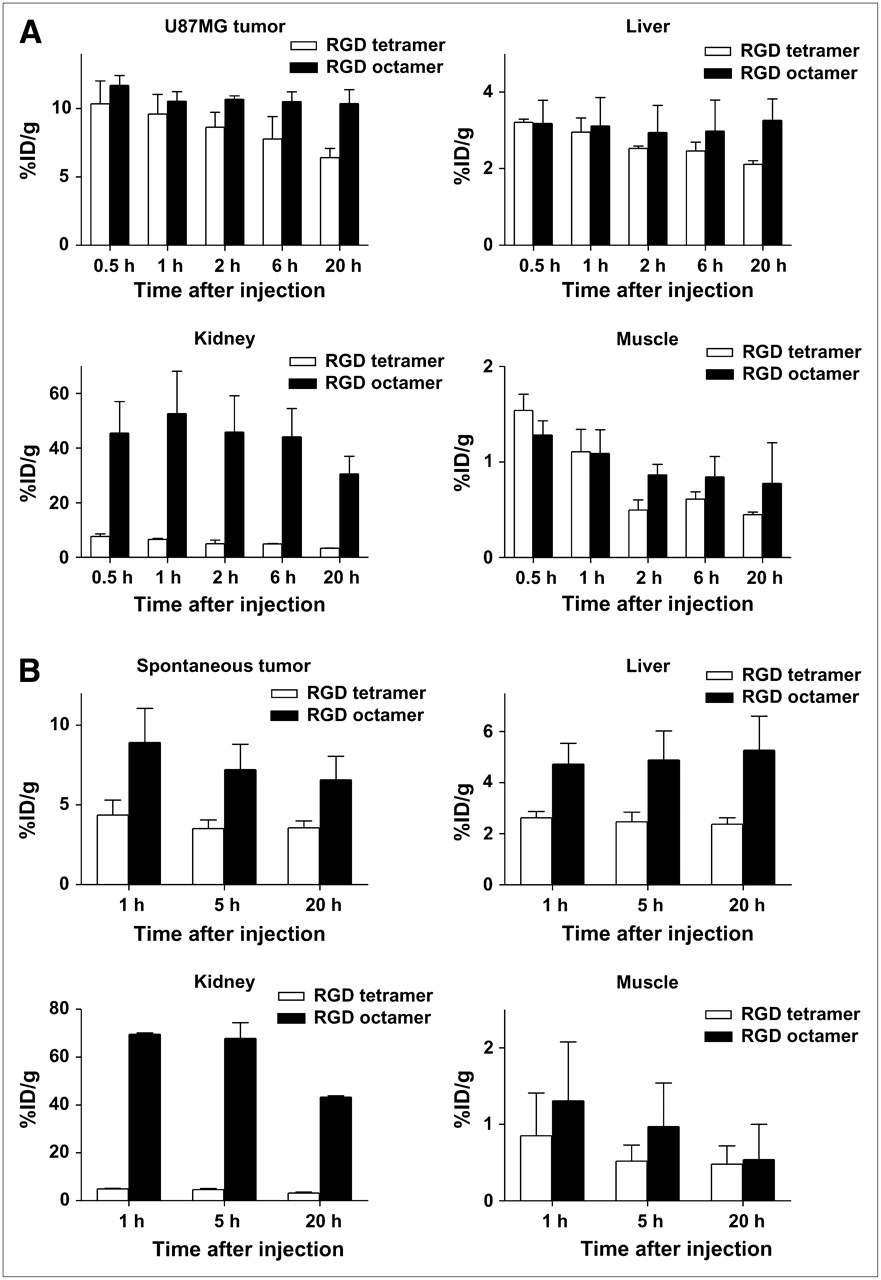
Quantitative analyses of small-animal PET data. (A) Comparison between 64Cu-DOTA-RGD tetramer and 64Cu-DOTA-RGD octamer uptake in U87MG tumor, liver, kidney, and muscle over time in U87MG xenograft model (n = 3). (B) Comparison between 64Cu-DOTA-RGD tetramer and 64Cu-DOTA-RGD octamer uptake in spontaneous tumor, liver, kidney, and muscle over time in c-neu oncomice (n = 3).
Uptake of 64Cu-DOTA-RGD octamer was higher than of 64Cu-DOTA-RGD tetramer in U87MG tumors at all time points examined, reaching 11.7 ± 0.7, 10.6 ± 0.7, 10.6 ± 0.3, 10.5 ± 0.7, and 10.3 ± 1.0 %ID/g at 0.5, 1, 2, 6, and 20 h after injection, respectively (Fig. 4A). Washout from the tumor during the experimental time span was minimal (20 h). Activity accumulation in the liver, kidneys, and muscle is also shown in Figure 4A. Uptake of the 2 tracers in the liver and muscle was similar, whereas uptake in kidney of 64Cu-DOTA-RGD octamer was much higher than of 64Cu-DOTA-RGD tetramer. Representative coronal images of U87MG tumor–bearing mice with and without coinjection of a blocking dose of c(RGDyK) (10 mg/kg) are shown in Figure 3B. Tracer uptake in the U87MG tumor was significantly reduced in the presence of c(RGDyK) in both cases (2.2 ± 0.1 %ID/g vs. 8.6 ± 1.0 %ID/g for 64Cu-DOTA-RGD tetramer and 1.7 ± 0.2 %ID/g vs. 10.6 ± 0.3 %ID/g for 64Cu-DOTA-RGD octamer at 2 h after injection), indicating the in vivo integrin αvβ3-binding specificity of both tracers. The uptake of both tracers in all other organs was also significantly lower, similar to that observed for other RGD peptide-based tracers (33).
The c-neu oncomouse model has been characterized with radiometal-labeled RGD peptides other than 64Cu. 111In-DOTA-E[c(RGDfK)]2 and 90Y-DOTA-E[c(RGDfK)]2 had, respectively, approximately 3.0 %ID/g at 2 h and approximately 1.5 %ID/g at 24 h after injection, whereas their monomeric counterparts had, respectively, approximately only 1.3 %ID/g at 2 h and approximately only 0.5 %ID/g at 24 h after injection (34). Uptake by tumors of our newly developed 64Cu-DOTA-RGD tetramer and 64Cu-DOTA-RGD octamer in this spontaneous mammary carcinoma model was studied. Decay-corrected coronal PET images are shown in Figure 3C, and quantitative data are shown in Figure 4B. Uptake of 64Cu-DOTA-RGD tetramer reached 4.4 ± 0.9 %ID/g (n = 3) at 1 h after injection, with slow clearance (3.6 ± 0.4 %ID/g at 20 h after injection). For 64Cu-DOTA-RGD octamer, uptake was 8.9 ± 2.1 %ID/g (n = 3) at 1 h after injection, almost twice as high as for 64Cu-DOTA-RGD tetramer. Washout from tumor was also slow, with the uptake being 6.6 ± 1.5 %ID/g at 20 h after injection.
Uptake in the liver of oncomice was significantly higher for 64Cu-DOTA-RGD octamer than for 64Cu-DOTA-RGD tetramer. This finding may be attributed to a possible liver metastasis (Fig. 4B). At 7 mo old, all mice had multiple tumors. Because the spontaneous tumor had much higher uptake of 64Cu-DOTA-RGD octamer, the liver metastasis is expected to follow the same trend. Uptake in muscle was similar for both tracers. In the kidney of c-neu oncomice, uptake of 64Cu-DOTA-RGD octamer is also much higher than uptake of 64Cu-DOTA-RGD tetramer, similar to the observation in athymic nude mice.
Biodistribution Studies and Blocking Experiment
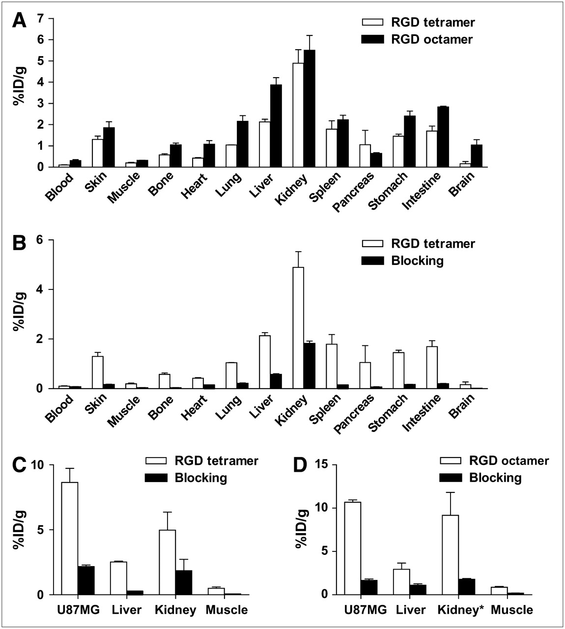
To investigate the localization of 64Cu-DOTA-RGD tetramer and 64Cu-DOTA-RGD octamer in normal athymic nude mice, we performed biodistribution studies at 20 h after injection. As can be seen in Figure 5A, uptake of 64Cu-DOTA-RGD tetramer in kidney was 5.0 ± 0.7 %ID/g (n = 3), whereas uptake was almost 5-fold higher for 64Cu-DOTA-RGD octamer (27.0 ± 3.5 %ID/g, n = 3). Because of the slower clearance, uptake of 64Cu-DOTA-RGD octamer was also slightly higher than of 64Cu-DOTA-RGD tetramer in most organs. The biodistribution of 64Cu-DOTA-RGD tetramer in female athymic nude mice with and without a blocking dose of c(RGDyK) is shown in Figure 5B; radioactivity in the kidney and all other dissected tissues was significantly decreased. Quantitative data of the PET scans shown in Figure 3B are presented in Figures 5C and 5D. An excess amount of c(RGDyK) successfully reduced the uptake of both 64Cu-DOTA-RGD tetramer and 64Cu-DOTA-RGD octamer in the U87MG tumor and reduced uptake in the kidney to the background level, confirming the integrin αvβ3–binding specificity of both tracers in vivo.

Biodistribution and receptor blocking experiments. (A) Biodistribution of 64Cu-DOTA-RGD tetramer and 64Cu-DOTA-RGD octamer in female athymic nude mice at 20 h after injection (n = 3). Plotted kidney uptake of 64Cu-DOTA-RGD octamer is one fifth of actual value (*). (B) Biodistribution of 64Cu-DOTA-RGD tetramer in female athymic nude mice at 20 h after injection with and without coinjection of 10 mg of c(RGDyK) per kilogram (n = 3). (C) Comparison of 64Cu-DOTA-RGD tetramer uptake at 2 h after injection in U87MG tumor, kidneys, liver, and muscle over time with and without coinjection of 10 mg of c(RGDyK) per kilogram (n = 3). (D) Comparison of 64Cu-DOTA-RGD octamer uptake in U87MG tumor, kidneys, liver, and muscle over time with and without coinjection of 10 mg of c(RGDyK) per kilogram (n = 3). Plotted kidney uptake of 64Cu-DOTA-RGD octamer is one fifth of actual value (*).
DISCUSSION
This article has described the synthesis of 64Cu-labeled RGD tetramer and RGD octamer based on the RGDyK sequence and their use for PET of tumor integrin αvβ3 expression. These RGD multimers showed high integrin αvβ3-binding affinity and specificity as determined by a cell adhesion assay and a cell-binding assay. The binding affinity and specificity of the newly developed tracers (64Cu-DOTA-RGD tetramer and 64Cu-DOTA-RGD octamer) in vivo was also confirmed by biodistribution studies and quantitative small-animal PET experiments.
A variety of radiolabeled peptides has been evaluated for tumor localization and therapy (15,16,20,21,23,32,35). Radiolabeled RGD peptides are of particular interest because they bind to integrin αvβ3, which is overexpressed on newly formed blood vessels and the cells of many common cancer types. However, most RGD peptide-based tracers developed so far have fast blood clearance accompanied by relatively low tumor uptake and rapid tumor washout, presumably because of the suboptimal receptor-binding affinity/selectivity and inadequate contact with the binding pocket located in the extracellular segment of integrin αvβ3.
We and others have previously applied the concept of bivalency to develop dimeric RGD peptides for tumor targeting (13,23,32,35,36). The introduction of the dimeric RGD peptide system resulted in increased receptor-binding affinity/specificity for integrin αvβ3 in vitro and enhanced tumor uptake and retention in vivo, compared with those of RGD monomer. Recently, we reported that 64Cu-labeled tetrameric RGDfK peptide had significantly higher affinity and specificity than either RGD dimer or RGD monomer in the integrin αvβ3-positive U87MG tumor model because of the synergistic effect of polyvalency (20). Previously, we also found that replacing D-Phe (f) with D-Tyr (y) increased the hydrophilicity of the RGD peptides and resulted in increased integrin αvβ3-mediated tumor uptake and more favorable biokinetics in an orthotopic MDA-MB-435 breast cancer model (32). On the basis of these findings and an incremental improvement in tumor targeting and pharmacokinetics, compared with the previous RGD peptide analogs, we then devoted our efforts to the synthesis of tetrameric and octameric RGD peptides with repeating c(RGDyK) units connected through glutamate linkers.
With the RGD/integrin system, polyvalency has been shown to be able to significantly improve integrin-binding affinity and selectivity (22). The minimum linker length between the 2 RGD moieties has been reported to be about 3.5 nm (∼25 bond distances) for simultaneous integrin αvβ3-binding in the immobilized integrin αvβ3 assay (37). For our RGD tetramer (E{E[c(RGDyK)]2}2 (Fig. 1A), the longest distance between the 2 RGD motifs is about 30 bond lengths, long enough for simultaneous binding to adjacent integrin αvβ3. For RGD octamer, the distance is about 40 bond lengths, and simultaneous binding to 2 or more receptors is possible.
We used 2 types of assays to examine the interaction between RGD multimers and αvβ3 integrin. We first used a cell adhesion assay to assess the antiadhesion effect of RGD multimers against integrin αvβ3. RGD octamer showed a significantly better inhibition ability than the monomer/dimer/tetramer counterparts, as could be attributed to the multiple binding sites or significantly increased local concentration. To evaluate the effect of polyvalency, we calculated the multivalent enhancement ratio (MVE), which was obtained by dividing the IC50 value for the RGD monomer by the IC50 of the RGD multimer (27). The antiadhesion MVE of RGD tetramer and RGD octamer was 8.4 and 25.6, respectively (Table 1). We then performed a cell-binding assay, an often-used method to determine the receptor-binding affinity of a given ligand. Again, the integrin αvβ3-binding affinity followed the order of RGD octamer > RGD tetramer > RGD dimer > RGD monomer (Fig. 2C; Table 1). DOTA conjugation had a minimal effect on the binding affinity of the RGD peptides. The receptor-binding MVE for RGD tetramer and RGD octamer was calculated to be 5.9 and 20.3, respectively. On the basis of both the cell adhesion assay and the cell-binding assay, RGD octamer showed a stronger multivalent effect than did RGD tetramer.
MVE of RGD Multimers Based on Cell Adhesion and Cell-Binding Assays Using Human Glioblastoma U87MG Cells
When applied to the U87MG glioblastoma xenograft model, which has been well established to have a high integrin αvβ3 expression (20,38), 64Cu-DOTA-RGD tetramer showed prominent uptake in tumor and primarily renal clearance (Figs. 3A and 4A). 64Cu-DOTA-RGD octamer had slightly higher initial tumor uptake and much longer tumor retention. The initially rapid and high tumor uptake might be attributed to the high integrin αvβ3-binding affinity of both tracers. The larger molecular size of 64Cu-DOTA-RGD octamer, along with the stronger MVE, may be attributed to its longer circulation time and slower tumor washout, compared with those of 64Cu-DOTA-RGD tetramer. We also tested these 2 tracers in the c-neu oncomouse model. Both tracers showed significantly higher uptake in the spontaneous tumor (medium integrin expression) than did the dimeric and monomeric analogs. The difference between 64Cu-DOTA-RGD tetramer and 64Cu-DOTA-RGD octamer is more substantial in this model than in the U87MG xenograft model. Uptake of 64Cu-DOTA-RGD octamer in tumor was almost twice as high as uptake of 64Cu-DOTA-RGD tetramer (Fig. 4B). A similar pattern has also been observed in the orthotopic MDA-MB-435 (medium integrin expression) breast cancer model. The advantage of higher integrin αvβ3-binding affinity and selectivity of RGD octamer over RGD tetramer appears to be more obvious in medium integrin αvβ3-expressing tumor models (e.g., MDA-MB-435 and c-neu oncomice) than in high integrin-expressing tumor models (e.g., U87MG). The mechanism underlying such a phenomenon remains to be elucidated.
Compared with 64Cu-DOTA-RGD tetramer, 64Cu-DOTA-RGD octamer exhibited significantly higher renal uptake in both subcutaneous U87MG xenografts and mammary adenocarcinoma–bearing c-neu oncomice. We initially proposed that the high renal uptake of 64Cu-DOTA-RGD octamer, compared with other RGD oligomers, might be caused by the overall difference in molecular charge. If we assign a value of −1 to each acidic residue (Asp (D) and Glu (E)) and the C-terminal -COOH, and a value of +1 to each basic residue (Arg (R) and Lys (K)) and the N-terminal -NH2, the overall charge of the peptide can be determined by adding up the charges. For both RGD tetramer and RGD octamer, the overall molecular charges are +1 although RGD octamer has a higher number of charged amino acid residues. Positively charged radiolabeled peptides or metabolites are usually retained in the kidney after resorption by renal tubular cells and lysosomal proteolysis. Blocking cationic binding sites in the kidneys with cationic amino acid infusion has been reported to reduce renal uptake without compromising the accumulation of activity in the tumor in both mice and humans (39). We tried the blocking experiment for 64Cu-DOTA-RGD octamer by coinjecting an excess amount of D-lysine; uptake in the kidney was only marginally reduced, suggesting that the overall molecular charge does not contribute significantly to the high renal uptake.
We noticed that even though uptake of 64Cu-DOTA-RGD octamer in the kidney was high, no appreciable activity was excreted to the urinary bladder over time. Such a phenomenon suggests that receptor-mediated binding might be involved. Integrins play important roles in renal development, and integrin αvβ3, in particular, has been identified in many parts of the developing kidney. Integrin αvβ3 is expressed in the renal endothelium of adults and, to a lesser extent, in all tubular epithelium (40). Effective blocking of activity accumulation in the kidney in the presence of an excess amount of c(RGDyK) also confirmed the integrin αvβ3 specificity of both 64Cu-DOTA-RGD tetramer and 64Cu-DOTA-RGD octamer (Figs. 2B and 5B). Immunohistochemical staining showed that mouse kidneys have high β3 expression on the endothelial cells of small glomerulus vessels (Supplemental Fig. 2), further confirming that renal uptake of both tracers is integrin-specific. The trend of increased kidney uptake from RGD monomer to dimer to tetramer to octamer would thus be due, in part, to the increased αvβ3-binding affinity and the molecular size.
It is important to have high tumor-to-kidney ratios, as well as high absolute tumor uptake and longer retention, for both imaging and therapeutic applications. For imaging purposes, renal accumulation of radiolabeled peptides will reduce detection sensitivity in the vicinity of the kidneys. For therapeutic applications, renal accumulation of radiolabeled peptides limits the maximum tolerated doses that can be administered without inducing radiation nephrotoxicity. Thus, further modification is needed to improve the pharmacokinetics of RGD peptide-based radiopharmaceuticals. First, high αvβ3-binding affinity is needed to afford high uptake and retention in tumors. For RGD octamer, the density of RGD units is rather high, and not all RGD units are amenable to effective binding to integrin αvβ3 located on the same cell surface. Our future work will focus on the structure–activity relationship to develop various dendritic and polymeric scaffolds for attaching RGD peptides, thereby further enhancing the multivalency effect. Second, appropriate modification of the DOTA-RGD multimers is needed to reduce renal uptake. By inserting a bifunctional linker between the DOTA chelator and the RGD multimer as a pharmacokinetic modifier, we may be able to modulate the overall molecular charge, hydrophilicity, and molecular size, thus possibly improving in vivo pharmacokinetics without compromising the tumor-targeting efficacy of the resulting radioconjugates.
CONCLUSION
64Cu-DOTA-RGD tetramer and 64Cu-DOTA-RGD octamer were developed for PET of tumor integrin αvβ3 expression. RGD octamer showed significantly higher integrin αvβ3-binding affinity in vitro than did RGD tetramer. In the noninvasive small-animal PET studies, both tracers showed rapid and high uptake in tumor, slow washout, and good tumor-to-background contrast in U87MG xenografts and c-neu oncomice. Overall, polyvalency has a profound effect on the receptor-binding affinity and in vivo kinetics of 64Cu-DOTA-RGD multimers. The information obtained here may guide future development of integrin αvβ3-targeted imaging and internal radiotherapy agents. These RGD peptide-based radiopharmaceuticals may also have promising applications in other angiogenesis-related diseases such as rheumatoid arthritis, myocardial infarction, and stroke.
Acknowledgments
This work was supported by the National Institute of Biomedical Imaging and Bioengineering (R21 EB001785), the National Cancer Institute (R21 CA102123, P50 CA114747, U54 CA119367, and R24 CA93862), the Department of Defense (W81XWH-04-1-0697, W81XWH-06-1-0665, W81XWH-06-1-0042, and DAMD17-03-1-0143), and a Benedict Cassen Postdoctoral Fellowship from the Education and Research Foundation of the Society of Nuclear Medicine. We thank Dr. Hui Wang and Dr. Gang Niu for their excellent technical support. We also thank the cyclotron team at the University of Wisconsin–Madison, for 64Cu production.
Footnotes
-
COPYRIGHT © 2007 by the Society of Nuclear Medicine, Inc.
References
- Received for publication January 21, 2007.
- Accepted for publication March 20, 2007.


Jump to section
Related Articles
Cited By...
- Development of FAPI Tetramers to Improve Tumor Uptake and Efficacy of FAPI Radioligand Therapy
- Development of FAPI Tetramers to Improve Tumor Uptake and Efficacy of FAPI Radioligand Therapy
- Synthesis, Preclinical Evaluation, and a Pilot Clinical PET Imaging Study of 68Ga-Labeled FAPI Dimer
- Small-Animal PET Imaging of Pancreatic Cancer Xenografts Using a 64Cu-Labeled Monoclonal Antibody, MAb159
- Cytoreductive Chemotherapy Improves the Biodistribution of Antibodies Directed against Tumor Necrosis in Murine Solid Tumor Models
- PET Imaging of Colorectal and Breast Cancer by Targeting EphB4 Receptor with 64Cu-Labeled hAb47 and hAb131 Antibodies
- RGD Peptide-Conjugated Multimodal NaGdF4:Yb3+/Er3+ Nanophosphors for Upconversion Luminescence, MR, and PET Imaging of Tumor Angiogenesis
- Imaging: Guiding the Clinical Translation of Cardiac Stem Cell Therapy
- Novel 64Cu- and 68Ga-Labeled RGD Conjugates Show Improved PET Imaging of {alpha}{nu}{beta}3 Integrin Expression and Facile Radiosynthesis
- 18F-FPPRGD2 and 18F-FDG PET of Response to Abraxane Therapy
- PET Imaging of Tumor Neovascularization in a Transgenic Mouse Model with a Novel 64Cu-DOTA-Knottin Peptide
- Noninvasive Imaging of {alpha}V{beta}3 Function as a Predictor of the Antimigratory and Antiproliferative Effects of Dasatinib
- Noninvasive De novo Imaging of Human Embryonic Stem Cell-Derived Teratoma Formation
- Engineered Knottin Peptides: A New Class of Agents for Imaging Integrin Expression in Living Subjects
- Multimodality Molecular Imaging of Tumor Angiogenesis
- Preparation of a Promising Angiogenesis PET Imaging Agent: 68Ga-Labeled c(RGDyK)-Isothiocyanatobenzyl-1,4,7-Triazacyclononane-1,4,7-Triacetic Acid and Feasibility Studies in Mice
- Advances in Anatomic, Functional, and Molecular Imaging of Angiogenesis
- 18F-Labeled BBN-RGD Heterodimer for Prostate Cancer Imaging
- Dual-Function Probe for PET and Near-Infrared Fluorescence Imaging of Tumor Vasculature