


Abstract
Arg-Gly-Asp (RGD) derivatives have been labeled with various radioisotopes for the imaging of angiogenesis in ischemic tissue, in which αvβ3 integrin plays an important role. In this study, cyclic Arg-Gly-Asp-d-Tyr-Lys [c(RGDyK)] was conjugated with 2-(p-isothiocyanatobenzyl)-1,4,7-triazacyclononane-1,4,7-triacetic acid (SCN-Bz-NOTA) and then labeled with 68Ga. The labeled RGD so produced was subjected to an in vitro binding assay and in vivo biodistribution and PET studies. Methods: A mixture of SCN-Bz-NOTA (660 nmol) and c(RGDyK) (600 nmol) in 0.1 M sodium carbonate buffer (pH 9.5) was allowed to react for 20 h at room temperature in the dark for thiourea bond formation. The conjugate obtained was purified by semipreparative high-performance liquid chromatography (HPLC). The purified c(RGDyK)–SCN-Bz-NOTA (NOTA-RGD) was then labeled with 68Ga from a 68Ge/68Ga generator and purified by semipreparative HPLC. A competitive binding assay for c(RGDyK) and NOTA-RGD was performed with 125I-c(RGDyK) as a radioligand and αvβ3 integrin–coated plates as a solid phase. 68Ga-NOTA-RGD (0.222 MBq/100 μL) was injected, through a tail vein, into mice with hind limb ischemia and into mice bearing human colon cancer SNU-C4 xenografts. Biodistribution and imaging studies were performed at 1 and 2 h after injection. Results: The labeling of NOTA-RGD with 68Ga was straightforward. The Ki values of c(RGDyK) and NOTA-RGD were 1.3 and 1.9 nM, respectively. In the biodistribution study, the mean ± SD uptake of 68Ga-NOTA-RGD by ischemic muscles was 1.6 ± 0.2 percentage injected dose per gram (%ID/g); this uptake was significantly blocked by cold c(RGDyK) to 0.6 ± 0.3 %ID/g (P < 0.01). Tumor uptake was 5.1 ± 1.0 %ID/g, and the tumor-to-blood ratio was 10.3 ± 4.8. Small-animal PET revealed rapid excretion through the urine and high levels of tumor and kidney uptake. Conclusion: Stable 68Ga-NOTA-RGD was obtained in a straightforward manner at a high yield and showed a high affinity for αvβ3 integrin, specific uptake by angiogenic muscles, a high level of uptake by tumors, and rapid renal excretion. 68Ga-NOTA-RGD was found to be a promising radioligand for the imaging of angiogenesis.
Angiogenesis is related to various diseases, such as cancer, arthritis, and psoriasis (1). The binding of 125I-labeled 3-iodo-y5–cyclic Arg-Gly-Asp-d-Tyr-Lys [c(RGDyK)] to αvβ3 integrin has been described in the literature for the targeting of angiogenesis (2). However, it has the shortcoming of high gut activity because of hepatobiliary excretion. Various other radiolabeled Arg-Gly-Asp (RGD) derivatives have been developed to improve pharmacokinetics, and the introduction of sugar residues has been found to increase renal excretion and to significantly improve pharmacokinetics (3,4).
Dimerization and multimerization have also been used to improve pharmacokinetics. Dimeric RGD derivatives labeled with 99mTc, which has a 6-h half-life, showed an increased affinity for αvβ3 integrin and favorable pharmacokinetics for imaging (5,6). In another study, dimeric RGD derivatives labeled with 111In and 90Y showed potential as cancer imaging and therapeutic agents, respectively (7). In addition, a quantitative study of an 18F-labeled dimeric RGD derivative with a short half-life proved its suitability for PET (8). A further study with multimeric (e.g., tetrameric and octameric) RGD derivatives revealed their feasibility as imaging or therapeutic agents after labeling with a radionuclide with a long half-life, such as 64Cu (half-life = 12.7 h, 40% β− [656 keV], 19% β+ [600 keV], 38% electron capture [EC]) (9).
The importance of 68Ga for clinical PET has increased recently (10). 68Ga is a positron emitter with a short half-life (67.6 min) and can be produced by use of a 68Ge/68Ga generator (11–13). Its short half-life and hydrophilic nature are adequate for the labeling of small peptides with rapid renal clearance for PET. A highly effective concentration method for generator-eluted 68Ga was reported for efficient peptide labeling (14,15). In addition, 68Ga has a great advantage for PET over other cyclotron-produced positron emitters because it can be obtained economically by use of a commercially available 68Ge/68Ga generator. The parent nuclide 68Ge has a long half-life (270.8 d), allowing its use as a generator for more than 1 year.
In the present study, we used a 1,4,7-triazacyclononane-1,4,7-triacetic acid (NOTA)–based bifunctional chelating agent to label an RGD peptide with 68Ga. NOTA is a 9-member cyclic compound and has been reported to form a highly stable neutral complex with gallium, which is inert even in 6N nitric acid (16). Functionalized NOTA derivatives suitable for conjugation with proteins or peptides have been reported by many researchers (17–20), and the feasibility of NOTA derivatives as bifunctional chelating agents for the labeling of monoclonal antibodies or somatostatin with 67Ga has been demonstrated by a biodistribution study in mice (21,22).
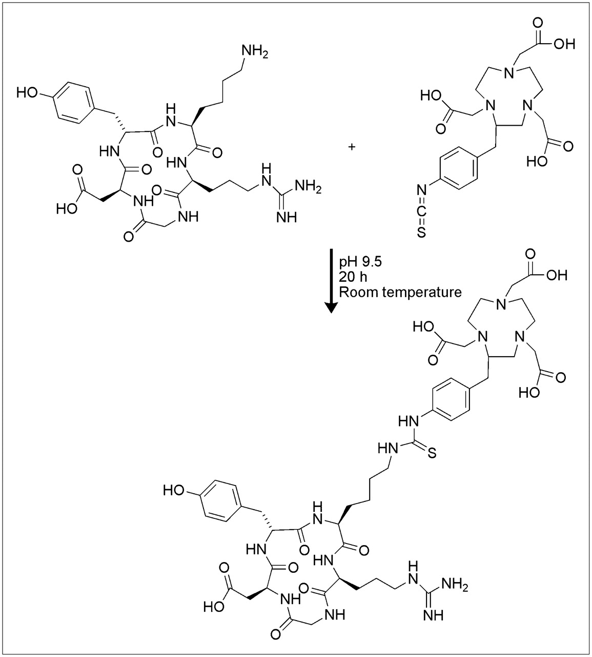
We conjugated c(RGDyK) with 2-(p-isothiocyanatobenzyl)-NOTA (SCN-Bz-NOTA) to produce a c(RGDyK)–SCN-Bz-NOTA (NOTA-RGD) conjugate (Fig. 1). We then labeled this conjugate with 68Ga and tested the feasibility of 68Ga-NOTA-RGD for the imaging of angiogenesis in mice with induced hind limb hypoxia and in mice bearing tumor xenografts.

Synthesis of NOTA-RGD. Molar equivalents of c(RGDyK) and SCN-Bz-NOTA were allowed to react in sodium carbonate buffer (pH 9.5) for 20 h at room temperature.
MATERIALS AND METHODS
General
An Agilent 1100 series high-performance liquid chromatography (HPLC) apparatus was used for purifying synthesized and labeled compounds. An XTerra column was purchased from Waters. Mass spectra were obtained with an API-3000 spectrometer (Applied Biosystems), and 1H nuclear magnetic resonance (NMR) spectra were obtained with a 300-MHz, AL 300 FT NMR spectrometer (JEOL Ltd.). A 68Ge/68Ga generator was purchased from Cyclotron Co. A DU650 spectrophotometer was obtained from Beckman Coulter Inc. A Bio-Scan system 200 imaging scanner was used to scan radioactivity distributions on instant thin-layer chromatography (ITLC) plates. A Cobra II γ-scintillation counter (Packard Canberra Co.) was used for the detection of radioactivity. A rodent R4 microPET scanner (Concorde Microsystems Inc.) was used for animal PET.
SCN-Bz-NOTA and c(RGDyK) were purchased from Futurechem. ITLC-silica gel [SG] was purchased from Gelman. Falcon 96-well polyvinyl chloride (PVC) plates were purchased from Becton Dickinson. 125I-NaI was purchased from NEN. All of the other reagents were purchased from Sigma-Aldrich.
Synthesis of NOTA-RGD and Cold Gallium-NOTA-RGD
A mixture of SCN-Bz-NOTA (660 nmol, 0.3 mg) and cRGDyK (600 nmol, 0.37 mg) in 0.1 M sodium carbonate buffer (pH 9.5) was allowed to react for 20 h at room temperature in the dark (Fig. 1). The reaction mixture was purified by HPLC (XTerra preparative column RP18; 10 × 250 mm; 0%–100% ethanol gradient in 0.01% trifluoroacetic acid from 0 to 30 min; flow rate, 3 mL/min), and the NOTA-RGD peak sample was collected at a retention time of 13.2 min. The pooled NOTA-RGD fractions were analyzed by liquid chromatography–electrospray ionization-mass spectrometry: m/z, [M+H]+ = 1,071 (calculated for molecular formula C47H67N13O14S; 1,069.5). The 1H NMR spectrum was obtained after drying under vacuum and redissolving in D2O: δ 0.78–1.90 (m, 9H), 2.57–3.65 (m, 34H), 4.00–4.05 (d, 2H), 4.18 (bs, 2H), 4.37 (bs, 2H), 6.57 (bs, 2H), 6.89 (d, 2H), 7.07 (d, 2H), and 7.19 (bs, 2H).
The molar absorptivity of NOTA-RGD at 240 nm in water was measured and found to be 14,300 M−1.
Gallium(III) chloride (110 nmol, 19.4 μg in 10 μL of water) was added to NOTA-RGD (20 nmol, 21.4 μg in 0.1 mL of water), and the mixture was incubated for 10 min at room temperature. The reaction mixture was analyzed with the same HPLC conditions as those described earlier.
Labeling with 68Ga
68Ga was eluted from the 68Ge/68Ga generator with 0.1N HCl. 68Ga (740 MBq in 1 mL of 0.1N HCl) was added to a NOTA-RGD solution (30 nmol in 0.1 mL of water) prepared as described earlier, and the pH was adjusted to 6.0 with a 7% sodium bicarbonate solution. After incubation for 10 min at room temperature, labeling efficiencies were checked by ITLC-SG with 0.1 M citric acid and 0.1 M sodium carbonate as eluting solvents, in which the Rf values of 68Ga-NOTA-RGD were 0.0 and 1.0, respectively, whereas those of free 68Ga were 1.0 and 0.0, respectively. The radiolabeled product was purified by HPLC (XTerra preparative column RP18; 10 × 250 mm; 0%–100% ethanol gradient in 0.01% trifluoroacetic acid from 0 to 30 min; flow rate, 3 mL/min), and the 68Ga-NOTA-RGD peak sample was collected at a retention time of 12.5 min. The radioactivity and the optical density at 240 nm of the peak fraction were measured, and the specific activity was calculated from the data. The stability was checked by ITLC after incubation for 4 h at room temperature.
In Vitro Binding Assay
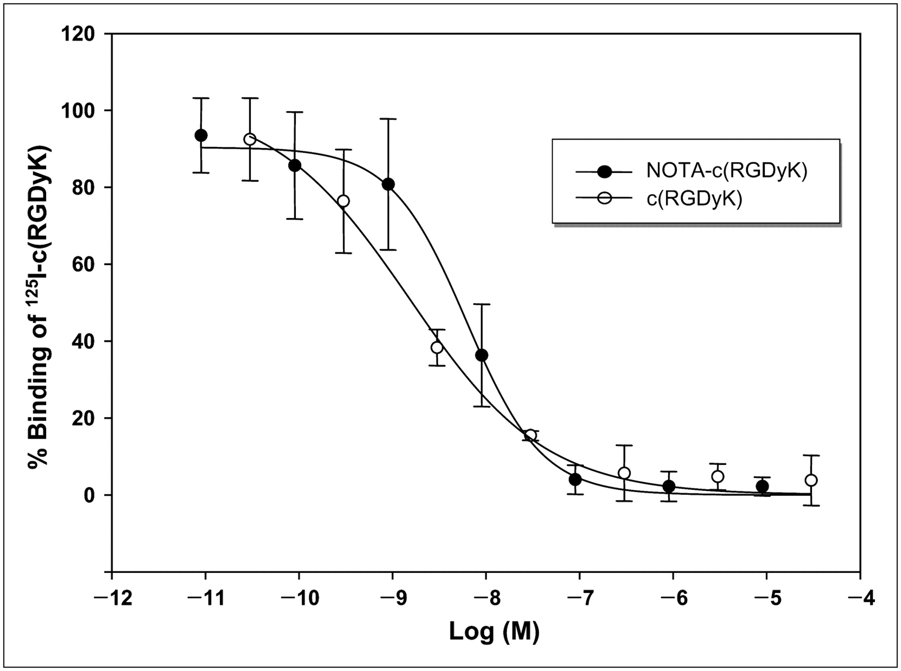
Human αvβ3 integrin was obtained from human placenta by affinity chromatography according to a published method (23,24). Preparation and dissociation constant determination for 125I-iodo-c(RGDyK) were reported previously (25). In brief, 125I-iodo-c(RGDyK) was prepared with the chloramine-T method at a yield of 92.6% and a radiochemical purity (after HPLC purification) of greater than 99%. The specific activity of 125I-iodo-c(RGDyK) was 80.7 GBq/μmol. Its dissociation constant and maximum binding values for human αvβ3 integrin (as a coating on plates), as determined by saturated-binding experiments, were 1.013 and 0.093 nM, respectively (25). The Ki of NOTA-RGD was determined as previously described (25). In brief, an αvβ3 integrin solution (1 μg/100 μL; purified from human placenta) in 50 mM sodium carbonate buffer (pH 9.6) was used to coat 96-well PVC plates. The radioligand 125I-c(RGDyK) (3.7 kBq) was then used in a competitive binding assay with 9 pM–9 μM c(RGDyK) and NOTA-RGD in 20 mM Tris-HCl buffer (pH 7.4) containing 150 mM NaCl, 1 mM CaCl2, 1 mM MnCl2, and 1 mM MgCl2 for 2 h at 4°C. Wells were washed 4 times with 200 μL of phosphate-buffered saline containing 1% bovine serum albumin. Wells were cut out with scissors, and counts were obtained with a γ-scintillation counter. The experiment was repeated 3 times in duplicate.
Biodistribution in Mice with Hind Limb Ischemia
All animal experiments were performed with the approval of the Institutional Animal Care and Use Committee of the Clinical Research Institute at Seoul National University Hospital (an Association for Assessment and Accreditation of Laboratory Animal Care–accredited facility). In addition, National Research Council guidelines for the care and use of laboratory animals (revised in 1996) were observed throughout.
For the creation of an angiogenesis model, unilateral hind limb ischemia was induced in male ICR mice (8–10 wk old) by left femoral artery ablation (25,26). On day 7 after the induction of ischemia, 68Ga-NOTA-RGD (0.22 MBq/0.1 mL) was injected through a tail vein. For blocking studies, a mixture of c(RGDyK) (3 mg/kg) and 68Ga-NOTA-RGD (0.15 MBq/0.1 mL) was injected intravenously into mice with hind limb ischemia (31.9 ± 2.5 [mean ± SD] g, n = 9). Mice were sacrificed by cervical dislocation at 1 h after injection; bilateral hind limb thigh muscles, blood, and other organs were separated immediately and weighed; and counts were obtained with a γ-scintillation counter. Results are expressed as the percentage injected dose per gram of tissue (%ID/g).
Biodistribution in Mice Bearing Colon Cancer Xenografts
The human colon cancer cell line SNU-C4 was grown in RPMI 1640 medium containing 10% fetal bovine serum and harvested with trypsin. Cells were washed with 10 mL of phosphate-buffered saline by centrifugation (3,000 rpm). Each nude mouse (male, 19.4 ± 1.3 g) was injected subcutaneously with 5 × 10⁶ SNU-C4 cells in the right shoulder. After 17 d, 68Ga-NOTA-RGD (0.15 MBq/0.1 mL) was injected intravenously into each xenograft-bearing mouse. Mice were sacrificed at 1 h after injection, and biodistribution was investigated as described earlier.
PET of Mice Bearing Xenografts
Nude mice received xenografts of SNU-C4 cells as described earlier, and tumors were grown for 14 d. 68Ga-NOTA-RGD (16 MBq/0.1 mL) was then injected either with or without cold c(RGDyK) (60 μg) through a tail vein. Mice were anesthetized with 2% isoflurane at 2 h after injection, and PET images were obtained for 20 min (static images). PET studies were performed with a dedicated small-animal PET scanner (rodent R4 microPET scanner; Concorde Microsystems Inc.). The acquired 3-dimensional emission data were reconstructed to temporally framed sinograms by use of Fourier rebinning and an ordered-subsets expectation maximization reconstruction algorithm without attenuation correction. Image visualization was performed with ASIPro software (Concorde Microsystems Inc.). For the PET scans, mice were kept under 2% isoflurane anesthesia and temperature was maintained at 30°C with a carbon pad as described previously (27).
RESULTS
Synthesis
The synthesis of NOTA-RGD is shown in Figure 1. The conjugated product NOTA-RGD was confirmed by mass spectrometry and NMR spectroscopy. HPLC purification of the reaction mixture was straightforward because of large differences between the retention times of the starting materials and the product; the retention times of c(RGDyK), NOTA-RGD, and SCN-Bz-NOTA were 9.7, 13.2, and 18.4 min, respectively. The NOTA-RGD peak moved forward to 12.4 min after chelation with cold gallium (Supplemental Fig. 1; supplemental materials are available online only at http://jnm.snmjournals.org).
Radiolabeling
The entire labeling procedure, including the purification step, was completed within 30 min. The 68Ga-NOTA-RGD produced was analyzed by ITLC. For this analysis, ITLC-SG plates were eluted with 0.1 M citric acid; the Rf values of free 68Ga and 68Ga-NOTA-RGD were 1.0 and 0.0, respectively. On the other hand, the Rf values changed markedly when ITLC-SG plates were eluted with 0.1 M sodium carbonate; free 68Ga remained at the origin, and 68Ga-NOTA-RGD moved with the solvent front. The labeling efficiency was 89%, and no free 68Ga was found after purification (Supplemental Fig. 2).
68Ga-NOTA-RGD showed an earlier retention time (12.6 min) than unlabeled NOTA-RGD in preparative HPLC (Supplemental Fig. 3). The free 68Ga peak appeared at 4.2 min, and the specific activity of the purified 68Ga-NOTA-RGD was 17.4 GBq/μmol. Moreover, the 68Ga-NOTA-RGD preparation was found to be stable for more than 4 h at room temperature.
The net charge of 68Ga-NOTA-RGD was determined to be positive by paper electrophoresis (Supplemental Fig. 4).
In Vitro Binding Assay
125I-Iodo-c(RGDyK) was used in a competitive binding assay with c(RGDyK) and NOTA-RGD, and the resulting inhibition curves showed similar patterns (Fig. 2). The Ki values of c(RGDyK) and NOTA-RGD were 1.4 ± 0.3 and 3.6 ± 2.4 nM, respectively, indicating that the conjugation of NOTA to c(RGDyK) did not substantially affect the binding affinity for c(RGDyK) and αvβ3 integrin.

Inhibition of 125I-c(RGDyK) binding to human αvβ3 integrin immobilized on PVC plates by c(RGDyK) and by NOTA-RGD. Ki values of c(RGDyK) and NOTA-RGD were 1.3 and 1.9 nM, respectively. Experiment was repeated 3 times in duplicate.
Biodistribution in Hind Limb Ischemia Model
Biodistribution was investigated with the mouse hind limb ischemia model at 7 d after left femoral artery ablation (Fig. 3). The highest level of 68Ga-NOTA-RGD uptake was found in the kidneys (5.1 ± 1.8 %ID/g at 1 h); the levels of uptake in the liver (2.6 ± 0.6 %ID/g at 1 h) and intestine (2.0 ± 0.3 %ID/g) were significantly lower (P < 0.01), indicating the predominance of the renal excretion route. Ischemic hind limb muscle (1.6 ± 0.2 %ID/g at 1 h) showed a significantly higher level of uptake (P < 0.01) than normal hind limb muscle (0.5 ± 0.1 %ID/g at 1 h). The uptake of 68Ga-NOTA-RGD by hind limb ischemic muscle decreased significantly (P < 0.01) when cold c(RGDyK) was coinjected, indicating that 68Ga-NOTA-RGD binding was specifically blocked by cold c(RGDyK).

Biodistribution of 68Ga-NOTA-RGD without and with cold c(RGDyK) coinjection in mice with induced hind limb ischemia. Values represent mean %ID/g, and error bars represent SD.
Biodistribution in Tumor Xenograft Model
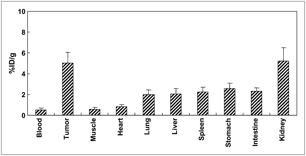
Biodistribution was investigated with human colon cancer SNU-C4 xenograft–bearing nude mice at 17 d after the xenograft procedure (Fig. 4). The highest levels of 68Ga-NOTA-RGD uptake were found in the kidneys (5.3 ± 1.3 %ID/g at 1 h) and then in tumors (5.1 ± 1.0 %ID/g at 1 h) (Fig. 4). Again, significantly lower levels of uptake in the liver (2.1 ± 0.5 %ID/g at 1 h) and intestine (2.3 ± 0.3 %ID/g at 1 h) than in the kidneys indicated the predominance of the renal excretion route (Fig. 4). The tumor-to-blood ratio (10.3 ± 4.8) and the tumor-to-muscle ratio (9.3 ± 3.9) were very high.

Biodistribution of 68Ga-NOTA-RGD in mice bearing SNU-C4 xenografts at 1 h after injection through tail vein. Values represent mean %ID/g, and error bars represent SD.
Imaging in Tumor Xenograft Model
A small-animal PET study was performed at 14 d after nude mice received human colon cancer SNU-C4 xenografts (Fig. 5). Images were obtained at 1 and 2 h after 68Ga-NOTA-RGD was injected either with or without cold c(RGDyK) through a tail vein. A high level of bladder activity at 1 h confirmed rapid renal excretion, which disappeared after urination on 2-h images (Fig. 5A). High levels of uptake were found in tumors and kidneys on 2-h images. The levels of uptake in the liver and intestine were not high, indicating minor hepatobiliary excretion (Fig. 5A). Tumor uptake was significantly blocked by cold c(RGDyK) (Fig. 5B).
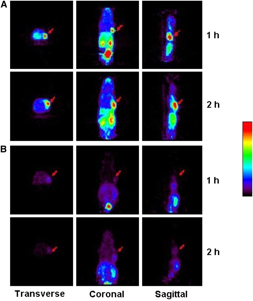
Small-animal PET of 68Ga-NOTA-RGD injected into mice bearing SNU-C4 xenografts at 1 and 2 h after injection without cold c(RGDyK) (A) or with cold c(RGDyK) (60 μg) (B). Images at 1 h were taken before micturition, and images at 2 h were taken after micturition. Arrows indicate tumor positions. Acquisition time was 20 min.
DISCUSSION
In the present study, a c(RGDyK) derivative was investigated as a potential ligand for imaging angiogenesis, because it was previously shown that cyclic derivatives of RGD, such as c(RGDfK) and c(RGDyK), have a high affinity for αvβ3 integrin (28–30). In terms of preparation, c(RGDyK) and SCN-Bz-NOTA were conjugated by thiourea bond formation, leaving all 3 carboxyl residues of NOTA intact and available for forming a complex with 68Ga. It was previously shown that Ga3+ complexes with the 3 carboxyl and 3 tertiary amine residues of NOTA to form an octahedral chelate with some trigonal–prismatic properties (31).
After purification by preparative HPLC, NOTA-RGD was labeled with 68Ga, which was eluted from a titanium dioxide–based 68Ge/68Ga -generator. More than 70% of the 68Ga activity was eluted in the second fraction of 1-mL samples from the generator. Labeling was performed at room temperature, whereas in previous studies, 90°C was used for the 68Ga labeling of 1,4,7,10-tetraazacyclododecane-N,N′,N″,N″′-tetraacetic acid (DOTA)–conjugated peptides (14,32). Nevertheless, because of the high levels of stability of gallium and the NOTA complex, this reaction was possible at room temperature over only 10 min; this reaction time is an attractive feature, especially for the labeling of heat-labile compounds. Although DOTA is more widely used as a bifunctional chelating agent, it is evident that NOTA is more suitable for complex formation with Cu2+ and Ga3+, which have relatively smaller ionic diameters than In3+ and Y3+ (the latter 2 form stable complexes with DOTA). Although a 68Ga-NOTA chelate should be neutral, paper electrophoresis showed that 68Ga-NOTA-RGD was positively charged, presumably because of its strongly basic arginine residue (Supplemental Fig. 4). This finding is supported by its lack of movement on acidic ITLC-SG plates when eluted with a citric acid solution. On the other hand, it could move with the solvent front when it was eluted with an Na2CO3 solution because the positive charge disappeared in the basic solution. Free 68Ga moved with the solvent front when eluted with a citric acid solution and remained at the origin when eluted with Na2CO3 because of colloid formation.
18F is one of the most important positron-emitting candidates for the labeling of RGD derivatives, and various RGD derivatives have been labeled with 18F for PET. 18F has a longer half-life than 68Ga, a property that increases the quality of PET images because a longer half-life allows more nonspecific activity washout. However, in general, 18F labeling is time-consuming and laborious. The most widely used conventional 18F-labeling method includes N-succinimidyl 4-18F-fluorobenzoic acid conjugation to amine residues of peptides (4,33–35). A straightforward and rapid 18F-labeling method using carrier-added 18F-F2 gas has also been described (36). However, its product had low specific activity (32.8 GBq/mmol) and showed a low level of tumor uptake and high levels of nonspecific uptake in the liver, intestine, and kidneys in a mouse biodistribution study. An efficient 18F-labeling method based on no-carrier-added 18F-fluoride has also been described. This method was based on oxime formation between 18F-fluorobenzaldehyde and aminooxy-modified galactosylated cyclic RGD derivatives. The adduct produced showed substantially higher levels of uptake in the liver, kidneys, and tumors than in other tissues (37). Another highly efficient and straightforward labeling method was based on the formation of hydrazone between 18F-fluorobenzaldehyde and hydrazinonicotinic acid–conjugated peptides (38). RGD derivatives labeled with this hydrazone formation method showed high specific activity (20.5 GBq/μmol) and substantially higher levels of uptake in angiogenic ischemic tissues than in normal tissues (25). However, they also showed high levels of uptake in the liver and intestine because of the hydrophobicity of the fluorobenzene moiety, which is therefore not ideal for whole-body PET because of high nonspecific abdominal activity. Moreover, a comparative study of 18F-labeled and 64Cu-labeled RGD derivatives clearly showed that 18F-labeled compounds were taken up by the liver and excreted into the intestine much faster than 64Cu-labeled compounds (39). These literature searches led us to conclude that high levels of liver and intestinal uptake of unglycosylated monomeric RGD derivatives labeled with the 18F-fluorobenzene moiety are attributable to hydrophobicity. In addition, 18F labeling requires a cyclotron system, 18O-enriched water, a long irradiation time for 18F production, and a complicated and time-consuming multistep procedure, all of which incur substantial costs.
Two animal biodistribution models were used in the present study, that is, hind limb ischemia and SNU-C4 (human colon cancer cell line) xenografts. Angiogenesis is known to occur after ischemia lesions as well as during cancer development. The mouse hind limb ischemia model has been used as a means of causing angiogenesis for experimental purposes because of its convenience and reliability (25,26). SNU-C4 xenograft–bearing mice have been used because the solid tumors produced induce angiogenesis. In the present study, we found a significantly higher level of 68Ga-NOTA-RGD uptake in ischemic muscle than in nonischemic muscle; this uptake was found to be blocked by cold c(RGDyK), indicating specific binding by 68Ga-NOTA-RGD (Fig. 3). 68Ga-NOTA-RGD uptake in tumors also was blocked by cold c(RGDyK) in PET studies, further evidence of its specific binding to angiogenic tissue (Fig. 5).
These results concur with those of a previously reported study, except that the liver and intestinal activities significantly decreased when 18F-labeled c(RGDyK) was used (25). The high liver and intestinal activities were the results of rapid hepatobiliary excretion because of hydrophobicity. The introduction of hydrophilic sugar residues significantly increased renal excretion and decreased hepatobiliary excretion (3,4). In the present study, increased renal excretion and decreased hepatobiliary excretion were attributable to the hydrophilicity of 68Ga-NOTA residues, which resembled the hydrophilic nature of sugar residues in a previous report (40).
In the present study, high levels of uptake in tumors and kidneys predominated in biodistribution and imaging studies with human colon cancer SNU-C4 xenograft–bearing nude mice at 1 and 2 h after injection. SNU-C4 cells inoculated into the right shoulder were grown for 14–17 d. Although the level of uptake in the kidneys was high, it is acceptable for a PET agent because it can be easily kept under a hazardous radiation level and will seldom disturb image reading or analysis.
CONCLUSION
A novel NOTA-RGD conjugate was synthesized, labeled with generator-eluted 68Ga, and then investigated in vitro and in vivo. First, 68Ga-NOTA-RGD was produced rapidly at a high-yield with a straightforward method. Second, it was found to be a promising agent for PET imaging of angiogenesis, with high stability, high affinity, high specificity, and excellent pharmacokinetic properties. Finally, it offers significant progress for PET imaging of angiogenesis because of the rapidly growing distribution of 68Ge/68Ga generators.
Acknowledgments
This work was supported by a Korea Research Foundation grant funded by the Korean Government (MOEHRD, Basic Research Promotion Fund) (KRF-2006-E00414). This work also was supported by a Korea Science and Engineering Foundation grant (KOSEF-2008-00610). This study was presented in part at the 17th International Symposium on Radiopharmaceutical Sciences, April 30–May 4, 2007, Aachen, Germany.
Footnotes
-
COPYRIGHT © 2008 by the Society of Nuclear Medicine, Inc.
References
- Received for publication September 18, 2007.
- Accepted for publication January 30, 2008.


Jump to section
Related Articles
Cited By...
- The Potential of PET in the Management of Peripheral Arterial Disease
- Novel PET Imaging of Atherosclerosis with 68Ga-Labeled NOTA-Neomannosylated Human Serum Albumin
- 68Ga-NOTA-PRGD2 PET/CT for Integrin Imaging in Patients with Lung Cancer
- Radiotracer Imaging of Peripheral Vascular Disease
- State-of-the-Art Methods for Evaluation of Angiogenesis and Tissue Vascularization: A Scientific Statement From the American Heart Association
- In Vivo Evaluation of Angiogenic Activity and Its Correlation with Efficacy of Indirect Revascularization Surgery in Pediatric Moyamoya Disease
- Radiotracer Imaging of Peripheral Vascular Disease
- First Experience of 18F-Alfatide in Lung Cancer Patients Using a New Lyophilized Kit for Rapid Radiofluorination
- Nanoparticles Modified by Encapsulation of Ligands with a Long Alkyl Chain to Affect Multispecific and Multimodal Imaging
- Gene Expression Profiles for Genotoxic Effects of Silica-Free and Silica-Coated Cobalt Ferrite Nanoparticles
- A Universally Applicable 68Ga-Labeling Technique for Proteins
- Development of Small-Animal PET Prototype Using Silicon Photomultiplier (SiPM): Initial Results of Phantom and Animal Imaging Studies
- A Nucleolin-Targeted Multimodal Nanoparticle Imaging Probe for Tracking Cancer Cells Using an Aptamer