
Main menu

  • Home
  • Content
    • Current
    • Ahead of print
    • Past Issues
    • JNM Supplement
    • SNMMI Annual Meeting Abstracts
    • Continuing Education
    • JNM Podcasts
  • Subscriptions
    • Subscribers
    • Institutional and Non-member
    • Rates
    • Journal Claims
    • Corporate & Special Sales
  • Authors
    • Submit to JNM
    • Information for Authors
    • Assignment of Copyright
    • AQARA requirements
  • Info
    • Reviewers
    • Permissions
    • Advertisers
  • About
    • About Us
    • Editorial Board
    • Contact Information
  • More
    • Alerts
    • Feedback
    • Help
    • SNMMI Journals
  • SNMMI
    • JNM
    • JNMT
    • SNMMI Journals
    • SNMMI

User menu

  • Subscribe
  • My alerts
  • Log in
  • My Cart

Search

  • Advanced search
Journal of Nuclear Medicine
  • SNMMI
    • JNM
    • JNMT
    • SNMMI Journals
    • SNMMI
  • Subscribe
  • My alerts
  • Log in
  • My Cart
Journal of Nuclear Medicine

Advanced Search

  • Home
  • Content
    • Current
    • Ahead of print
    • Past Issues
    • JNM Supplement
    • SNMMI Annual Meeting Abstracts
    • Continuing Education
    • JNM Podcasts
  • Subscriptions
    • Subscribers
    • Institutional and Non-member
    • Rates
    • Journal Claims
    • Corporate & Special Sales
  • Authors
    • Submit to JNM
    • Information for Authors
    • Assignment of Copyright
    • AQARA requirements
  • Info
    • Reviewers
    • Permissions
    • Advertisers
  • About
    • About Us
    • Editorial Board
    • Contact Information
  • More
    • Alerts
    • Feedback
    • Help
    • SNMMI Journals
  • View or Listen to JNM Podcast
  • Visit JNM on Facebook
  • Join JNM on LinkedIn
  • Follow JNM on Twitter
  • Subscribe to our RSS feeds
Research ArticleCLINICAL INVESTIGATIONS

The Effects of Carbidopa on Uptake of 6-18F-Fluoro-l-DOPA in PET of Pheochromocytoma and Extraadrenal Abdominal Paraganglioma

Henri J.L.M. Timmers, Mohiuddin Hadi, Jorge A. Carrasquillo, Clara C. Chen, Lucia Martiniova, Millie Whatley, Alexander Ling, Graeme Eisenhofer, Karen T. Adams and Karel Pacak
Journal of Nuclear Medicine October 2007, 48 (10) 1599-1606; DOI: https://doi.org/10.2967/jnumed.107.042721
Henri J.L.M. Timmers
  • Find this author on Google Scholar
  • Find this author on PubMed
  • Search for this author on this site
Mohiuddin Hadi
  • Find this author on Google Scholar
  • Find this author on PubMed
  • Search for this author on this site
Jorge A. Carrasquillo
  • Find this author on Google Scholar
  • Find this author on PubMed
  • Search for this author on this site
Clara C. Chen
  • Find this author on Google Scholar
  • Find this author on PubMed
  • Search for this author on this site
Lucia Martiniova
  • Find this author on Google Scholar
  • Find this author on PubMed
  • Search for this author on this site
Millie Whatley
  • Find this author on Google Scholar
  • Find this author on PubMed
  • Search for this author on this site
Alexander Ling
  • Find this author on Google Scholar
  • Find this author on PubMed
  • Search for this author on this site
Graeme Eisenhofer
  • Find this author on Google Scholar
  • Find this author on PubMed
  • Search for this author on this site
Karen T. Adams
  • Find this author on Google Scholar
  • Find this author on PubMed
  • Search for this author on this site
Karel Pacak
  • Find this author on Google Scholar
  • Find this author on PubMed
  • Search for this author on this site
  • Article
  • Figures & Data
  • Info & Metrics
  • PDF
Loading

Article Figures & Data

Figures

  • Tables
  • FIGURE 1. 
    • Download figure
    • Open in new tab
    • Download powerpoint
    FIGURE 1. 

    CT scan (A) and 18F-DOPA PET scans without carbidopa (B) and after carbidopa (C) in patient 9. In PET scans, anterior reprojected images are at left, details of anterior reprojected images are at top right, and transverse sections at tumor level are at bottom right. CT scan shows (recurrent) tumor in left adrenal bed, near tail of pancreas, consistent with abnormal left adrenal focus on 18F-DOPA PET after carbidopa. On baseline 18F-DOPA PET, left adrenal bed is masked by physiologic uptake by pancreas.
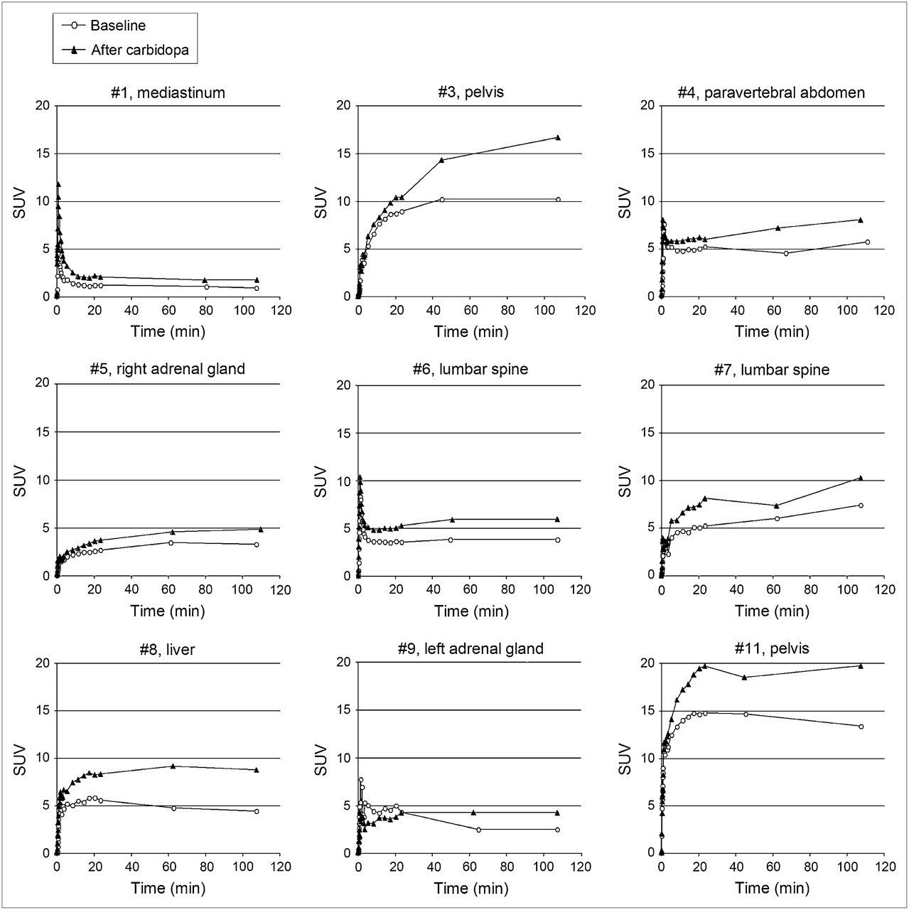
  • FIGURE 2. 
    • Download figure
    • Open in new tab
    • Download powerpoint
    FIGURE 2. 

    Dynamics of 18F-DOPA tumor uptake. Time–activity curves of 18F-DOPA uptake by index lesions at baseline (no carbidopa) vs. after carbidopa. Patient numbers and locations of index lesions are indicated in graph titles.

Tables

  • Figures
    • View popup
    TABLE 1

    Patient Characteristics

    Patient no.SexMutationAge at diagnosis (y)Location of primary tumorBiochemical phenotypeTime to metastases* (y)Locations of metastasesPrevious treatment
    1MSDHB27L extraadrenal abdominalNone0Abdominal LN, mediastinum, bonePrimary tumor resection; CVD
    2FSDHB51R paraaortic abdominalNE——MIBG
    3FSDHB23Paraaortic abdominalNE0.3Abdominal LN, bonePrimary tumor resection; CVD
    4MSDHD22R adrenalNE10.3Abdominal LN, liver, mediastinum, bonePrimary tumor resection; MIBG
    5FPending53R adrenalNE———
    6MSDHB10R paraadrenalNE + DA8.4Abdominal LN, lung, liver, bone, neckMIBG
    7MPending39R adrenalNE + DA14.7Abdominal LN, mediastinum, bonePrimary tumor resection
    8FRET33R + L adrenalNE + E6.2Abdominal LN, liver, bonePrimary tumor resection; MIBG
    9FRET33R + L adrenalE——Primary tumor resection
    10MSDHB40L paraaorticNE3.9LiverPrimary tumor resection
    11MPending40R adrenalNE + DA5.4Lung, bonePrimary tumor resection; CVD; MIBG
    • ↵* Time between diagnosis of primary tumor and metastatic disease.

    • SDHB/D = succinate dehydrogenase subunit B/D; LN = lymph nodes; CVD = cyclophosphamide/vincristine/dacarbazine; NE = norepinephrine; MIBG = 131I-methyliodobenzylguanidine; DA = dopamine; RET = rearranged during transfection; E = epinephrine.

    • View popup
    TABLE 2

    Tumor Detection

    18F-DOPA PET
    ParameterCT or MRIBaselineCarbidopa
    Number of lesions
     Head—77
     Neck445
     Chest161717
     Abdomen/pelvis5833*35*
     Total, except head7854*57*
     Total, including head—6164
    Number of positive regions
     Head—44
     Neck322
     Chest544
     Abdomen/pelvis1079
     Total, except head181315
     Total, including head—1719
    • ↵* P < 0.01 vs. CT or MRI.

    • View popup
    TABLE 3

    Dynamics of 18F-DOPA Tumor Uptake

    Time to peak (min)Peak SUV
    Patient no.Location of index lesionBaselineCarbidopaBaselineCarbidopa
    1Mediastinum24241.22.1
    3Pelvis779010.817.2
    4Paravertebral abdomen107*107*5.58.0
    5R adrenal gland791013.54.8
    6Lumbar spine80813.96.2
    7Lumbar spine107*107*7.310.2
    8Liver20745.99.2
    9L adrenal gland19764.74.3
    11Pelvis32107*14.719.7
    Mean ± SD60.6 ± 34.785.2 ± 25.1†6.4 ± 3.99.1 ± 5.6†
    • ↵* Uptake curve was still rising at last imaging time point.

    • ↵† P < 0.05 vs. baseline.

    • View popup
    TABLE 4

    Physiologic Distribution of 18F-DOPA

    Maximum SUVMean SUV
    LocationBaselineCarbidopaBaselineCarbidopa
    Basal ganglia2.43 ± 0.553.73* ± 0.811.28 ± 0.312.04* ± 0.43
    Myocardium2.53 ± 0.573.18* ± 0.640.75 ± 0.161.04* ± 0.22
    Lungs0.69 ± 0.240.88* ± 0.240.22 ± 0.080.29* ± 0.07
    Liver3.13 ± 0.753.42 ± 0.641.3 ± 0.341.48* ± 0.28
    Kidneys5.33 ± 1.515.32 ± 1.472.51 ± 0.662.43 ± 0.71
    Pancreas6.21 ± 1.79—3.03 ± 1.04—
    • ↵* P < 0.01 vs. baseline.

PreviousNext
Back to top

In this issue

Journal of Nuclear Medicine: 48 (10)
Journal of Nuclear Medicine
Vol. 48, Issue 10
October 2007
  • Table of Contents
  • Table of Contents (PDF)
  • About the Cover
  • Index by author
Print
Download PDF
Article Alerts
Sign In to Email Alerts with your Email Address
Email Article

Thank you for your interest in spreading the word on Journal of Nuclear Medicine.

NOTE: We only request your email address so that the person you are recommending the page to knows that you wanted them to see it, and that it is not junk mail. We do not capture any email address.

Enter multiple addresses on separate lines or separate them with commas.
The Effects of Carbidopa on Uptake of 6-18F-Fluoro-l-DOPA in PET of Pheochromocytoma and Extraadrenal Abdominal Paraganglioma
(Your Name) has sent you a message from Journal of Nuclear Medicine
(Your Name) thought you would like to see the Journal of Nuclear Medicine web site.
Citation Tools
The Effects of Carbidopa on Uptake of 6-18F-Fluoro-l-DOPA in PET of Pheochromocytoma and Extraadrenal Abdominal Paraganglioma
Henri J.L.M. Timmers, Mohiuddin Hadi, Jorge A. Carrasquillo, Clara C. Chen, Lucia Martiniova, Millie Whatley, Alexander Ling, Graeme Eisenhofer, Karen T. Adams, Karel Pacak
Journal of Nuclear Medicine Oct 2007, 48 (10) 1599-1606; DOI: 10.2967/jnumed.107.042721

Citation Manager Formats

  • BibTeX
  • Bookends
  • EasyBib
  • EndNote (tagged)
  • EndNote 8 (xml)
  • Medlars
  • Mendeley
  • Papers
  • RefWorks Tagged
  • Ref Manager
  • RIS
  • Zotero
Share
The Effects of Carbidopa on Uptake of 6-18F-Fluoro-l-DOPA in PET of Pheochromocytoma and Extraadrenal Abdominal Paraganglioma
Henri J.L.M. Timmers, Mohiuddin Hadi, Jorge A. Carrasquillo, Clara C. Chen, Lucia Martiniova, Millie Whatley, Alexander Ling, Graeme Eisenhofer, Karen T. Adams, Karel Pacak
Journal of Nuclear Medicine Oct 2007, 48 (10) 1599-1606; DOI: 10.2967/jnumed.107.042721
Twitter logo Facebook logo LinkedIn logo Mendeley logo
  • Tweet Widget
  • Facebook Like
  • Google Plus One
Bookmark this article

Jump to section

  • Article
    • Abstract
    • MATERIALS AND METHODS
    • RESULTS
    • DISCUSSION
    • CONCLUSION
    • Acknowledgments
    • Footnotes
    • References
  • Figures & Data
  • Info & Metrics
  • PDF

Related Articles

  • This Month in JNM
  • PubMed
  • Google Scholar

Cited By...

  • Imaging of Pheochromocytoma and Paraganglioma
  • Comparison of the Amino Acid Tracers 18F-FET and 18F-DOPA in High-Grade Glioma Patients
  • Molecular and Therapeutic Advances in the Diagnosis and Management of Malignant Pheochromocytomas and Paragangliomas
  • Characterization of Neuroblastic Tumors Using 18F-FDOPA PET
  • Correlation of the Genotype of Paragangliomas and Pheochromocytomas with Their Metabolic Phenotype on 3,4-Dihydroxy-6-18F-Fluoro-L-Phenylalanin PET
  • Modern Nuclear Imaging for Paragangliomas: Beyond SPECT
  • Complementary Roles of 18F-DOPA PET/CT and 18F-FDG PET/CT in Medullary Thyroid Cancer
  • 18F-FDOPA PET and PET/CT Accurately Localize Pheochromocytomas
  • The clinical value of [18F]fluoro-dihydroxyphenylalanine positron emission tomography in primary diagnosis, staging, and restaging of neuroendocrine tumors
  • 18F-DOPA PET and PET/CT
  • Google Scholar

More in this TOC Section

  • Feasibility of Ultra-Low-Activity 18F-FDG PET/CT Imaging Using a Long–Axial-Field-of-View PET/CT System
  • Cardiac Presynaptic Sympathetic Nervous Function Evaluated by Cardiac PET in Patients with Chronotropic Incompetence Without Heart Failure
  • Validation and Evaluation of a Vendor-Provided Head Motion Correction Algorithm on the uMI Panorama PET/CT System
Show more Clinical Investigations

Similar Articles

SNMMI

© 2025 SNMMI

Powered by HighWire