


Article Figures & Data
Figures
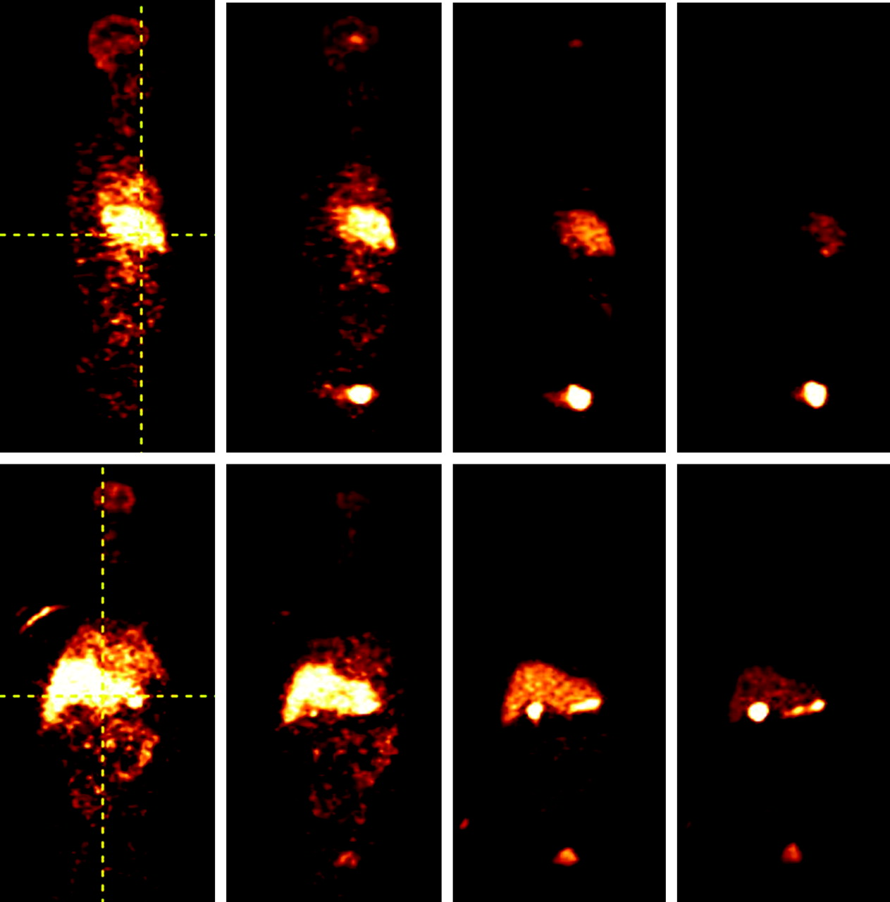
- FIGURE 1.
Sagittal (top row) and coronal (bottom row) views of whole-body images in 1 healthy human subject at 1, 10, 20, and 40 min after injection of 11C-raclopride. Vertical crosshair in sagittal view shows the slice level of coronal images. Vertical crosshair in coronal view shows the slice level of sagittal images. Initial uptake is predominantly in liver but eventually settles in gallbladder and urinary bladder (compare with Fig. 2). Note prominent uptake in striatum (brain) at 10 and 20 min.
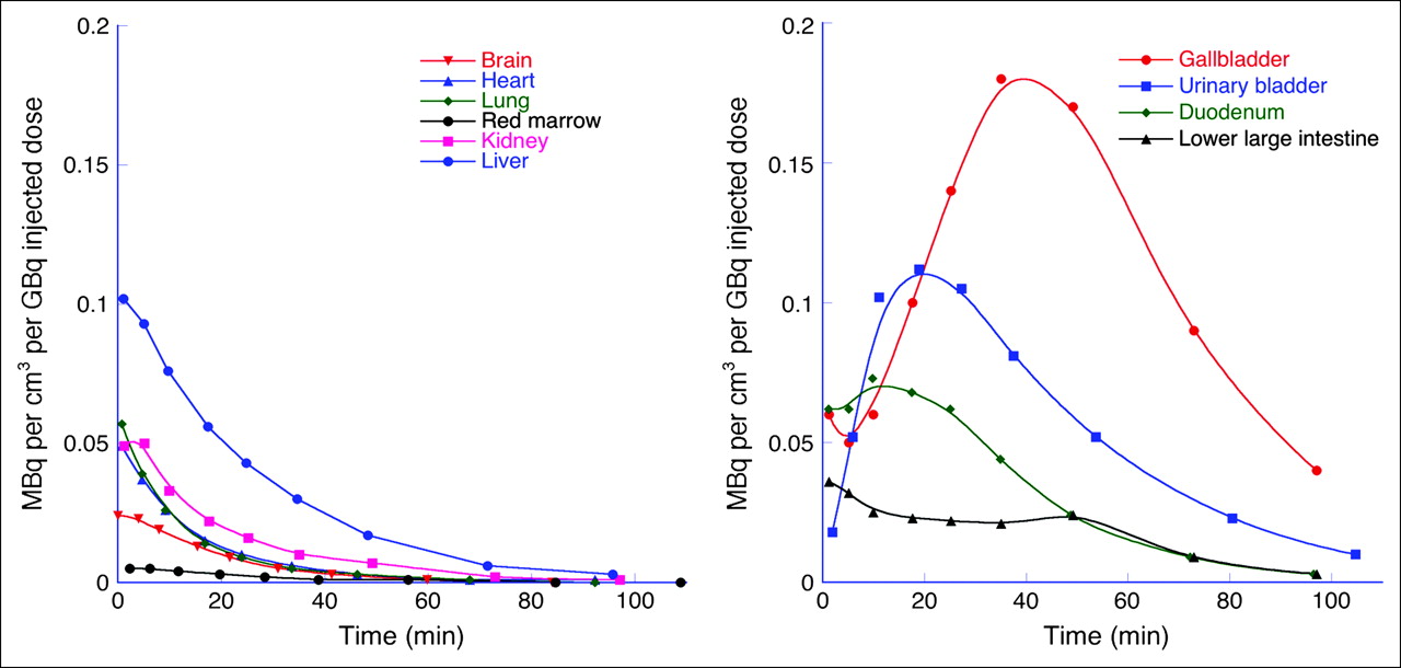
- FIGURE 2.
Time–activity curves for all organs. Units are activity concentration (MBq/cm3) normalized to injected dose (GBq). (A) Low- and moderate-uptake organs. (B) High-uptake organs (urinary bladder contents concentration is taken from a central subsample for comparability with other organs). Markers are average of measured data, interpolated to the same time for each organ (timing varied slightly from subject to subject). Fitted curves are for display and do not represent a physiologic model.
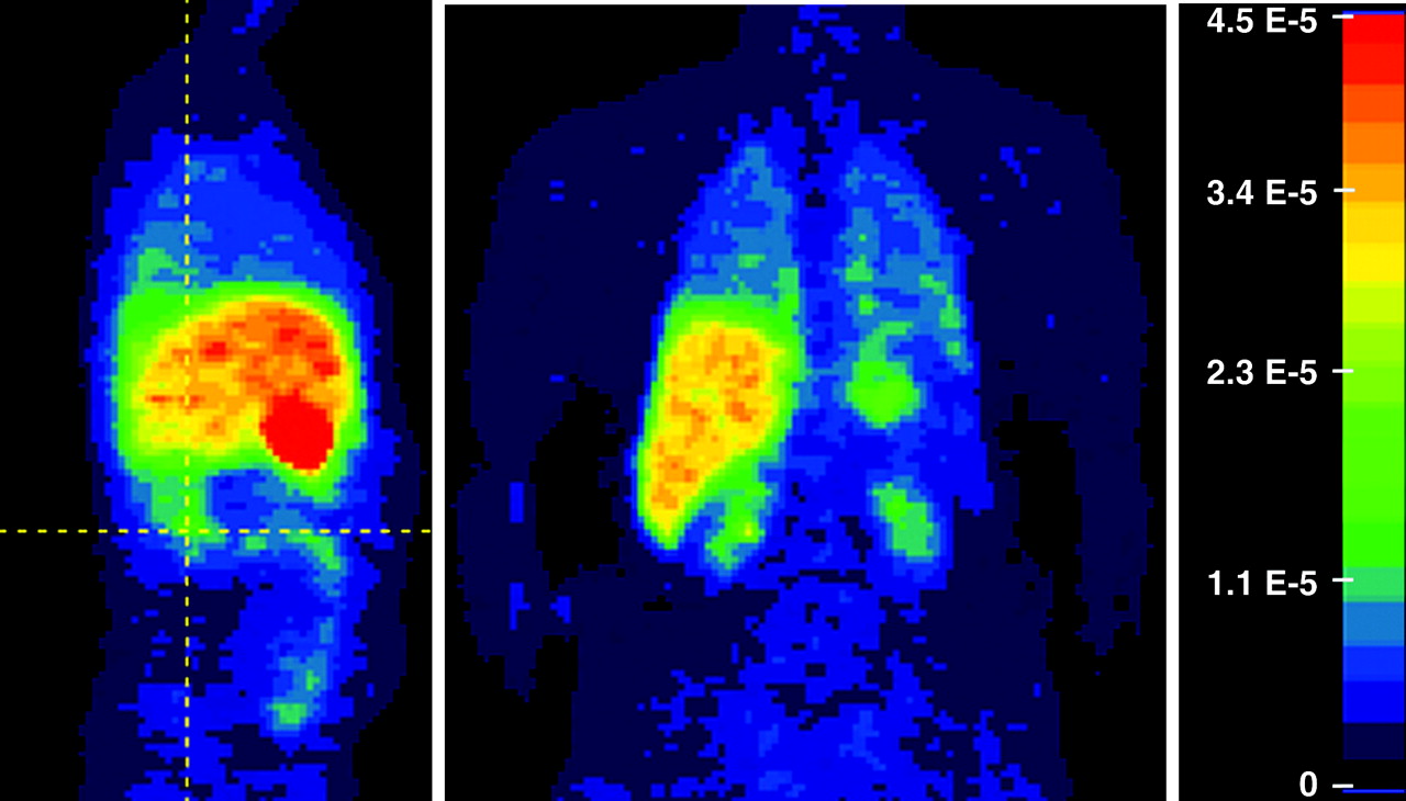
- FIGURE 3.
Weighted summation of dynamic data from 1 subject. Frames were weighted by the pass durations so the summation represents an approximation to a residence time density image. Units are h/cm3. Color scale was truncated at 4.5 × 10⁻⁵ h/cm3 (right) so that high-uptake (gallbladder) and low-uptake (kidney) organs could be visualized in same image—that is, all values ≥4.5 × 10⁻⁵ h/cm3 are mapped to same color (red). Vertical gridline on sagittal (left, facing to right) view shows slice level of coronal view (center), at level of kidney.
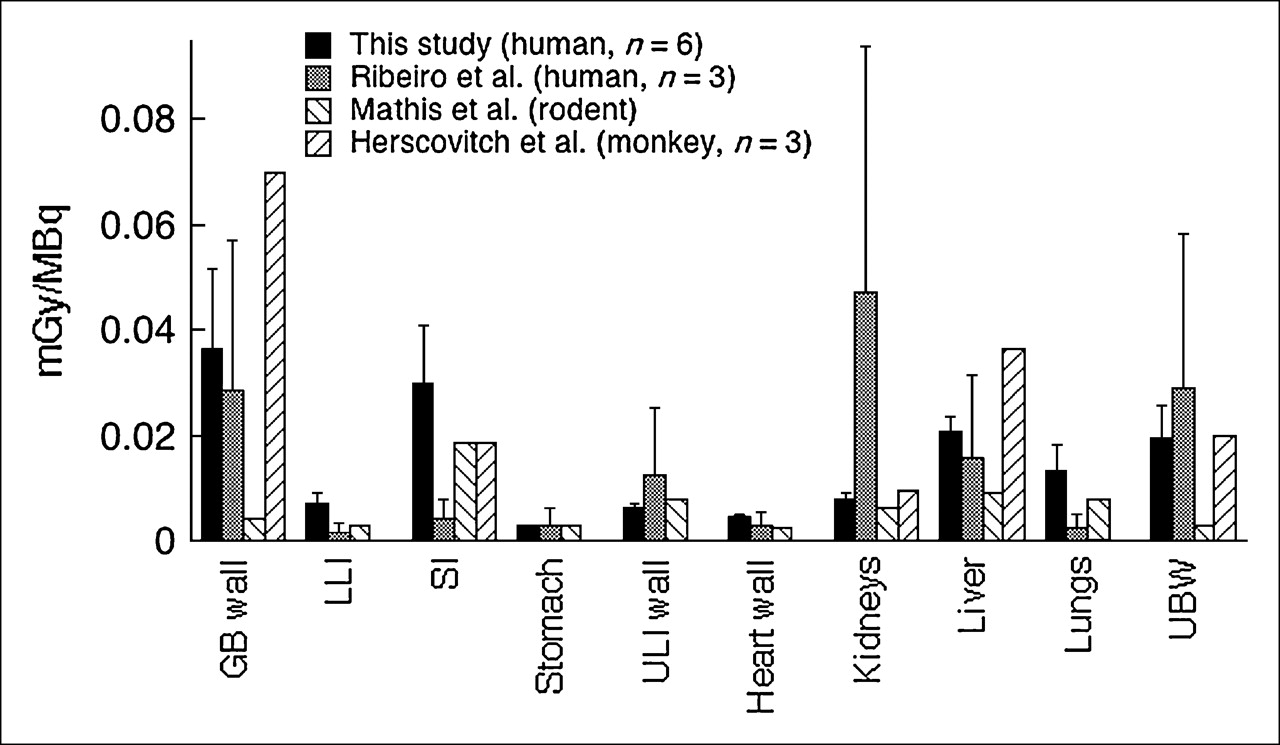
- FIGURE 4.
Comparison of radiation-absorbed doses from this study with other studies (18–20). Units are mGy/MBq. Error bars are SD (not available for rodent and monkey studies). GB = gallbladder; LLI = lower large intestine; SI = small intestine; ULI = upper large intestine; UBW = urinary bladder wall.


Tables
Subject Sex Age (y) Height (m) Mass (kg) Injected dose (MBq) 1 F 34 1.68 74.84 650.83 2 F 21 1.60 62.60 577.61 3 F 30 1.73 72.57 487.66 4 M 35 1.73 103.87 477.67 5 M 34 1.83 81.65 373.33 6 M 26 1.75 68.95 619.01 Mean ± SD 30 ± 5.55 1.72 ± 0.08 77.41 ± 14.42 531.02 ± 103.79 - TABLE 2
Residence Times ± SD (n = 6) of 11C-Raclopride for 11 Measured Organs and Remainder of Body
Organ Residence time (h) Liver 0.099 ± 0.019 Small intestine 0.081 ± 0.039 Lung 0.040 ± 0.020 Urinary bladder 0.018 ± 0.005 Cortical bone 0.014 ± 0.003 Brain 0.014 ± 0.002 Gallbladder 0.012 ± 0.006 Heart 0.006 ± 0.001 Kidney 0.005 ± 0.001 Lower large intestine 0.004 ± 0.003 Red marrow 0.003 ± 0.001 Remainder of body 0.195 ± 0.039 Organ mGy/MBq rad/mCi Adrenal 3.17E−03 1.17E−02 Brain 3.44E−03 1.27E−02 Breast 1.70E−03 6.28E−03 Gallbladder wall 3.15E−02 1.17E−01 Lower large intestine wall 6.15E−03 2.28E−02 Small intestine 2.58E−02 9.55E−02 Stomach 2.59E−03 9.59E−03 Upper large intestine wall 5.22E−03 1.93E−02 Heart wall 4.06E−03 1.50E−02 Kidney 6.78E−03 2.51E−02 Liver 1.77E−02 6.56E−02 Lung 1.14E−02 4.21E−02 Muscle 2.00E−03 7.40E−03 Ovary 3.91E−03 1.45E−02 Pancreas 3.23E−03 1.19E−02 Red marrow 2.26E−03 8.35E−03 Bone surface 2.00E−03 7.39E−03 Skin 1.42E−03 5.25E−03 Spleen 2.17E−03 8.04E−03 Testis 1.60E−03 5.93E−03 Thymus 1.97E−03 7.28E−03 Thyroid 1.57E−03 5.79E−03 Urinary bladder wall 1.35E−02 5.00E−02 Uterus 3.90E−03 1.44E−02 Total body 2.83E−03 1.05E−02 mSv/MBq rem/mCi Effective dose equivalent 8.73E−03 3.23E−02 Effective dose 6.26E−03 2.31E−02 - TABLE 4
Comparison of Instrumentation and Several Scanning Parameters Between This Study and the Study of Ribeiro et al. (20)
Comparison This study Study of Ribeiro et al. (20) Scanner Accel HR+ Acquisition/reconstruction 2-dimensional 3-dimensional Transmission scan duration 5 min per bed 3 min per bed Frame duration per bed 15 s to 2 min 1 min Scan starting time 1 min after injection Not specified


Jump to section
Related Articles
Cited By...
- Human Radiation Dosimetry for the N-Methyl-D-Aspartate Receptor Radioligand 11C-CNS5161
- Whole-Body Distribution and Radiation Dosimetry of 11C-(+)-PHNO, a D2/3 Agonist Ligand
- Radiation Dosimetry and Biodistribution of the TSPO Ligand 11C-DPA-713 in Humans
- Human Biodistribution and Dosimetry of the D2/3 Agonist 11C-N-Propylnorapomorphine (11C-NPA) Determined from PET
- 1-11C-Methyl-4-Piperidinyl-N-Butyrate Radiation Dosimetry in Humans by Dynamic Organ-Specific Evaluation