


Article Figures & Data
Figures
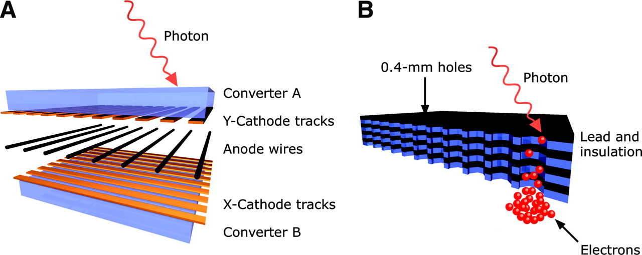
- FIGURE 1.
(A) Construction of a detector module in 3 layers: 2 converters connected by a MWPC. An incoming photon is converted into an electron that is amplified and accelerated toward the anode wires. (B) Each converter contains interleaved lead and insulation sheets, mechanically drilled with a dense matrix of small holes. A photon interacts with the lead, resulting in an electron that avalanches in a strong electrical field and accelerates toward the MWPC.
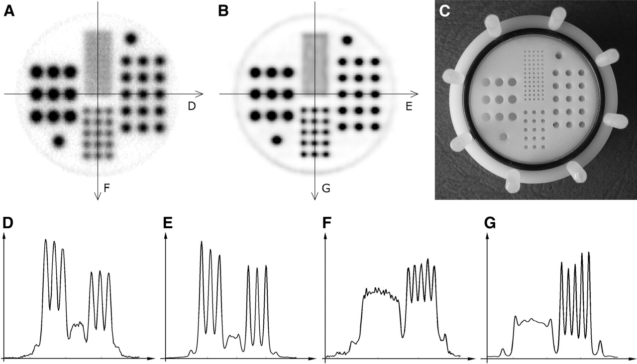
- FIGURE 2.
Reconstruction results of a 18F-filled high-resolution phantom, constructed from a plastic cylinder, in which holes of different sizes (0.5, 1, 1.5, and 2 mm) were drilled (C). Holes are clearly visible in FBP (A) and OPL-EM (B) reconstructed images down to a size of 1 mm with sharp signals in the profile lines (D and F, horizontal and vertical profiles FBP; E and G, horizontal and vertical profiles OPL-EM).
- FIGURE 3.
Absolute sensitivity of quadHIDAC for different line source lengths. Measurements were performed using method of Bailey et al. (14).
- FIGURE 4.
Count rate performance measured with line source (A), mouse phantom (B), and rat phantom (C). Denoted by × is the rate of the total number of detected coincidences. Subtracting randoms (denoted by ○), estimated from the single rate yields the set of stars (*). True unscattered coincidence rate (denoted by +) was obtained by subtracting scattered events (▵).
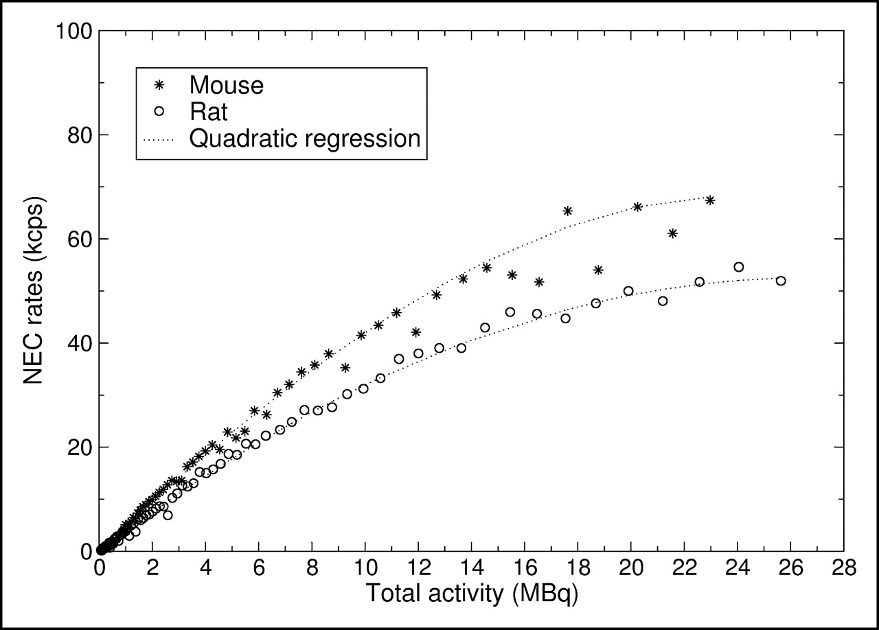
- FIGURE 5.
NEC rates for mouse phantom and rat phantom.
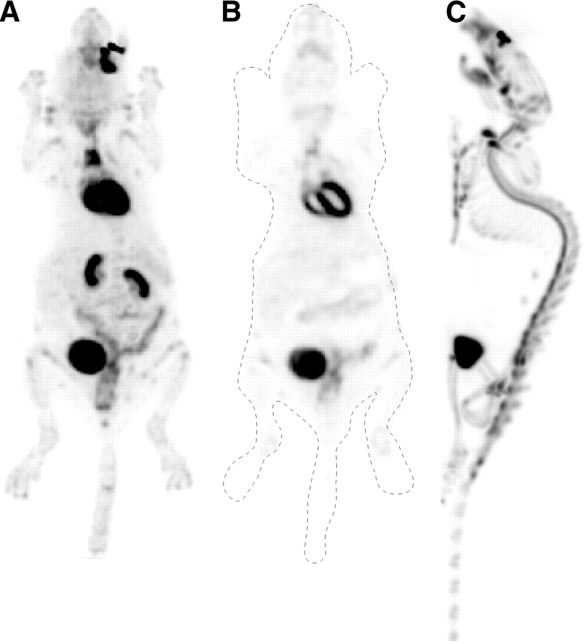
- FIGURE 6.
(A and B) Images (OPL-EM) of 22-g mouse, acquired in 15 min, 1 h after injection of 18F-FDG show maximum intensity projection (A) and a single central slice (B). (C) Maximum intensity projection of 27-g mouse, 1 h after injection of 18F fluoride.


Tables
- TABLE 2
Count Rate Measurements of Line Source and Mouse Phantom Experiment Normalized to Computed Coincidence Decay Rate
Parameter Total Trues Scatter Scatter fraction (%) Attenuation fraction (%) Line source 0.0238 0.0149 0.0076 32 — Mouse scatter phantom 0.0203 0.0115 0.0075 37 22
In this issue


Jump to section
Related Articles
Cited By...
- Short-Term Colony-Stimulating Factor 1 Receptor Inhibition-Induced Repopulation After Stroke Assessed by Longitudinal 18F-DPA-714 PET Imaging
- A Longitudinal PET/MRI Study of Colony-Stimulating Factor 1 Receptor-Mediated Microglia Depletion in Experimental Stroke
- Combined PET Imaging of the Inflammatory Tumor Microenvironment Identifies Margins of Unique Radiotracer Uptake
- Preclinical Evidence That 3'-Deoxy-3'-[18F]Fluorothymidine PET Can Visualize Recovery of Hematopoiesis after Gemcitabine Chemotherapy
- Gemcitabine Mechanism of Action Confounds Early Assessment of Treatment Response by 3'-Deoxy-3'-[18F]Fluorothymidine in Preclinical Models of Lung Cancer
- Variability of Proliferation and Diffusion in Different Lung Cancer Models as Measured by 3'-Deoxy-3'-18F-Fluorothymidine PET and Diffusion-Weighted MR Imaging
- PET with 18F-FDG-Labeled T Lymphocytes for Diagnosis of Acute Rat Renal Allograft Rejection
- Translational 18F-FDG PET/CT Imaging to Monitor Lesion Activity in Intestinal Inflammation
- National Electrical Manufacturers Association NU-4 Performance Evaluation of the PET Component of the NanoPET/CT Preclinical PET/CT Scanner
- Potential of Noninvasive Serial Assessment of Acute Renal Allograft Rejection by 18F-FDG PET to Monitor Treatment Efficiency
- Molecular Imaging of Cardiac Sympathetic Innervation by 11C-mHED and PET: From Man to Mouse?
- Isochronous Assessment of Cardiac Metabolism and Function in Mice Using Hybrid PET/MRI
- NOS2 Gene Deficiency Protects from Sepsis-Induced Long-Term Cognitive Deficits
- Quantification of Left Ventricular Volumes and Ejection Fraction in Mice Using PET, Compared with MRI
- Clinical molecular imaging in intestinal graft-versus-host disease: mapping of disease activity, prediction, and monitoring of treatment efficiency by positron emission tomography
- Performance Evaluation of the GE Healthcare eXplore VISTA Dual-Ring Small-Animal PET Scanner
- Accurate Noninvasive Measurement of Infarct Size in Mice with High-Resolution PET
- Age- and Training-Dependent Development of Arrhythmogenic Right Ventricular Cardiomyopathy in Heterozygous Plakoglobin-Deficient Mice
- Monitoring Left Ventricular Dilation in Mice with PET