

FIGURE 3.
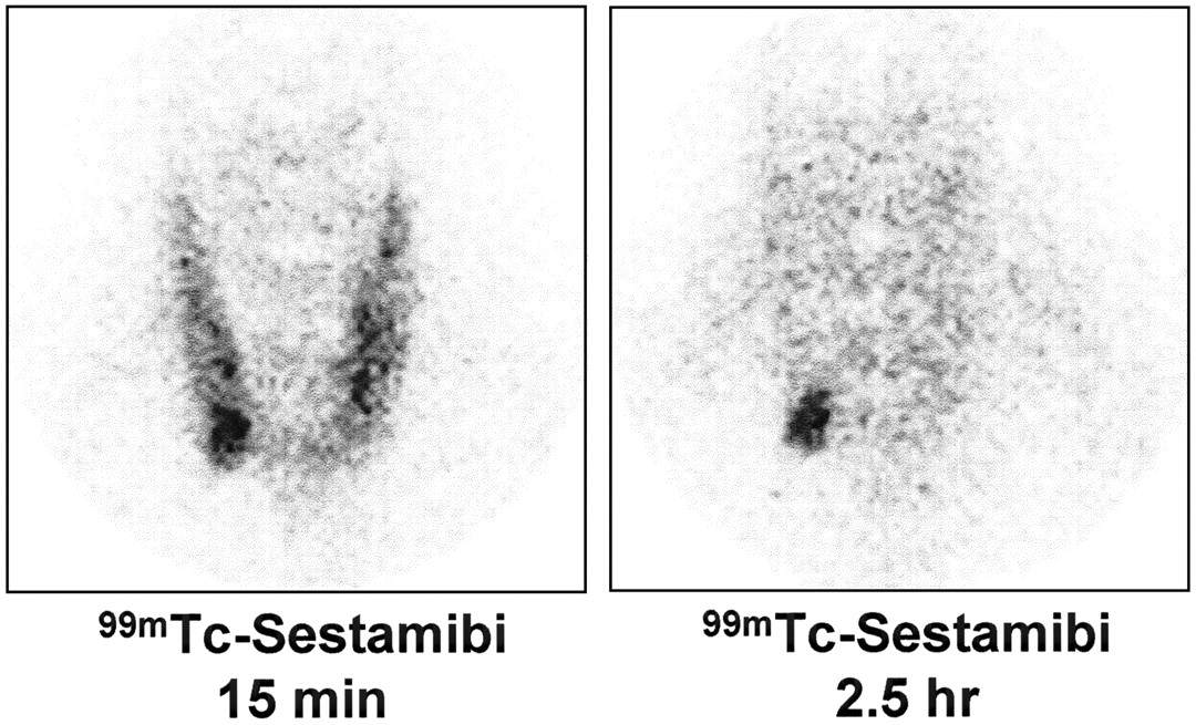
FIGURE 3.
Classical single-tracer, double-phase parathyroid scintigraphy with 99mTc-sestamibi. (Left) Image at 15 min shows physiologic early uptake in thyroid gland, with clear focus of moderately increased accumulation at lower pole of right thyroid lobe. (Right) Late scan shows almost complete washout of 99mTc-sestamibi from thyroid gland, with obvious focal retention of radioactivity at lower pole of right thyroid lobe. Minimally invasive radioguided surgery confirmed presence of parathyroid adenoma.
In this issue


Related Articles
Cited By...
- Imaging of the Thyroid and Parathyroid Using a Cardiac Cadmium-Zinc-Telluride Camera: Phantom Studies
- Radioguided parathyroidectomy in forearm graft for recurrent hyperparathyroidism
- Parathyroid Imaging and Localization Using SPECT/CT: Initial Results
- Preoperative 123I/99mTc-Sestamibi Subtraction SPECT and SPECT/CT in Primary Hyperparathyroidism
- Pinhole Versus Parallel-Hole Collimators for Parathyroid Imaging: An Intraindividual Comparison
- Differences in Accuracy of 99mTc-Sestamibi Scanning Between Severe and Mild Forms of Primary Hyperparathyroidism
- Subtraction SPECT for Parathyroid Scintigraphy Based on Maximization of Mutual Information
- Is the "Ideal" {gamma}-Probe for Intraoperative Radioguided Surgery Conceivable?
- Radioguided Surgery of Primary Hyperparathyroidism Using the Low-Dose 99mTc-Sestamibi Protocol: Multiinstitutional Experience from the Italian Study Group on Radioguided Surgery and Immunoscintigraphy (GISCRIS)
- The Value of 99mTc-Sestamibi SPECT/CT over Conventional SPECT in the Evaluation of Parathyroid Adenomas or Hyperplasia