


Article Figures & Data
Figures
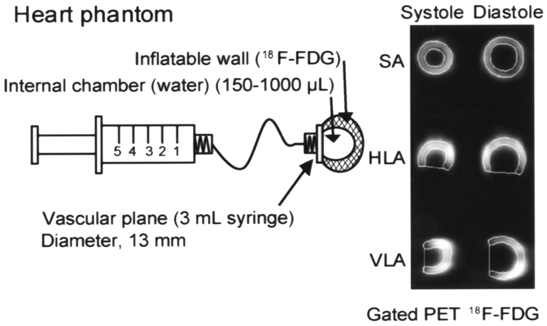
- FIGURE 1.
Schematic of heart phantom showing variable-volume internal chamber filled with water and sealed, fixed-volume dual-layer wall filled with 18F-FDG. Phantom can accommodate endocardial volume ranging from 150 to 1,000 μL. Shown on right is example of systolic (300 μL) and diastolic (900 μL) images extracted by QGS analysis of simulated series of gated PET images of phantom. SA = short axis; HLA = horizontal long axis; VLA = vertical long axis.
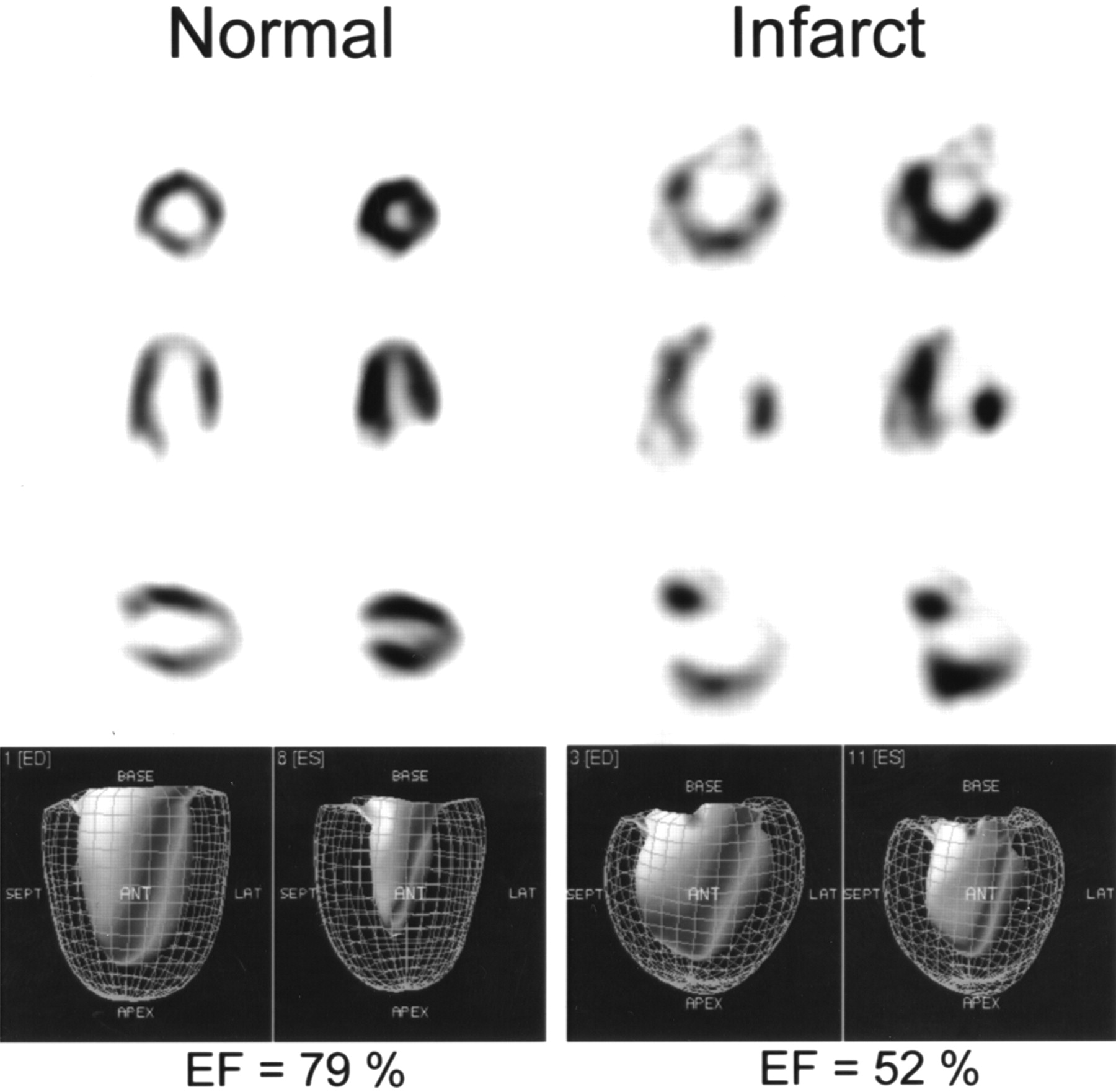
- FIGURE 2.
Results of QGS-analyzed, ECG-gated 18F-FDG PET of normal and infarcted rats using Sherbrooke small-animal PET scanner. EF = ejection fraction.
- FIGURE 3.
Correlation between endocardial volume of cardiac phantom and volume determined using QGS: linear regression analysis of measurements performed in triplicate (A) and Bland–Altman plot (B). Sy.x = SEE.
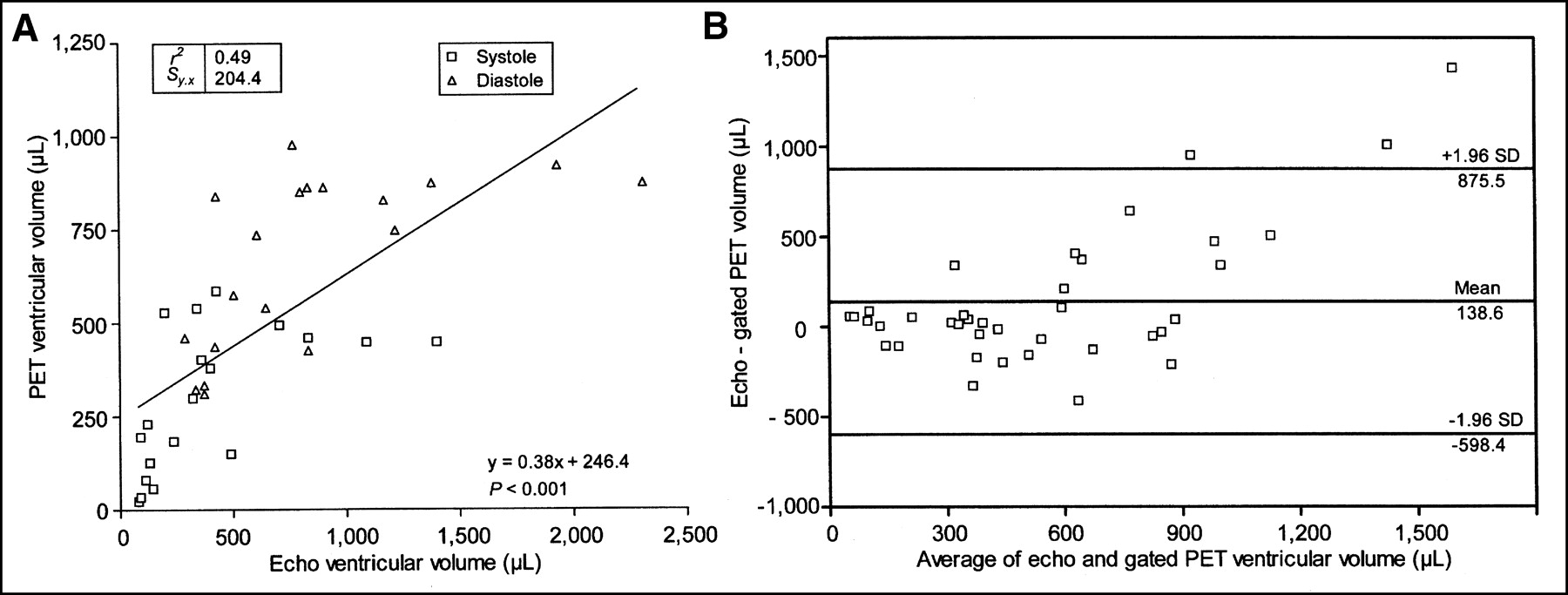
- FIGURE 4.
Correlation between ventricular end-systolic and end-diastolic volumes measured by gated PET and echocardiography in healthy rats (n = 11): linear regression (A) and Bland–Altman plot (B). Echo = echocardiography; Sy.x = SEE.
- FIGURE 5.
Correlation between ventricular end-systolic and end-diastolic volumes measured by gated PET and echocardiography in infarcted (n = 15) and septic (n = 4) rats: linear regression (A) and Bland–Altman plot (B). Echo = echocardiography; Sy.x = SEE.
- FIGURE 6.
Correlation between LVEF measured by gated PET and echocardiography for normal (n = 11), infarcted (n = 15), and septic (n = 4) rats: linear regression (A) and Bland–Altman plot (B). Echo = echocardiography; Sy.x = SEE.


Tables
In this issue


Jump to section
Related Articles
Cited By...
- Resident and recruited macrophages differentially contribute to cardiac healing after myocardial ischemia
- Resident and recruited macrophages differentially contribute to cardiac healing after myocardial ischemia
- Angiotensin II-Converting Enzyme Inhibition Improves Survival, Ventricular Remodeling, and Myocardial Energetics in Experimental Aortic Regurgitation
- Small-Animal PET: What Is It, and Why Do We Need It?
- Reproducibility of Serial Left Ventricle Perfusion, Volume, and Ejection Fraction Measurements Using Multiplexed Multipinhole SPECT in Healthy Rats and Rats After Myocardial Infarction
- Evaluation of a Novel 18F-Labeled Positron-Emission Tomography Perfusion Tracer for the Assessment of Myocardial Infarct Size in Rats
- Quantification of Left Ventricular Volumes and Ejection Fraction in Mice Using PET, Compared with MRI
- A New Tool for Molecular Imaging: The Microvolumetric {beta} Blood Counter
- Noninvasive Measurement of Cardiovascular Function in Mice with High-Temporal-Resolution Small-Animal PET
- Monitoring Left Ventricular Dilation in Mice with PET
- Assessment of Left Ventricular Perfusion, Volumes, and Motion in Mice Using Pinhole Gated SPECT
- Gated PET and Ventricular Volume