


Article Figures & Data
Figures
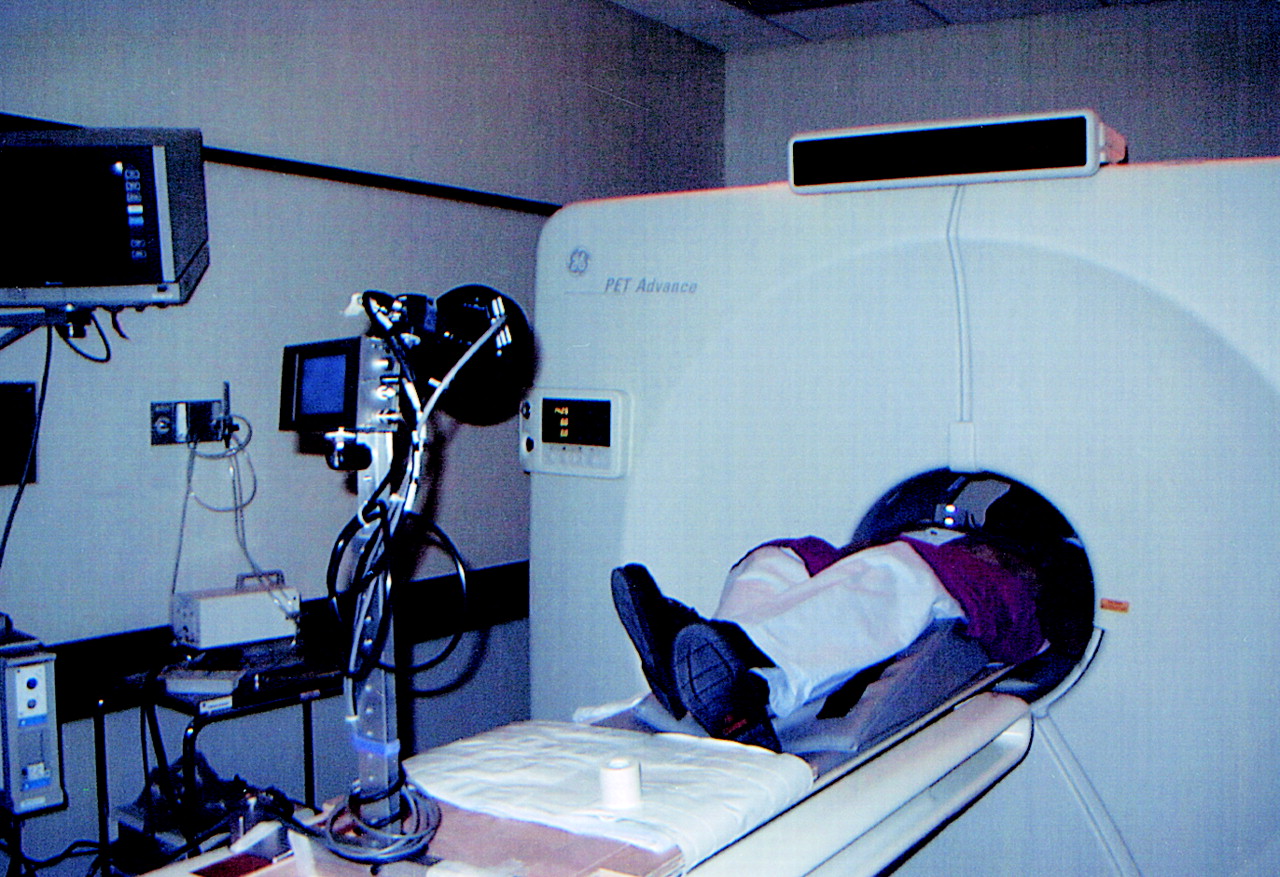
- FIGURE 1.
Patient setup in gated mode.
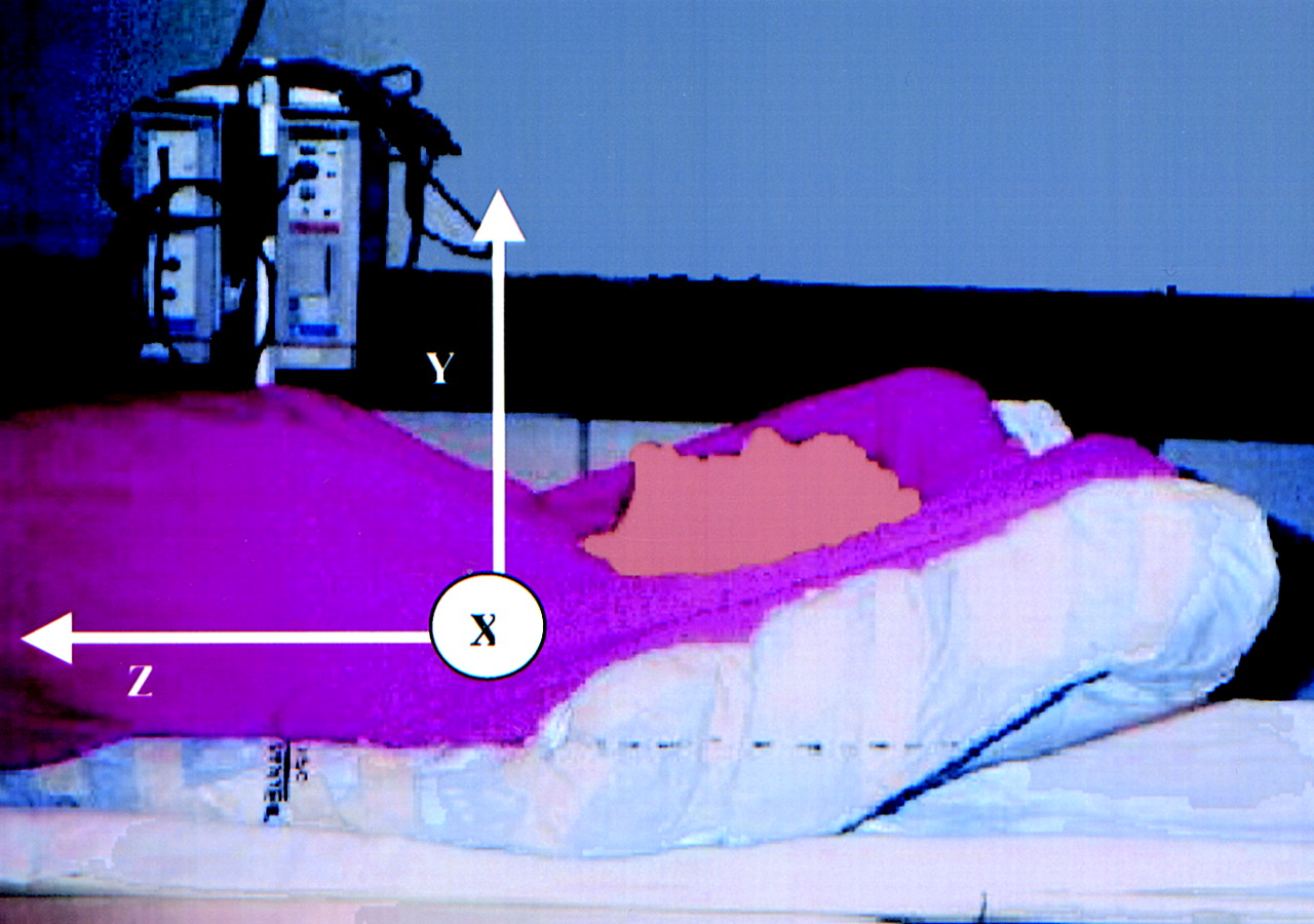
- FIGURE 2.
Patient coordinate system.
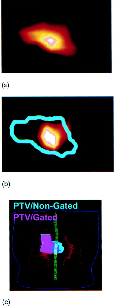
- FIGURE 3.
Transaxial 18F-FDG PET image through 1 patient’s lesion in nongated mode (A) and corresponding image in gated mode acquired in first bin (B). (C) Planning target volume in nongated (light blue) and gated (pink) modes. Note that light blue extends under whole pink area. Gating, in this particular case, has mainly spared left lung tissues from high doses.
- FIGURE 4.
Comparison between lesion volumes in nongated and gated modes. Gating shows consistency in reducing apparent lesion volume.
- FIGURE 5.
Comparison between SUVmax in nongated and gated modes. Gating shows consistency in improving accuracy in SUVmax measurements.
- FIGURE 6.
Maximum TLG (TLGmax) measurements in gated mode show linear dependence on those measured in nongated mode because increase in SUVmax should result in reduction in lesion volume by same factor.
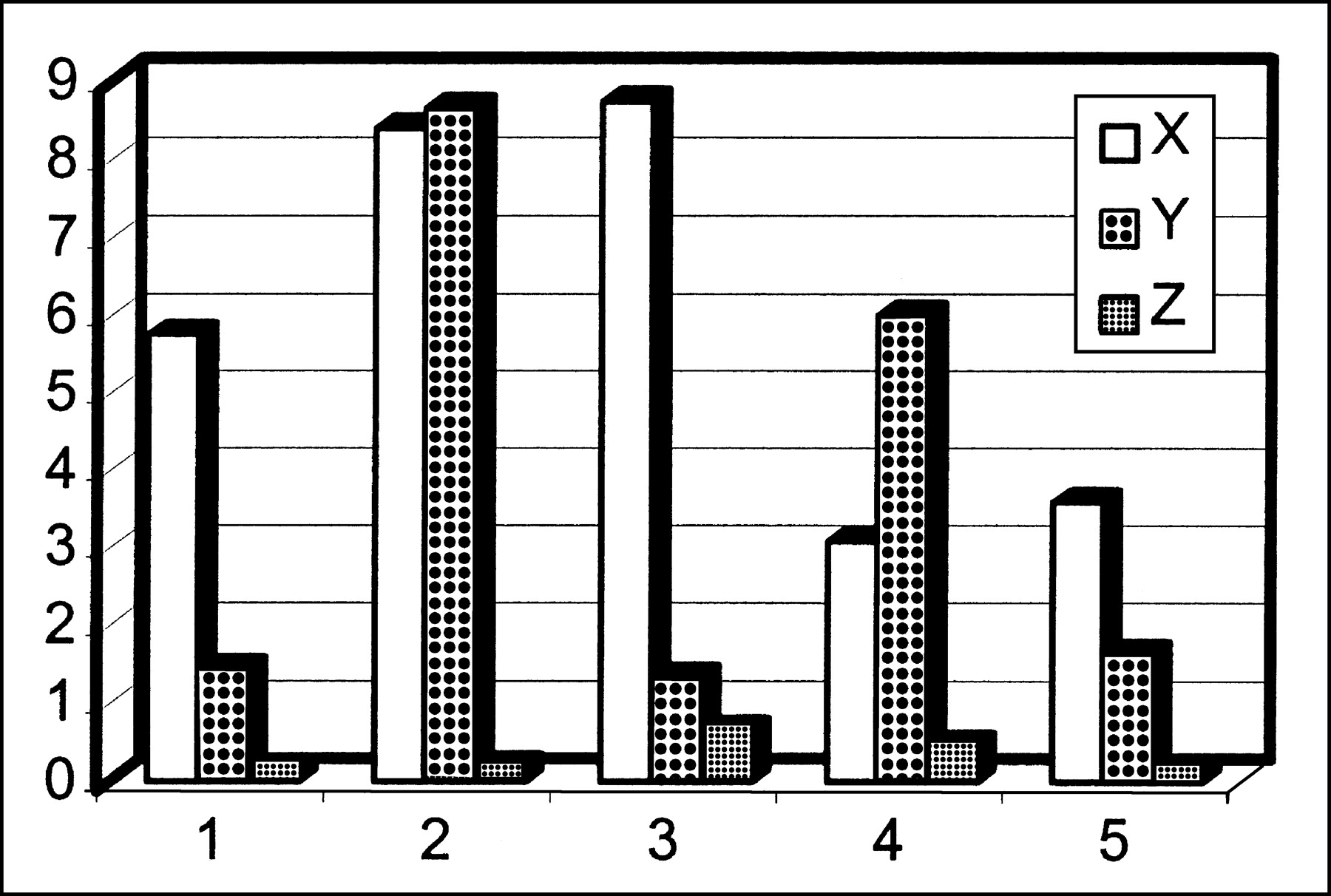
- FIGURE 7.
Maximum deviations of lesion centroids in x-, y-, and z-directions.


Tables
Patient no. Sex Age (y) Tumor site Tumor histology 1 F 59 Left hilum NSCLC 2 F 83 Left lower lobe NSCLC 3 F 40 Left peribronchial/mediastinal NSCLC 4 M 69 Right upper lobe NSCLC 5 F 69 Left hilar Squamous cell NSCLC = non-small cell lung cancer.
- TABLE 2
Summary of Percentage Reduction in Lesion Volume and Percentage Increase in SUVmax
Patient no. Volume (%) SUVmax (%) 1 27.65633 159.1610 2 34.59119 70.4774 3 20.19324 7.4646 4 13.79567 20.8655 5 27.93120 56.5025
In this issue


Jump to section
Related Articles
Cited By...
- Effectiveness of Data-Driven Gating FDG PET/CT for Abdominal Region
- Evaluation of Data-Driven Respiration Gating in Continuous Bed Motion in Lung Lesions
- Gated 18F-FDG PET/CT of the Lung Using a Respiratory Spirometric Gating Device: A Feasibility Study
- Respiratory Motion Compensation for PET/CT with Motion Information Derived from Matched Attenuation-Corrected Gated PET Data
- Repeatability of 18F-FDG PET/CT in Advanced Non-Small Cell Lung Cancer: Prospective Assessment in 2 Multicenter Trials
- Practical PET Respiratory Motion Correction in Clinical PET/MR
- Motion Correction Strategies for Integrated PET/MR
- Interobserver Agreement of Qualitative Analysis and Tumor Delineation of 18F-Fluoromisonidazole and 3'-Deoxy-3'-18F-Fluorothymidine PET Images in Lung Cancer
- Respiratory Motion Correction in Oncologic PET Using T1-Weighted MR Imaging on a Simultaneous Whole-Body PET/MR System
- MRI-Based Nonrigid Motion Correction in Simultaneous PET/MRI
- 4'-[Methyl-11C]-Thiothymidine PET/CT for Proliferation Imaging in Non-Small Cell Lung Cancer
- Value of 4-Dimensional 18F-FDG PET/CT in the Classification of Pulmonary Lesions
- Measurement of Regional Specific Lung Volume Change Using Respiratory-Gated PET of Inhaled 13N-Nitrogen
- Implementation of an Automated Respiratory Amplitude Gating Technique for PET/CT: Clinical Evaluation
- Phased Versus Midventilation Attenuation-Corrected Respiration-Correlated PET for Patients with Non-Small Cell Lung Cancer
- Nonrigid Versus Rigid Registration of Thoracic 18F-FDG PET and CT in Patients with Lung Cancer: An Intraindividual Comparison of Different Breathing Maneuvers
- Deep-Inspiration Breath-Hold PET/CT of Lung Cancer: Maximum Standardized Uptake Value Analysis of 108 Patients
- Use of H215O-PET and DCE-MRI to Measure Tumor Blood Flow
- Quantitative PET Comparing Gated with Nongated Acquisitions Using a NEMA Phantom with Respiratory-Simulated Motion
- Deep-Inspiration Breath-Hold PET/CT: Clinical Findings with a New Technique for Detection and Characterization of Thoracic Lesions
- Postacquisition Detection of Tumor Motion in the Lung and Upper Abdomen Using List-Mode PET Data: A Feasibility Study
- Deep-Inspiration Breath-Hold PET/CT of the Thorax
- Tissue Characterization of Solitary Pulmonary Nodule: Comparative Study Between Helical Dynamic CT and Integrated PET/CT
- Comparison of Different Methods for Delineation of 18F-FDG PET-Positive Tissue for Target Volume Definition in Radiotherapy of Patients with Non-Small Cell Lung Cancer
- On the Use of Positioning Aids to Reduce Misregistration in the Head and Neck in Whole-Body PET/CT Studies
- The CT Motion Quantitation of Lung Lesions and Its Impact on PET-Measured SUVs
- Respiratory Gating for 3-Dimensional PET of the Thorax: Feasibility and Initial Results
- Implementing Biologic Target Volumes in Radiation Treatment Planning for Non-Small Cell Lung Cancer
- Software Approach to Merging Molecular with Anatomic Information
- Reduction of Respiratory Motion Artifacts in PET Imaging of Lung Cancer by Respiratory Correlated Dynamic PET: Methodology and Comparison with Respiratory Gated PET
- Automated 3-Dimensional Registration of Stand-Alone 18F-FDG Whole-Body PET with CT