


Abstract
The aim of this study was to compare cardiac volume and function assessment using PET with the reference technique of cardiovascular magnetic resonance (CMR). Methods: Left ventricular (LV) and right ventricular (RV) end-diastolic volume (EDV), end-systolic volume (ESV), stroke volume (SV), and ejection fractions (EF) were measured in 9 patients using both CMR and PET with inhaled C15O. Results: Correlation between the techniques was generally reasonable (r values ranged from 0.63 to 0.99). Best agreement was seen for ESV (LV and RV). With PET, there was a tendency to underestimate LV EF and EDV, and RV EDV and SV. Agreement was worst for LV SV. Percentage difference between CMR and PET measurements ranged from −2% to 15%; Bland-Altman limits of agreement ranged from 24% to 75%. Conclusion: Although small systematic differences exist, the agreement between PET and CMR suggests useful information regarding function, and volumes may be obtained from a standard PET protocol.
The prognostic and therapeutic implications of cardiac volumes and function are well established in the study of cardiac disease (1). Cardiovascular magnetic resonance (CMR) provides accurate, reproducible assessment of cardiac function through acquisition of tomographic images of high spatial and temporal resolution, free of exposure to ionizing radiation. CMR is now the recognized reference standard for determining parameters of left ventricular (LV) and right ventricular (RV) function. Information on ventricular function may also be obtained from cardiac PET (2), although this technique is more established for imaging and quantification of myocardial metabolism, perfusion, and receptor density. However, if a patient were undergoing a PET examination, it would be useful to simultaneously obtain ventricular function data that were comparable with data that would be obtained from CMR.
Therefore, the aim of this pilot study was to compare cardiac volumes and function assessment using PET blood-pool imaging and electrocardiographic (ECG) gating with CMR.
MATERIALS AND METHODS
The Research and Ethics Committees of participating hospitals approved the study, and patients gave informed written consent. Procedures were performed in accordance with local guidelines. Radiation exposure was approved by the U.K. Administration of Radioactive Substances Advisory Committee.
CMR Methodology
Nine patients were imaged with a Picker Edge 1.5-T scanner (Picker, Cleveland, OH), using the body coil, ECG triggering, and parameters previously described (3). Briefly, the cardiac short axis (SA) was determined from 3 scout images of the LV: the transverse, vertical long axis, and breath-hold diastolic horizontal long axis. The basal SA slice was positioned just forward of the atrioventricular ring, and all subsequent breath-hold cines were acquired in 1-cm steps toward the apex. A breath-hold segmented gradient-echo fast low-angle shot sequence was used for each of the contiguous SA slices encompassing the LV. Image analysis was performed on a personal computer using in-house developed software (CMRtools, Imperial College, London, U.K.). The reproducibility of this technique in our center has been previously published: The interstudy percentage variability was 2.5% for end-diastolic volume (EDV), 3.1% for end-systolic volume (ESV), and 4.8% for ejection fraction (EF) (3).
PET Methodology
PET studies were performed on the ECAT EXACT 3-dimensional positron tomograph (CTI, Knoxville, TN). A transmission scan was obtained using a 150-MBq 137Cs point source. The emission data were acquired using inhaled C15O (half-life, 2.06 min). Labeling of red blood cells occurs through the formation of carboxyhemoglobin, which remains limited to the intravascular space. The gas (1.5 MBq/mL) was delivered at a rate of 500 mL/min through a face mask for 4 min (effective dose equivalent, 1.15 mSv). Data acquisition commenced immediately and continued for 11 min. This scanner allows data acquisition in list mode synchronized with the ECG R wave, and thus retrospective (or after acquisition) off-line rebinning of data into datasets corresponding to time frames per R−R interval (gates). Eight gates per cardiac cycle were used. The data were corrected for attenuation and scatter and were reconstructed by dedicated array processors and a reprojection reconstruction algorithm using a Hann filter (cutoff at the Nyquist frequency).
A threshold-based edge detection algorithm was used to calculate LV and RV volumes (4). The gated images were averaged together to provide a static blood-pool image. This static image was used to define SA reslice parameters. Both the static and gated images were then resliced to the SA view. The static image was used to identify those planes contributing to the RV and LV; any planes inferior or superior to the ventricles were discarded from the gated image. The atrioventricular valve plane was identified as the plane where the gap between the LV and RV disappeared. LV and RV analysis was performed individually. The maximum value for the image was determined by applying 4 circular regions of interest to 4 image planes containing the ventricle being studied. The voxels within the 4 regions were treated as a volume of interest (VOI). The maximum voxel value of the VOI divided by 2 (half maximum value) was used as the threshold, which defined the cutoff value of the ventricular cavity boundary. Signal contribution of the myocardial vasculature was assumed to be 10% of the blood pool, and the thresholds were scaled accordingly: threshold = half maximum + (half maximum × background fraction). The threshold values obtained were used with an interactive edge detection program to automatically determine the ventricular chamber boundary for each gate. This area multiplied by the plane thickness results in a plane volume. EDV and ESV were calculated by summing all the plane volumes at end-diastole and end-systole, respectively. Stroke volume (SV) = EDV - ESV. EF = SV/EDV. Interobserver variability is 1.1% for EDV and 1.8% for ESV using this technique (4).
RESULTS
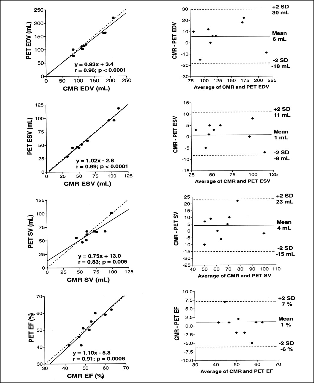
Values for LV and RV EDV, ESV, SV, and EF are shown in Table 1 for both CMR and PET. A small but significant difference was found between CMR and PET values for LV EF, with borderline differences for LV SV and LV EDV (Table 2). Correlation between the techniques was generally reasonable for all parameters measured (0.63−0.99). The scatterplots suggested agreement between the techniques for both LV (Fig. 1) and RV (Fig. 2) parameters. Best agreement was seen for ESV. With PET, the LV EF and EDV and the RV EDV and SV tended to be underestimated. Agreement was worst for LV SV. These findings were confirmed by the Bland-Altman plots and limits of agreement (Table 2; Figs. 1 and 2).

Scatterplots and Bland-Altman plots for left ventricular parameters measured with CMR and PET. Line of unity (dashed line) and linear regression line (solid line) with equation, r, and probability values are shown on each scatterplot.

Scatterplots and Bland-Altman plots for right ventricular parameters measured with CMR and PET. Line of unity (dashed line) and linear regression line (solid line) with equation, r, and probability values are shown on each scatterplot.
Mean Values (±SD) for Left and Right Ventricular Volumes and Function
Differences (CMR vs. PET), Correlation, and Bland-Altman Limits for All Parameters
DISCUSSION
Agreement was worst for SV, reflecting the fact that it is calculated from EDV and ESV. With PET, there was a trend to underestimate the EDV for both the LV and the RV, overestimate the ESV for the LV, and underestimate the ESV for the RV. Several factors may have accounted for these differences. Basal-plane identification and through-plane motion affect both techniques, causing discrepancy. Only 8 gates were used for the PET data, whereas on average 12 frames were used with CMR. Total count statistics dictated the use of 8 gates in this pilot study, but future work may use 12 to 16 gates. Summation of volumes with fewer frames causes overestimation of the LV ESV. Using fewer gates results in reconstruction of the EDV image fractionally later during the cardiac cycle with PET than with CMR, leading to a smaller measured EDV with PET. This effect has also been documented in gated SPECT, for which an underestimation of the EF derived from 8-gate studies compared with values measured on 16-gate studies has been reported (5). This systematic error may be addressed through reconstruction of the PET data with a shorter gate interval. EF is useful because it gives the clinician an appreciation of overall ventricular function. Both LV and RV EF were reasonably correlated with PET and CMR, despite a small but significant difference between the 2 techniques for LV EF. This reflects the fact that both techniques are freely tomographic and not hindered by geometric assumptions made in other techniques such as ECG.
Limitations of this pilot study include the fact that both scans were not completed on the same day for all patients (within 1 ± 2 d); the restriction of fluid intake before PET studies, which affects ventricular filling and hence volumes and function; and the relatively symmetric LV geometry in our patient population. Future studies require a larger number of patients with a range of volumes and function, including ventricles distorted by myocardial infarction.
CONCLUSION
Acceptable agreement was shown between ventricular function and volume measurements using gated cardiac PET and CMR. Therefore, useful information may be obtained regarding function and volumes as part of a standard PET protocol.
Footnotes
Received Sep. 4, 2001; revision accepted Jan. 22, 2002.
For correspondence or reprints contact: Dudley J. Pennell, MD, Cardiovascular MR Unit, Royal Brompton Hospital, Sydney St., London SW3 6NP, United Kingdom.
E-mail: d.pennell{at}ic.ac.uk
In this issue


Jump to section
Related Articles
Cited By...
- Measurement of LV Volumes and Function Using Oxygen-15 Water-Gated PET and Comparison With CMR Imaging
- Automatic Extraction of Myocardial Mass and Volume Using Parametric Images from Dynamic Nongated PET
- Micro-Positron Emission Tomography in the Evaluation of Trypanosoma cruzi-Induced Heart Disease: Comparison with Other Modalities
- Gated Cardiac 13N-NH3 PET for Assessment of Left Ventricular Volumes, Mass, and Ejection Fraction: Comparison with Electrocardiography-Gated 18F-FDG PET
- Monitoring Left Ventricular Dilation in Mice with PET
- Comparison of Gated PET with MRI for Evaluation of Left Ventricular Function in Patients with Coronary Artery Disease
- Validation of QGS and 4D-MSPECT for Quantification of Left Ventricular Volumes and Ejection Fraction from Gated 18F-FDG PET: Comparison with Cardiac MRI
- Model-Based Analysis of Electrocardiography-Gated Cardiac 18F-FDG PET Images to Assess Left Ventricular Geometry and Contractile Function
- Quantitative Gated PET for the Assessment of Left Ventricular Function in Small Animals
- Gated PET and Ventricular Volume