


Article Figures & Data
Figures
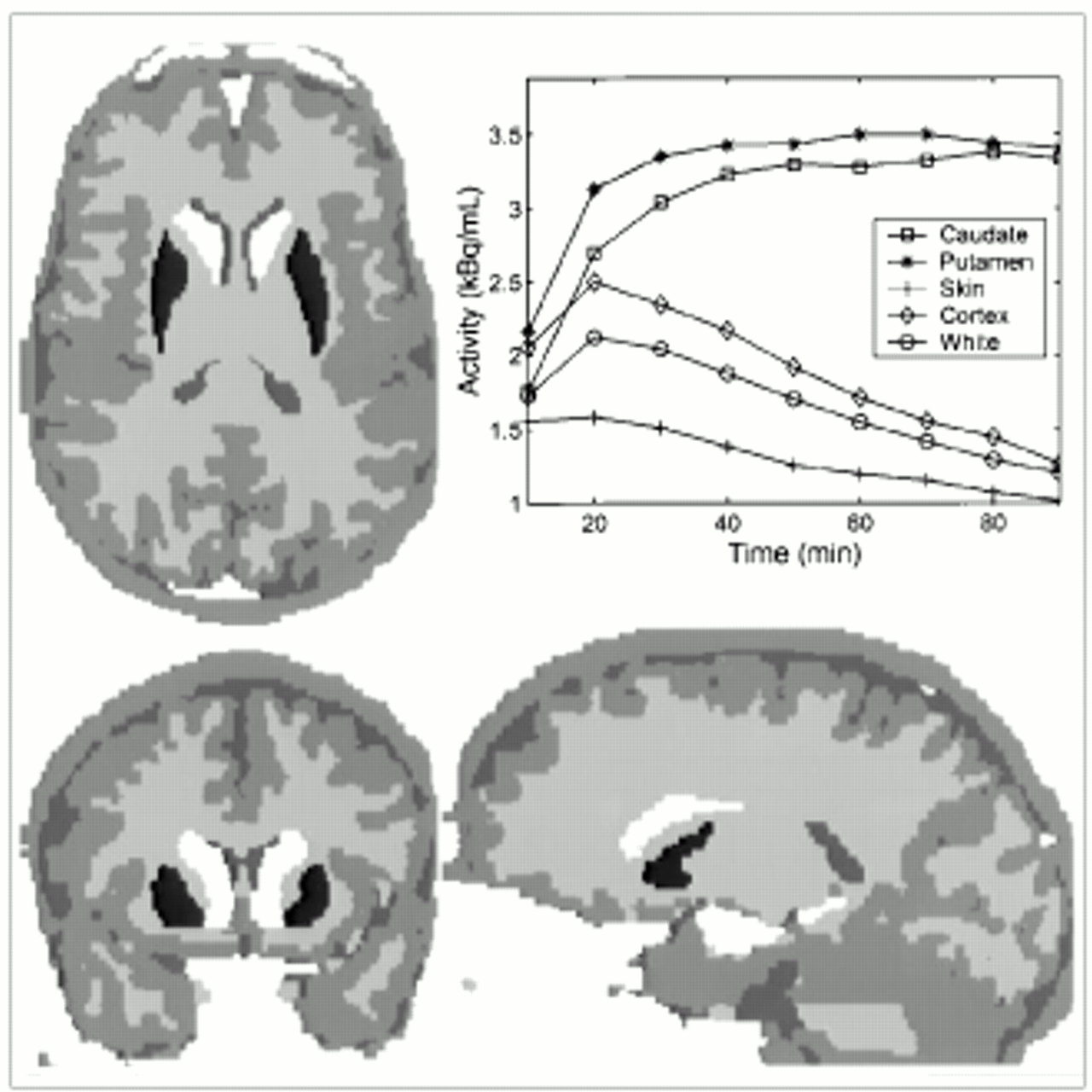
- FIGURE 1.
The 7 compartments considered in Monte Carlo simulations—skin/muscle, gray matter, white matter, right caudate, left caudate, right putamen, and left putamen—shown with arbitrary gray levels in axial, coronal, and sagittal views. Plot shows TACs used for each structure in 18F-l-dopa Monte Carlo simulation.
- FIGURE 2.
Flow chart illustrating building of family of missegmented right caudate. (A) Original caudate. On left, 1-voxel eroded core is shown in light gray. On right, 1-voxel-thick internal layer is shown in gray and 1-voxel-thick external layer, from original shape, is shown in dark gray. (B) Addition of one tile after another. (C) Tessellated caudate.
- FIGURE 3.
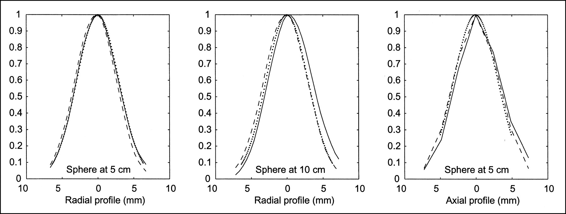
Profiles for 3-mm-diameter sphere at radial distances of 5 and 10 cm from center of FOV, including measured data (solid line), RSF for image GTM (gaussian) approach (dotted line), and RSF for sinogram GTM (simulation) approach (dashed line).
- FIGURE 4.
Plot of ARC with time that indexes PET dynamic simulations. Regional ARCs shown are uncorrected (solid line), sinogram GTM PVE corrected (dashed line), and image GTM PVE corrected (dotted line). First row displays curves on left caudate and second row on right putamen. First column displays curves from 18F-dopa simulation and second column from 11C-raclopride simulation.
- FIGURE 5.
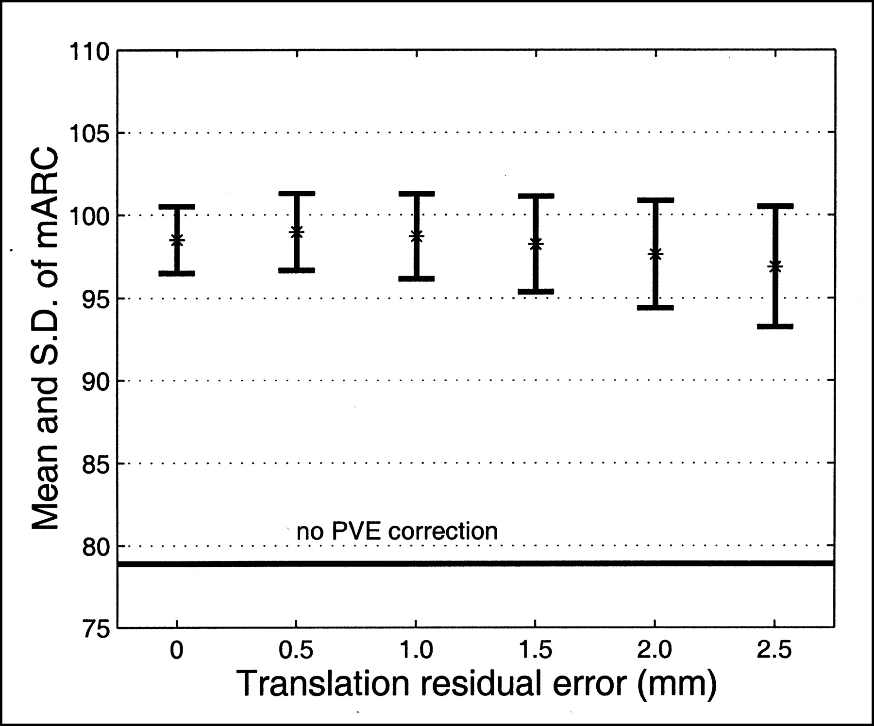
Robustness of GTM method with respect to translation error between PET and MRI: example of right caudate structure with 18F-dopa simulation study. Evolution of mean (%) and SD of mARC value is plotted against amplitude of translation residual error. One mARC value is obtained for each randomly generated translation error (21 drawings per amplitude).
- FIGURE 6.
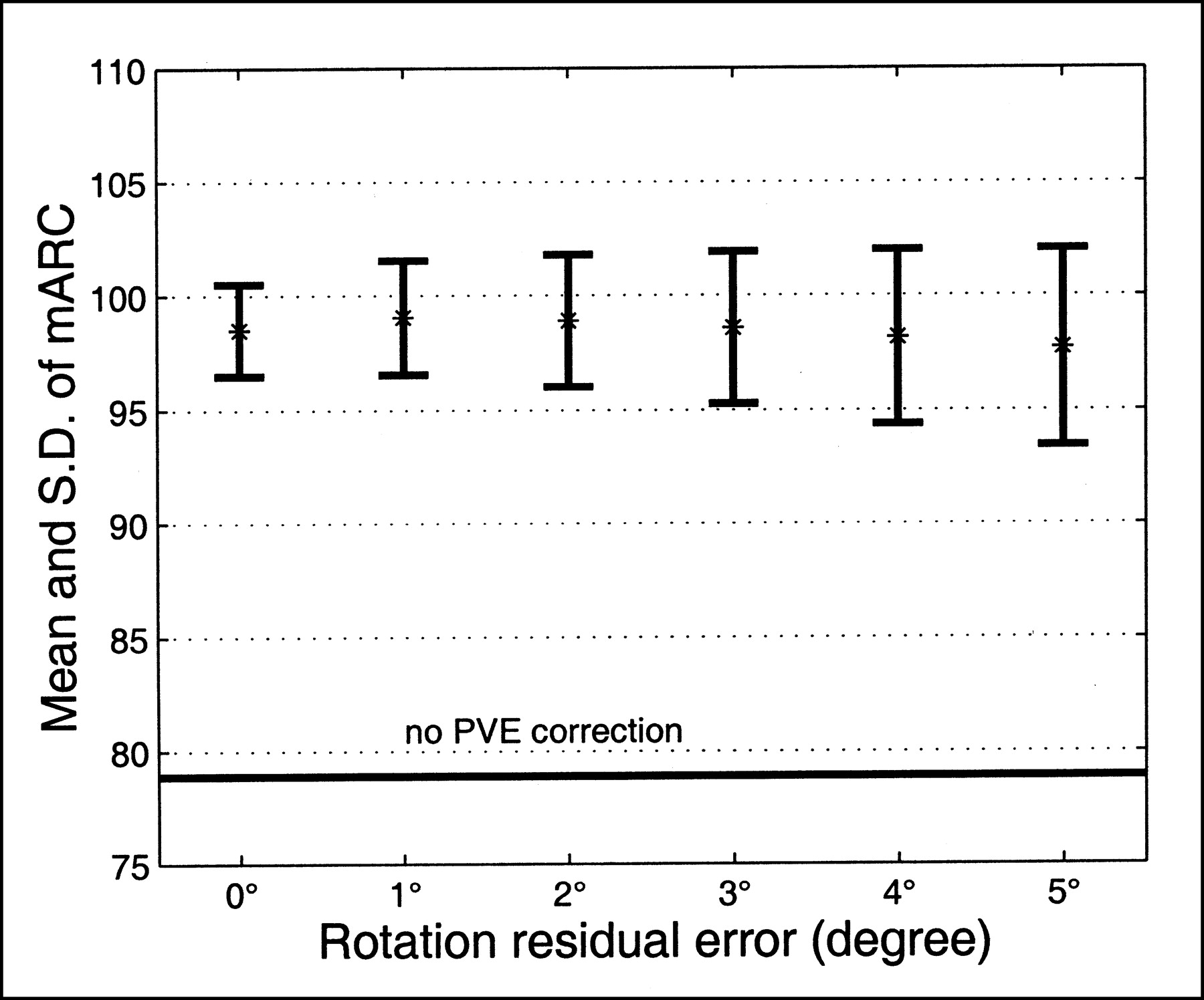
Robustness of GTM method with respect to rotation error between PET and MRI: example of right caudate structure with 18F-dopa simulation study. Evolution of mean (%) and SD of mARC value is plotted against amplitude of rotation residual error. One mARC value is obtained for each randomly generated rotation error (21 drawings per amplitude).
- FIGURE 7.
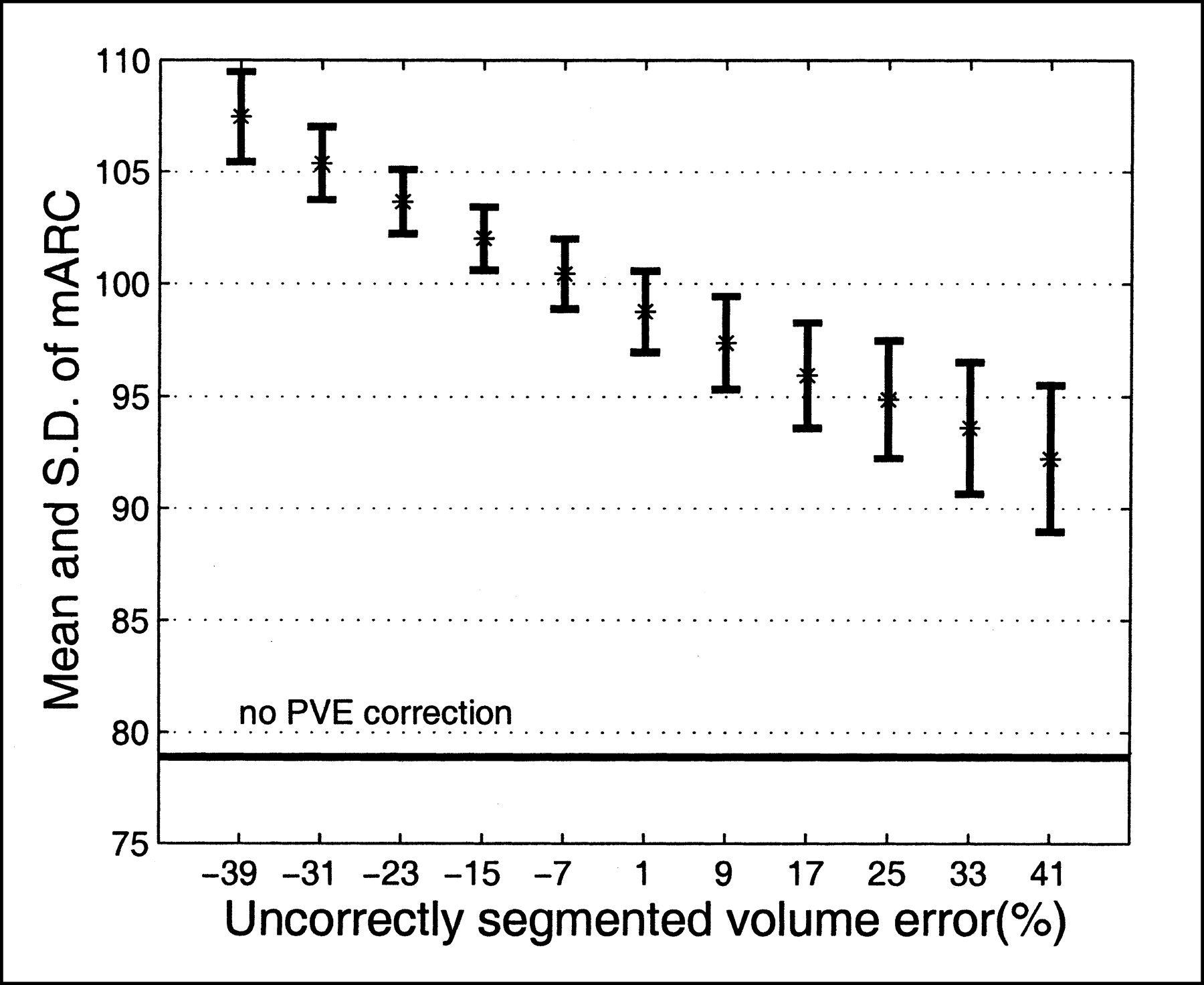
Robustness of GTM method with respect to segmentation error: example of right caudate structure with 18F-dopa simulation study. Evolution of mean (%) and SD of mARC value is plotted against amplitude of segmentation residual error (relative error size). One mARC value is obtained for each randomly generated segmentation error (30 drawings per amplitude).


Tables
- TABLE 1
Full Widths of Profile Obtained for 3-mm-Diameter Sphere at Radial Distance of 5 cm From Center of FOV
Approach Radial Tangential Axial FWHM FWTM FWHM FWTM FWHM FWTM Measured 6.83 12.73 6.73 12.76 7.38 14.84 Monte Carlo 6.77 12.48 6.63 12.20 6.95 13.23 Sinogram 6.77 12.25 6.63 12.10 7.56 13.85 Image 6.85 12.52 6.85 12.52 7.22 n.m. Units are millimeters
FWTM = full width at one tenth of maximum; n.m. = not measured.
Skin/muscle Gray matter White matter Right caudate Left caudate Right putamen Left putamen Skin/muscle 72.0 (73.6) 0.7 (0.5) 0.0 (0.0) 0.0 (0.0) 0.0 (0.0) 0.0 (0.0) 0.0 (0.0) Gray matter 0.6 (0.4) 70.4 (70.4) 17.7 (17.7) 0.0 (0.0) 0.0 (0.0) 0.0 (0.0) 0.0 (0.0) White matter 0.0 (0.0) 18.2 (18.4) 78.0 (77.4) 0.4 (0.4) 0.4 (0.4) 0.4 (0.4) 0.4 (0.4) Right caudate 0.0 (0.0) 1.5 (1.7) 28.2 (29.1) 61.6 (59.9) 0.0 (0.0) 1.4 (1.5) 0.0 (0.0) Left caudate 0.0 (0.0) 1.7 (1.9) 28.2 (29.2) 0.0 (0.0) 59.9 (58.4) 0.0 (0.0) 1.0 (1.2) Right putamen 0.0 (0.0) 1.8 (2.0) 31.7 (31.9) 0.7 (0.7) 0.0 (0.0) 66.3 (65.3) 0.0 (0.0) Left putamen 0.0 (0.0) 1.6 (1.8) 32.3 (33.2) 0.0 (0.0) 0.5 (0.6) 0.0 (0.0) 65.9 (64.5) Lightface indicates coefficients (%) estimated with sinogram implementation; boldface indicates coefficients (%) estimated with image implementation.
Compartment Approach 18F-Dopa simulation 11C-Raclopride simulation Skin/muscle Uncorrected 69.86 ± 0.11 73.63 ± 1.89 Sinogram 96.70 ± 0.11 97.92 ± 1.69 Image 95.78 ± 0.08 98.20 ± 2.09 Gray matter Uncorrected 82.58 ± 0.64 81.24 ± 5.00 Sinogram 95.45 ± 0.22 96.16 ± 0.59 Image 97.13 ± 0.13 97.75 ± 0.36 White matter Uncorrected 100.95 ± 0.50 117.52 ± 16.91 Sinogram 101.19 ± 0.23 104.02 ± 3.41 Image 100.75 ± 0.22 103.25 ± 2.75 Right caudate Uncorrected 76.52 ± 8.50 68.78 ± 1.90 Sinogram 97.00 ± 1.48 96.89 ± 1.36 Image 98.51 ± 1.93 97.49 ± 1.71 Left caudate Uncorrected 73.63 ± 7.56 65.75 ± 1.03 Sinogram 94.14 ± 1.71 93.67 ± 2.31 Image 95.35 ± 1.83 95.16 ± 1.75 Right putamen Uncorrected 79.02 ± 6.00 73.06 ± 2.04 Sinogram 96.15 ± 1.02 94.91 ± 1.46 Image 96.43 ± 1.02 95.67 ± 1.61 Left putamen Uncorrected 77.70 ± 6.20 72.39 ± 2.68 Sinogram 95.77 ± 1.42 96.19 ± 2.27 Image 95.34 ± 1.01 95.76 ± 2.74 Values are percentages (mean ± SD).
Compartment Approach Half-life* Activity† ARC‡ (mean ± SD) White matter Uncorrected 95.26 88.31 105.00 ± 4.57 Sinogram 99.39 98.81 103.55 ± 0.60 Image 99.75 96.60 102.43 ± 0.24 Right caudate Uncorrected 87.03 74.56 67.11 ± 1.79 Sinogram 93.28 90.18 104.37 ± 1.34 Image 95.30 98.74 95.35 ± 0.84 Left caudate Uncorrected 86.55 72.02 64.53 ± 1.79 Sinogram 92.70 94.35 99.92 ± 1.41 Image 96.18 97.03 94.34 ± 0.67 Right putamen Uncorrected 86.57 73.78 66.13 ± 1.83 Sinogram 91.85 96.37 97.33 ± 1.54 Image 94.89 96.29 92.69 ± 0.89 Left putamen Uncorrected 85.22 75.29 66.62 ± 2.07 Sinogram 88.97 95.53 95.71 ± 2.12 Image 93.02 98.30 93.22 ± 1.25
In this issue


Jump to section
Related Articles
Cited By...
- Lower Brain Glucose Metabolism in Normal Ageing is Predominantly Frontal and Temporal: A Systematic Review and Pooled Effect Size and Activation Likelihood Estimates Meta-Analyses
- An Anthropomorphic Phantom Study of Brain Dopamine Transporter SPECT Images Obtained Using Different SPECT/CT Devices and Collimators
- New Approach to Quantification of Molecularly Targeted Radiotracer Uptake from Hybrid Cardiac SPECT/CT: Methodology and Validation
- Regional variability of imaging biomarkers in autosomal dominant Alzheimer's disease
- Voxel-Based Analysis of Asymmetry Index Maps Increases the Specificity of 18F-MPPF PET Abnormalities for Localizing the Epileptogenic Zone in Temporal Lobe Epilepsies
- Muscarinic Receptor Upregulation in Patients With Myocardial Infarction: A New Paradigm
- Age-associated leukoaraiosis and cortical cholinergic deafferentation
- Design and Implementation of an Automated Partial Volume Correction in PET: Application to Dopamine Receptor Quantification in the Normal Human Striatum
- Estimation of the {beta}+ Dose to the Embryo Resulting from 18F-FDG Administration During Early Pregnancy
- Up-regulation of hippocampal serotonin metabolism in mild cognitive impairment
- Partial-Volume Effect in PET Tumor Imaging
- Partial-Volume Correction in PET: Validation of an Iterative Postreconstruction Method with Phantom and Patient Data
- Integrated Software for the Analysis of Brain PET/SPECT Studies with Partial-Volume-Effect Correction