


Abstract
The aim of this study was to determine the human biodistribution and radiation dosimetry of 99mTc-RP527, a promising radioligand for the visualization of gastrin-releasing peptide (GRP) receptor–expressing human malignancies. Methods: Whole-body scans were obtained up to 48 h after intravenous injection of 555 MBq 99mTc-RP527 in each of 6 subjects. Blood samples were taken at various times up to 48 h after injection. Urine was collected up to 48 h after injection for calculation of renal clearance and whole-body clearance. Time–activity curves were generated for the thyroid, heart, breasts in women, testes in men, and liver by fitting the organ-specific geometric mean counts, obtained from regions of interest, on the respective images as a function of the time after injection. The MIRD formulation was applied to calculate the absorbed radiation dose for various organs. Results: The serial whole-body images showed rapid hepatobiliary excretion, resulting in low background and potentially high-contrast imaging of the thoracic region. Imaging of abdominal tumors may prove problematic, however, because of the extensive bowel activity. 99mTc-RP527 was predominantly cleared by the kidneys and to a lesser extent by the gastrointestinal tract. The mean excretion in the urine (±SD) at 48 h after injection was 58.3 ± 5.4 percentage of the injected activity corrected for decay to the time of injection. The highest absorbed doses were received by the excretory organs (i.e., the urinary bladder and gallbladder wall). The average effective dose of 99mTc-RP527 was estimated to be 0.0095 mSv/MBq. Conclusion: The biodistribution of 99mTc-RP527 revealed low lung, myocardial, and liver uptake, which allowed early imaging of the supradiaphragmatic region with a favorable dosimetry (including effective dose) for administered activities required for SPECT imaging.
Gastrin-releasing peptide (GRP) belongs to the family of bombesin-like peptides (BLPs) that includes the amphibian peptide bombesin as well as the mammalian counterpart GRP and neuromedin B (1). Aside from their physiologic role (e.g., stimulation of secretion of gastrin and pancreatic enzymes and gallbladder contraction) (2,3), BLPs have been shown to play a role in various tumor models and human cancer (4–13).
In humans, both GRP and GRP antagonists mediate their action through the membrane-bound G-protein–coupled GRP receptor (GRP-R) (14). GRP receptors have been detected in various types of human carcinoma (15–17). The presence or absence of receptors on tumor cells, including GRP-R, as well as their level of expression have been correlated with cancer prognosis in several settings (18–22).
In analogy to the diagnosis of somatostatin receptor–expressing tumors in vivo with somatostatin receptor scintigraphy, in vivo GRP-R scintigraphy may be a potentially valuable tool to visualize and semiquantitate tumor GRP expression in a noninvasive way. This may allow prediction of therapy responsiveness to GRP antagonists and in vivo prognosis stratification. 99mTc-RP527 is a targeting peptide derived from bombesin linked at its N-terminus through a linker group to a peptide sequence that chelates 99mTc. 99mTc-RP527 binds GRP-Rs with similar affinity to bombesin and is taken up by prostate, pancreas, and breast tumor tissue in animal models as well as in humans in vivo (23,24). This article reports on its biodistribution and dosimetry in humans.
MATERIALS AND METHODS
Radiopharmaceutical Synthesis
99mTc-RP527 labeling was performed using a kit formulation; the reaction mixture contains 0.1 mL stannous chloride (2 mmol/L), 0.1 mL sodium gluconate (60 mmol/L), 1,850–2,035 MBq 99mTcO4 in 0.3 mL sodium chloride (0.9%), 0.5 mL sodium chloride (0.9%), and 100 μg RP527. After 35 min in a boiling water bath, the reaction mixture was allowed to cool to room temperature and injected on a high-performance liquid chromatography system using a gradient of ethanol, water, and acetic acid. The radiolabeled peptide was collected at 45 min, and the collected eluent was diluted with 10 mL sodium chloride (0.9%). The overall yield of the radiosynthesis was approximately 30% with a radiochemical purity of ≥90% and a specific activity of ≥4.32 TBq/Gmmol.
Subjects
This study was approved by the Medical Ethics Committee of the University Hospital Ghent and performed according to good clinical practice. Six individuals (3 men, 3 women; mean age, 46 y; age range, 24–73 y) were included in the study. All subjects gave their written informed consent for participation in the study. All volunteers were free from illness on the basis of screening by medical history, physical examination, serum chemical analyses, complete blood cell count, and urinalysis. Because of safety concerns, all chemical analyses were repeated during the experiment and the results were compared to their baseline (screening) values.
Imaging
Subjects were positioned supine with their arms alongside their body. Whole-body images were obtained using a triple-head gamma camera (Irix; Picker International, Cleveland Heights, OH) equipped with low-energy, high-resolution, parallel-hole collimators. The energy peak was centered at 140 keV with a 15% window. Whole-body planar images were acquired at approximately 30 min and 1, 3, 6, 24, and 48 h after injection. Acquisition was performed simultaneously in anterior and posterior positions with a scan speed of 11.4 cm/min. The matrix size was 256 × 1,024 pixels.
Urine Sampling
For all subjects, all voided urine from the time of injection until 48 h after injection was collected. The subjects were requested to collect urine before each emission scan and at home ad libitum. For each voiding, the urine was collected in a separate container, and the volume and time of voiding were recorded. For each void time, two 1-mL urine aliquots were sampled, and radioactivity was counted in a NaI(T1) counter (Cobra; Packard Instrument Co., Downers Grove, IL) after the counting efficiency of the system had been determined. The amount of radioactivity in the urine at each void time was expressed as percentage of the injected activity corrected for decay to the time of injection (%IA) of 99mTc-RP527.
Blood Sampling
Blood samples were taken at 15 s, 30 s, 45 s, 1 min, 1 min 15 s, 1 min 30 s, 2 min, 5 min, 7 min 30 s, 10 min, 12 min, 15 min, 20 min, 30 min, 55 min, 2 h, 4 h, 6 h, 24 h, and 48 h after injection of the tracer. Duplicate 1-mL aliquots from each blood sample were assayed for 99mTc radioactivity as described above. Total blood volume and, consequently, activity were calculated using a total blood volume based on body weight and height (25) and expressed as %IA.
Dosimetry
Image Analysis.
For quantification of radioactivity uptake after injection of 99mTc-RP527, regions of interest (ROIs) over the total body and organs of interest were drawn manually on the earliest images, and the shapes and sizes (i.e., number of pixels) were kept constant over all subsequent images. Correction for background counts was performed using a region over the shoulder and subtracting the mean counts per pixel in the background ROI from the mean counts per pixel in each organ’s ROI to yield net counts in each organ. For each ROI (i.e., each organ), the geometric mean, corrected for physical decay, of total anterior and posterior counts was calculated. The total-body geometric mean activity calculated on the first image (1 h after injection) was taken as the total injected activity, considering that no urine was excreted before the first whole-body scan. The activity in the total body and different organs was expressed as %IA calculated by the following equation: (geometric mean net counts in organ or total body)/(geometric mean counts in first total body) × 100.
Dosimetric Calculations.
For each individual, time–activity curves were generated for the thyroid, heart, breasts in women, testes in men, liver, and whole body. Using in-house curve-fitting software, time–activity curves were generated for these organs by biexponential fits. Two routes of excretion were considered: urinary and fecal excretion. The fecal excretion was predicted using data from the source organs, the urinary excretion, and the material excreted by the liver. Of the material excreted by the liver, 30% was assumed to flow directly in the small intestine. Activity in the small intestine was assumed to follow the gastrointestinal kinetic model in International Commission on Radiobiological Protection Publication 30 (26). Activity in the intestines was assumed to pass through the various segments at standard rates, with mean transit times of 4 h for the small intestine, 13 h for the upper large intestine, and 24 h for the lower large intestine. The transfer rate coefficient from the gallbladder to the small intestine was taken to be 1.8 h−1, derived by taking the mean of average emptying rates reported by others (27).
Source organ residence times were determined from analytic integration of the time–activity curves. Residence times were then used to determine target organ radiation doses based on the MIRD methodology (28) for the normal adult (29) using the MIRDOSE software package (30).
RESULTS
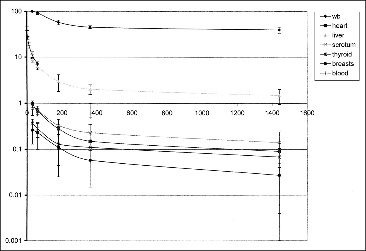
After injection of approximately 555 MBq 99mTc-RP527 (maximum, 3 ng/kg per subject), no short-term adverse or subjective effects were noted in any of the subjects. Their vital signs remained stable throughout the experiment. Moreover, no meaningful changes were observed in any of the clinical laboratory assays performed on the blood and urine specimens obtained at 1, 6, 24, and 48 h after administration of the radioligand when compared with baseline findings. Figure 1 presents whole-body images of a male and female subject showing the biodistribution of radioactivity on injection of 99mTc-RP527 at different time points after injection. The whole-body images obtained between 30 min and 24 h after injection show most of the activity distributed in the bladder, liver, gallbladder, and intestines, reflecting the known urinary and hepatobiliary excretion of peptides. Although the kidneys were not visualized in all subjects, there was activity in the urinary bladder in all subjects, indicating prompt excretion through the renal system. Uptake in the brain, myocardium, and lungs was low. There was a diffuse uptake and retention of radioactivity in the normal breast tissue in women as well as in the testes in men. Although images at 24 and 48 h after injection were of low quality (attributed to low counting statistics), these images showed most of the remaining activity distributed in the intestines. Figure 2 presents averaged ROI data, over all subjects (expressed as %IA), of the biodistribution of 99mTc-RP527 in the total body and in various organs at the different points in time. Clearance of radioactivity from blood and organs fitted a biexponential curve. Assays of blood samples showed that the elimination of activity from the blood was initially fast: by 20 min after injection, <20% of the injected dose remained in the blood. The mean calculated half-life was 1.5 min (first exponent).
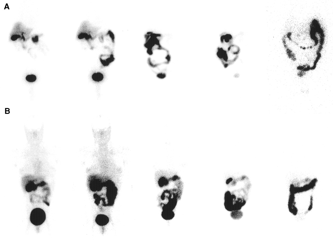
Anterior whole-body images obtained from man (A) and woman (B) at 30 min and 1, 3, 6, and 24 h after injection.

Time–activity curves for total body (wb), various organs, and blood, calculated from direct measurements of counts in organ-specific ROIs or in blood. Physical decay-corrected data (expressed as %IA) are averaged over 6 subjects, except for breasts (n = 3) and testes (n = 3). Error bars represent 1 SD. Units of abscissa and ordinate are time (h) and %IA, respectively. Respective biexponential functions obtained for various ROIs and whole body are whole body, y = 38.16e−0.00001×t + 65.90e−0.00569×t (r = 0.96528); heart, y = 0.18e−0.0005×t + 1.19e−0.0140×t (r = 0.99898); liver, y = 2.36e−0.0003×t + 10.17e−0.0154×t (r = 0.99893); scrotum, y = 0.19e−0.0002×t + 0.77e−0.0083×t (r = 0.99015); thyroid, y = 0.12e−0.0004×t + 0.41e−0.0156×t (r = 0.99881); breasts, y = 0.03e−0.0002×t + 0.28e−0.0069×t (r = 0.99705); and blood, y = 23.57e−0.1842×t + 18.44e−0.0163×t (r = 0.99963).
The average excretion fractions of the 6 subjects predicted for feces and measured for urine are given in Table 1. Most of the administered activity was eliminated by the renal system. The mean cumulative total measured urinary excretion (±SD) of the 6 subjects at 48 h after injection was 58.3 ± 5.4 %IA, whereas the mean predicted %IA excreted in the feces (±SD) was 3.9 ± 0.8 %IA at 48 h after injection.
Excretion Fractions (%IA) for 99mTc-RP527
The residence time was highest for the remainder of the body in all subjects. This value was followed by the urinary bladder and the liver (Table 2).
Residence Times (h) for 99mTc-RP527 for Each Source Organ
The mean radiation dose estimates (±SD) were calculated for each subject independently and then averaged (Table 3). The organs receiving the highest absorbed doses were involved in the excretion of 99mTc-RP527 from the body. On average, the highest dose was received by the urinary bladder (9.63 × 10⁻² ± 1.16 × 10⁻² mGy/MBq) followed by the gallbladder wall (1.69 × 10⁻² ± 4.32 × 10⁻³ mGy/MBq) and the testes (1.23 × 10⁻² ± 2.70 × 10⁻³ mGy/MBq). The estimated mean effective dose (±) for the adult subject was 9.53 × 10⁻³ ± 1.06 × 10⁻³ mSv/MBq.
Radiation Absorbed Dose Estimates (mGy/MBq) for 99mTc-RP527
DISCUSSION
This study shows the biodistribution and kinetics of 99mTc-RP527 in humans. Its dosimetry is comparable with that of other 99mTc-labeled diagnostic radiopharmaceuticals. Moreover, although limited in terms of safety, the results of this study indicate that 99mTc-RP527 is a pharmacologically safe radioligand because it did not produce any demonstrable pharmacologic effects in the short term. The lack of a pharmacologic effect was expected because the dose injected (±3 ng/kg) was only half of the dose of unlabeled bombesin reported to induce side effects (e.g., nausea, vomiting) (31).
In humans, 99mTc-RP527 was cleared rapidly from circulation: by 20 min after injection, <20% of the injected dose remained in the blood. The mean calculated blood clearance half-life of 1.5 min for 99mTc-RP527 is comparable with the 2.2 min for unlabeled bombesin reported in men (32). Low brain, lung, and myocardial uptake and rapid hepatobiliary excretion of 99mTc-RP527 resulted in excellent imaging conditions for the supradiaphragmatic region, even at early time points (1 h) after injection. On the other hand, in addition to a predominant urinary clearance, 99mTc-RP527 was also cleared enterohepatically, resulting in a high accumulation of radioactivity affecting interpretation of the abdominal region.
Of interest is the visualization of the breasts in women and the testes in men. Visualization of the breasts is in keeping with the known bombesin-like immunoreactivity shown in mammary glands of various species (33,34), indicating that bombesin or GRP may be involved in the regulation of mammary cell proliferation and differentiation. Data by Halmos et al. (35) showing a highly positive correlation (r = 0.671; P < 0.005) between the binding capacity of high-affinity (Tyr4)-bombesin binding sites and estrogen receptor levels and between the concentration of low-affinity (Tyr4)-bombesin binding sites and peptide receptor levels (r = 0.541; P < 0.005) in biopsies of human breast carcinoma suggest a possible interaction with other receptor systems.
In primates, a bombesin-like peptide resulting from alternate splicing of the GRP gene has been detected in testis. In vitro, GRP at a concentration of 100 nmol/L, added after ionophore treatment, enhanced significantly (P < 0.05) human sperm motility, capacitation, zona binding, and acrosome reaction, thus supporting a role for BLPs in the regulation of sperm cell proliferation and differentiation (36). In this regard, the visualization of the testes in all 3 male subjects after injection of 99mTc-RP527 suggests the presence of a currently unidentified human testicular BLP receptor. Because NMB-R gene expression is prominent in mouse testis and the BRS-3 receptor has been shown to be expressed in rat testis, it is possible that different subtypes of bombesin receptors mediate the same response in different species (37).
The MIRDOSE software provides a calculation of the effective dose as defined in International Commission on Radiological Protection (ICRP) Publication 60 (38). On the basis of the mean effective dose of 9.5 × 10⁻³ mSv/MBq obtained in this study, patients and volunteers could easily be investigated with 555 MBq 99mTc-RP527, allowing both planar and SPECT imaging. The corresponding effective dose of 5.27 mSv is equal to the reported average effective dose per patient from nuclear medicine procedures in Europe (39) and only half of the 10-mSv upper-limit average effective dose of category IIa of the World Health Organization and category IIb of the ICRP (40). Because the highest dose is received by the bladder wall, frequent voiding will reduce the absorbed dose to the urinary bladder. Given the 1% probability for severe hereditary disorders in the offspring per Sv received by the testis, the associated risk related to injection of 555 MBq 99mTc-RP527 is <1 per 10,000. This risk is low compared with the 1.6% prevalence of naturally occurring genetic disorders (38).
CONCLUSION
The biodistribution of 99mTc-RP527 showed low lung, myocardial, and liver uptake, which allowed early imaging of the supradiaphragmatic region with a dosimetry favorable for clinical SPECT imaging.
Acknowledgments
This work is supported by grant 0035.01 from the National Fund for scientific research.
Footnotes
Received Jan. 31, 2001; revision accepted Jul. 16, 2001.
For correspondence contact: Christophe Van de Wiele, MD, PhD, University Hospital Ghent, De Pintelaan 185, B-9000 Ghent, Belgium.
REFERENCES
In this issue


Jump to section
Related Articles
Cited By...
- Clinical Translation of a Dual Integrin {alpha}v{beta}3- and Gastrin-Releasing Peptide Receptor-Targeting PET Radiotracer, 68Ga-BBN-RGD
- 68Ga-NOTA-Aca-BBN(7-14) PET/CT in Healthy Volunteers and Glioma Patients
- Targeted Radiotherapy of Prostate Cancer with a Gastrin-Releasing Peptide Receptor Antagonist Is Effective as Monotherapy and in Combination with Rapamycin
- Radiopeptide Imaging and Therapy in Europe
- Generation of hyperpolarized substrates by secondary labeling with [1,1-13C] acetic anhydride
- Bombesin Receptor Antagonists May Be Preferable to Agonists for Tumor Targeting
- Gastrin-Releasing Peptide Receptor Imaging in Human Breast Carcinoma Versus Immunohistochemistry
- In Vivo Evaluation and Small-Animal PET/CT of a Prostate Cancer Mouse Model Using 64Cu Bombesin Analogs: Side-by-Side Comparison of the CB-TE2A and DOTA Chelation Systems
- [Lys40(Ahx-DTPA-111In)NH2]Exendin-4, a Very Promising Ligand for Glucagon-like Peptide-1 (GLP-1) Receptor Targeting
- 18F-Labeled Bombesin Analogs for Targeting GRP Receptor-Expressing Prostate Cancer
- Evaluation of [99mTc/EDDA/HYNIC0]Octreotide Derivatives Compared with [111In-DOTA0,Tyr3, Thr8]Octreotide and [111In-DTPA0]Octreotide: Does Tumor or Pancreas Uptake Correlate with the Rate of Internalization?
- Species Differences of Bombesin Analog Interactions with GRP-R Define the Choice of Animal Models in the Development of GRP-R-Targeting Drugs
- GRP Receptor-Targeted PET of a Rat Pancreas Carcinoma Xenograft in Nude Mice with a 68Ga-Labeled Bombesin(6-14) Analog
- Gastrin releasing peptide receptor expression is decreased in patients with Crohn's disease but not in ulcerative colitis
- Synthesis and Evaluation of Bombesin Derivatives on the Basis of Pan-Bombesin Peptides Labeled with Indium-111, Lutetium-177, and Yttrium-90 for Targeting Bombesin Receptor-Expressing Tumors
- microPET and Autoradiographic Imaging of GRP Receptor Expression with 64Cu-DOTA-[Lys3]Bombesin in Human Prostate Adenocarcinoma Xenografts