


Abstract
11C-Acetate can act as a probe of tissue metabolism through entry into catabolic or anabolic metabolic pathways as mediated by acetyl–coenzyme A. The uptake of 11C-acetate in prostate cancer was investigated to determine whether this tracer has potential in tumor identification. Methods: Twenty-two patients with prostate cancer underwent PET after intravenous administration of 740 MBq 11C-acetate. Eighteen of the 22 patients were also investigated with 18F-FDG PET. Standardized uptake values (SUVs) for each tumor were investigated for tracer activity at 10–20 min after 11C-acetate and 40–60 min after 18F-FDG administration. Results: Adenocarcinoma of the prostate showed variable uptake of 11C-acetate, with SUVs ranging from 3.27 to 9.87. In contrast, SUVs for 18F-FDG ranged from 1.97 to 6.34. By visual inspection, 11C-acetate accumulation in primary prostate tumors was positive in all patients, whereas 18F-FDG accumulation was positive in only 15 of 18 patients. 11C-Acetate PET in a patient with lymph node metastasis showed high intrapelvic accumulation corresponding to metastatic sites. Similarly, 2 patients with bone metastases were 11C-acetate avid. Conclusion: 11C-Acetate shows marked uptake in prostate cancer and is more sensitive in detection of prostate cancer than is 18F-FDG PET. 11C-Acetate represents a new tracer for detection of prostate cancer with PET, measuring radiopharmaceutical uptake pathways that are different from those measured by 18F-FDG.
In tumor imaging using 18F-FDG PET, 18F-FDG is avidly taken up by tumor cells because cancer tissue consumes a large amount of glucose as an energy source (1). In general, tumors exhibit increased expression of glucose transporters (gluts), especially glut1 (2), and increased activity of hexokinase (HK), especially HK2 (3). Gluts transport glucose into cells, where it is phosphorylated by HK; 18F-FDG enters tumor cells by the same transport route (4). 18F-FDG PET has significantly changed the ability to diagnose malignant tumors. Because of the high glucose use of tumors, 18F-FDG PET has been shown to be useful for the detection of many kinds of neoplasms, including those of the brain, head and neck, lung, and pancreas (5–8). 18F-FDG PET is now widely being accepted as a highly effective means for imaging a wide variety of cancers (9).
The success of 18F-FDG PET in many cancers has led to the evaluation of this radiopharmaceutical for use with prostate cancer. Prostate cancer is the most commonly diagnosed cancer in men and is the second leading cause of cancer death in men older than 40 y in the United States. Unfortunately, the primary disease within the prostate gland cannot be reliably imaged using 18F-FDG (10,11). The poor performance of PET using 18F-FDG is likely related to the low glucose metabolic rate that results from the relatively slow growth of most prostate cancers, as well as to other factors, including significant excretion of the tracer into the adjacent urinary bladder. In some cases, 18F-FDG has been shown to have a relatively high sensitivity for detecting prostate cancer lesions, but only when there is high tumor viability, such as a high histologic grade, a high clinical stage, or a tumor in a patient with a high serum prostate-specific antigen (PSA) value (11). Although 18F-FDG PET still has some value for staging prostate cancer when tumor viability is high, the limitations of 18F-FDG require development of better imaging radiopharmaceuticals.
A second imaging technique for prostate cancer, scintigraphy using the radiolabeled monoclonal antibody 111In-capromab pendetide (ProstaScint; Cytogen Corp., Princeton, NJ), has been introduced to aid in the diagnosis of prostate cancer. Although imaging with this radiopharmaceutical may be of value, the sensitivity and specificity still remain far from ideal, with most reports showing a range of 50%–70% for both measures (12–14).
Recently, PET using 11C-acetate has been introduced in diagnosing cancer disease. Shreve et al. (15,16) reported that different histologic types of renal cell carcinomas showed high uptake of 11C-acetate but differed markedly in the clearance of tissue tracer activity, which allows for the clear differentiation of the neoplasm from normal tissue on image frames beyond 10 min after tracer administration.
We have investigated the potential of 11C-acetate to image prostate cancer. The purpose of this study was to determine the feasibility of this tracer for detecting primary or metastatic prostate cancer lesions.
MATERIALS AND METHODS
Twenty-two patients (age range, 52–85 y; median age, 72.0 y) were enrolled in this study. Adenocarcinoma of the prostate was histologically diagnosed in these patients at Fukui Medical University between July 1997 and September 1999. Histologic diagnosis was through specimens obtained by a transrectal systematic sextant prostate biopsy. Clinical staging was according to the fifth edition of the TNM Classification of Malignant Tumors (17), and histologic grade was evaluated using the Gleason grading system (18). The serum PSA value was determined with a double monoclonal antibody radioimmunoassay (Tandem-R; Hybritech, Inc., San Diego, CA). PET studies were performed before the start of any type of treatment. In 8 patients, clinical stage T1/T2 was diagnosed; in 4, stage T3/T4; and in the remaining 10, stage N(+)/M(+). Five of the 22 patients ultimately underwent a radical prostatectomy, including 1 patient who received endocrine therapy as neoadjuvant therapy. The other 17 did not require surgery and received only endocrine therapy with luteinizing hormone-releasing hormone agonist (3.6 mg goserelin) over a 28-d period. The protocol was approved by the Ethics Committee of Fukui Medical University. All patients were informed of the purpose of this study, the method of scanning, the time required, and the necessary pretreatment. Each consented to participate.
Patient Preparation
All patients underwent 11C-acetate PET, and 18 patients also underwent 18F-FDG PET within 1 wk. Each patient underwent 18F-FDG PET after fasting for at least 4 h. During scanning, the bladder was irrigated continuously with 10 L physiologic saline through a 20 French 3-way balloon catheter, indwelt to prevent retention of 18F-FDG in the bladder, enabling accurate evaluation of 18F-FDG accumulation in the prostate.
PET Imaging Procedure
11C-Acetate was produced from carbon dioxide by Grignard’s reaction (19). 18F-FDG was produced with the method of Hamacher et al. (20), using an automated 18F-FDG synthesis system (NKK Corp., Tokyo, Japan) with a small cyclotron (OSCAR3; Oxford Instruments, Witney, U.K.). PET scanning was performed with an Advance system (General Electric Medical Systems, Milwaukee, WI). The physical characteristics of this scanner have been described in detail by DeGrado et al. (21). Two transmission scans covering the prostate and adjacent lower abdominal regions were obtained for 10 min each. A standard pin source of 68Ge/68Ga was used for attenuation corrections of the emission images.
A 740-MBq dose of 11C-acetate was administered through the cubital vein over 10 s. Static images covering the prostate gland were obtained by scanning at 10–20 min after injection. A 350-MBq dose of 18F-FDG was administered through the cubital vein over 10 s. Static scans covering the prostate were obtained at 40–60 min after injection.
Data Analysis
A circular region of interest (ROI) was placed on transaxial PET images at the location corresponding to the anatomic location of the prostate as shown on CT or MRI. The ROI location was determined in the area of highest accumulated radioactivity. As an index of 18F-FDG uptake, the standardized uptake value (SUV) was calculated for each patient according to the following formula: SUV = radioactivity in ROI (Bq/cm3)/injected dose (Bq)/body weight (g). We used the mean value of SUV within an ROI to represent 11C-acetate and 18F-FDG uptake in that particular region.
The PET images obtained were compared with images from other conventional modalities. The relationships between the accumulation of 11C-acetate or 18F-FDG and histologic grade, clinical stage, and serum PSA value were evaluated.
Statistical Evaluation
ANOVA was used to compare tracer uptake with the parameters of the prostate cancer. The correlation between SUV and serum PSA value was determined using Pearson’s correlation coefficient.
RESULTS
Cancer Detection
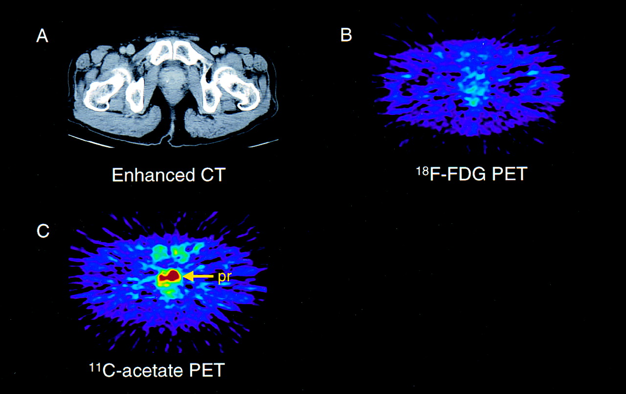
Figure 1 shows images of a 71-y-old patient (patient 5) with an increased serum PSA value of 8.4 ng/mL. A transrectal needle biopsy of the prostate revealed well-differentiated adenocarcinoma with a Gleason sum of 2. CT showed mild prostate swelling without apparent capsular or seminal invasion. Bone scintigraphy did not show any high uptake within the bone. Both 11C-acetate and 18F-FDG PET were performed on this patient. 18F-FDG PET showed only low uptake of the radiopharmaceutical in the prostate, with an SUV of 1.97, whereas 11C-acetate PET showed higher uptake within the prostate, with an SUV of 6.41. A clinical stage of T2a N0 M0 was diagnosed, and a radical prostatectomy was performed. Surgically resected specimens revealed no capsular invasion, and no lymph node metastasis was seen.

PET images of prostate obtained using 18F-FDG and 11C-acetate compared with CT image from 71-y-old man with well-differentiated (Gleason sum 2) adenocarcinoma of prostate. (A) CT shows only minimal enlargement of prostate. (B) 18F-FDG PET shows low uptake in prostate, with SUV of 1.97. (C) 11C-Acetate PET shows high uptake in primary prostate cancer lesion, with SUV of 6.41. pr = prostate
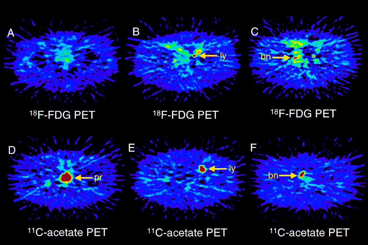
Figure 2 shows the PET images of a 73-y-old patient (patient 6) with an increased serum PSA value of 92.8 ng/mL. A transrectal needle biopsy of the prostate revealed poorly differentiated adenocarcinoma with a Gleason sum of 7. Bone scintigraphy showed high uptake at the right pubic bone. MRI revealed a mass (2.5 cm in diameter) to the left of the urinary bladder. 11C-Acetate PET and 18F-FDG PET were performed on this patient. 18F-FDG PET showed mild uptake of 18F-FDG in the prostate, with an SUV of 2.87, whereas 11C-acetate PET showed high uptake having an SUV of 5.45. 11C-Acetate PET also showed high uptake in the right pubic bone, as well as left pelvic mass lesions that were also detected by 18F-FDG PET. Both extraprostate lesions were considered to be bone and lymph node metastases. A clinical stage of T3c N2 M1 was diagnosed, and androgen ablation therapy was prescribed.

PET images of prostate, lymph node, and bone metastases obtained using 18F-FDG and 11C-acetate from 73-y-old man with poorly differentiated (Gleason sum 7) adenocarcinoma of prostate. (A) 18F-FDG PET shows low uptake in prostate, with SUV of 2.87. (B and C) 18F-FDG uptake was shown in left iliac lymph node metastatic lesion (B) and right pubic bone metastatic lesion (C). (D–F) 11C-Acetate PET shows high uptake in prostate (D), with SUV of 5.45; in left iliac lymph node metastatic lesion (E); and in right pubic bone metastatic lesion (F). bn = bone; ly = lymph node; pr = prostate.
In all 22 patients, 11C-acetate PET showed primary prostate cancer lesions and no 11C-acetate accumulation in the urine (sensitivity, 100%). Of the 18 patients who were imaged with 18F-FDG, primary lesions were seen in 15 of 18 (sensitivity, 83%). The remaining patient could not be evaluated because of high 18F-FDG retention in the bladder. Of the 5 patients who had lymph node metastases, 11C-acetate PET showed all to have high intrapelvic accumulations corresponding to metastatic sites. These intrapelvic accumulations were seen in only 2 of the 5 patients when 18F-FDG was used for imaging. Seven of the patients in the test group were found to have bone metastases, which were detected with 99mTc-hydroxymethylene diphosphonate bone scintigraphy (Table 1). Of these 7, 6 showed high 11C-acetate accumulation at the site of the bone metastases, compared with 4 in whom high accumulation was seen when 18F-FDG was used as the tracer compound (Table 2).
Patient Details and Uptake of 11C-Acetate and 18F-FDG
Detection of Prostate Cancer Lesions by PET
Evaluation of Tumor Viability
The relationship between the accumulation of 11C-acetate or 18F-FDG and clinical parameters was evaluated (Fig. 3). No correlations existed between the Gleason sum and 11C-acetate or 18F-FDG uptake. Patients with an advanced clinical stage of primary prostate cancer showed higher 18F-FDG uptake than did patients with earlier stages (P = 0.03). No correlation was seen between clinical stage and 11C-acetate uptake. A weak correlation was found between 18F-FDG uptake and the serum PSA value (r = 0.71).
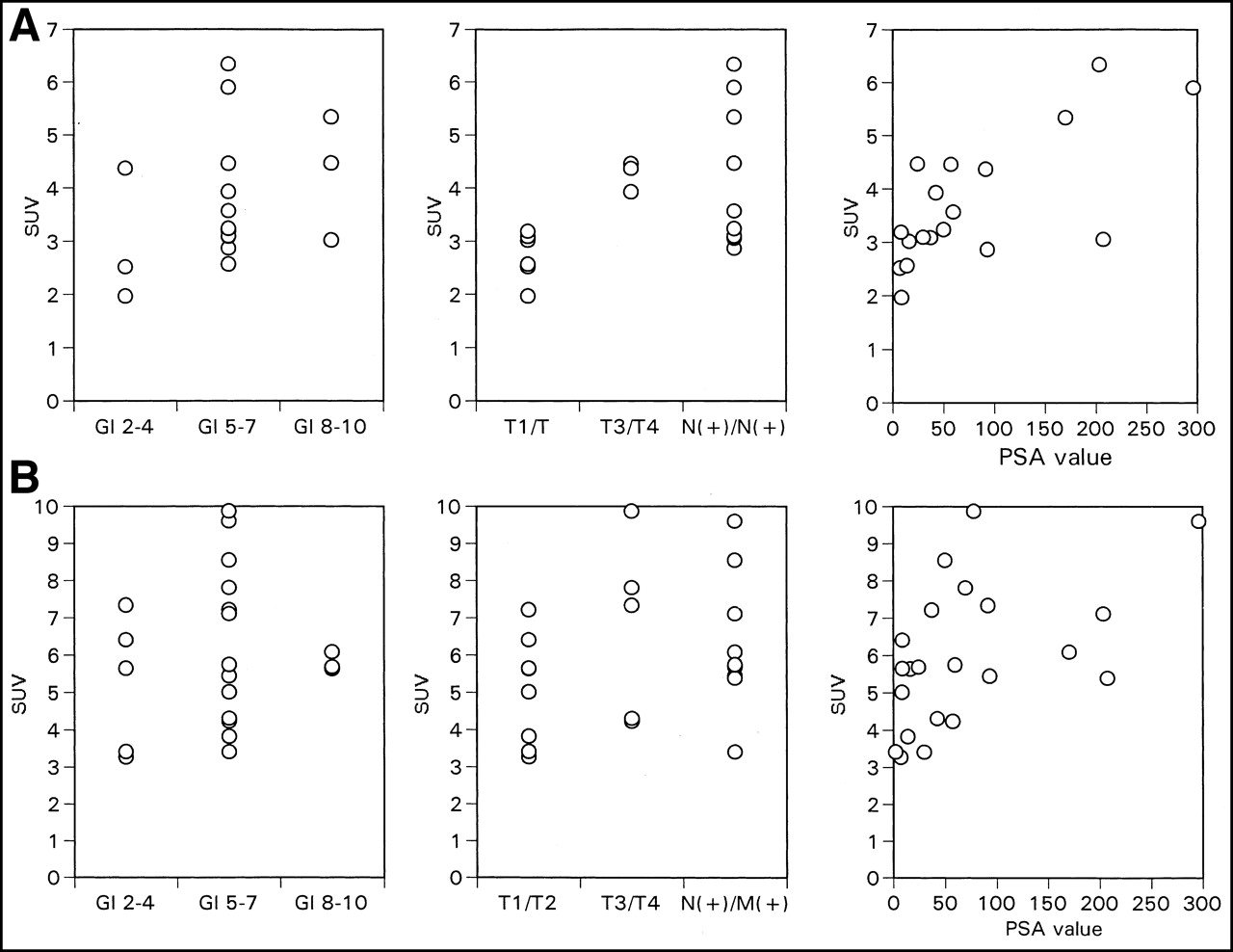
(A) No correlation was found between Gleason sum and 18F-FDG uptake (P = 0.42). Patients with advanced-stage disease showed higher 18F-FDG uptake than did those with earlier stages (P = 0.03). Weak correlation was found between 18F-FDG and serum PSA value (r = 0.71). (B) No correlation was found among clinical parameters and 11C-acetate uptake. Gl = Gleason sum.
DISCUSSION
Conventional imaging modalities such as sonography, CT, and MRI are used for the anatomic evaluation of prostate cancer. However, visible anatomic changes are not always present in early stages, making use of these imaging modalities difficult in early detection of prostate cancer sites.
18F-FDG PET for cancer diagnosis has been introduced as a metabolic imaging technique on the basis of the large amount of glucose that cancer tissue, in general, consumes as an energy source. However, for the diagnosis of prostate cancer, 18F-FDG PET has no value in comparison with conventional diagnostic modalities such as PSA, CT, or MRI. The 60%–70% sensitivity previously shown for cancer detection is not high enough to justify the clinical application of 18F-FDG PET for the detection of prostate cancer, given the expense of 18F-FDG PET over conventional methods (10,11).
11C-Acetate has been known as a positron-emitting tracer for measuring oxidative metabolism in the myocardium. As soon as the myocardium takes in 11C-acetate, it is converted to acetyl–coenzyme A in the mitochondria, followed by rapid clearance as carbon dioxide through the citric acid cycle. Recently, 11C-acetate has been reported to show high uptake in tumor tissue (15,16). The mechanism of high 11C accumulation in tumor cells, although yet unknown, is thought to be different from that of myocardium uptake. Yoshimoto et al. (22) studied uptake of 14C-acetate in 4 different tumor cell lines and a fibroblast cell line to investigate the metabolic pathway of 11C-acetate in tumor cells. 14C accumulation in each of the 4 tumor lines was higher than that in the fibroblast cells, and this accumulation in tumor cells was shown to be caused by enhanced lipid synthesis. Given the highly active basal lipid metabolism associated with the cell membrane because of tumor growth, 11C-acetate may be an important probe of this anabolic pathway of metabolism in cancer tissue.
The University of Michigan group (23), using 18F-FDG and 11C-acetate PET imaging, studied 18 prostate cancer patients who had rising PSA levels and bone scan or CT evidence of locally recurrent or regional metastatic disease. 11C-Acetate PET showed high sensitivity for detecting both primary and nodal metastatic prostate cancer lesions. Tumor uptake of 11C-acetate, as compared with 18F-FDG, was found to be moderately high, and 11C-acetate had higher sensitivity for tumor detection, without the confounding bladder activity of 18F-FDG PET.
11C-Acetate PET has been introduced as a new modality for imaging prostate cancer and its metastases (23,24). We found primary prostate cancer to be positive for 11C-acetate accumulation in all patients imaged. In the meantime, the 83% sensitivity for detection of primary prostate cancer using 18F-FDG was higher than previously reported (10,11). The high sensitivity found for 18F-FDG PET may be the result of the relatively high proportion of patients with advanced-stage disease studied. 18F-FDG PET has been reported to show higher sensitivity for detecting prostate cancer lesions of higher clinical stages (11). Indeed, in our 19 patients who underwent 18F-FDG PET, the sensitivity was lower for localized disease than for advanced-stage disease (67% vs. 92%).
11C-Acetate PET also showed high sensitivity for metastatic prostate cancer lesions. We investigated patients with confirmed metastases from prostate cancer; 11C-acetate PET detected all known lymph node metastases and all bone metastases except 1. The high sensitivity of 11C-acetate in prostate imaging will greatly help in detecting prostate cancer and extended metastases or sites of local recurrence, which are difficult to detect by conventional imaging modalities.
We also confirmed a previously reported (11) positive correlation between clinical stage and 18F-FDG uptake in prostate cancer. This finding indicates that glucose use, shown by 18F-FDG PET, is associated with progression of prostate cancer. On the contrary, this study found no relationship between the accumulation of 11C-acetate and clinical parameters such as Gleason sum, clinical stage, and serum PSA value. This result cannot be explained by only the enhanced lipid metabolism that is associated with the cell membrane because of tumor growth. A further clinical study with a larger number of patients, as well as basic studies, is needed to clarify the mechanism of 11C-acetate uptake in prostate cancer.
CONCLUSION
We evaluated 11C-acetate as a potential PET tracer for imaging prostate cancer and found a marked uptake of 11C-acetate into primary prostate cancer and metastatic sites, with higher sensitivity than that of 18F-FDG PET. 11C-Acetate is a promising tracer for imaging prostate cancer and its metastases. Further studies using 11C-acetate PET with a larger number of patients are needed to determine its ultimate clinical utility.
Acknowledgments
The authors thank Katsuya Sugimoto and Shingo Kasamatsu for expert technical support and Drs. Yasuhisa Fujibayashi, Kouichi Ishizu, and Tatsuro Tsuchida for helpful discussions. The authors also thank Dr. Yasutaka Kawamura for CT images and Drs. Yoshiji Miwa, Harutoshi Tsuka, Bunya Miyaji, Hirokazu Ishida, Yasuhiko Ito, Kazuya Tanase, Masakatsu Tawada, Yosuke Matsuta, Rikiya Shioyama, Masanobu Maekawa, and Masaharu Nakai for support in patient recruitment. The authors strongly thank Dr. Michael J. Welch for critically reading the manuscript and Michelle E. Weber for expertly editing the manuscript.
Footnotes
Received Jun. 18, 2001; revision accepted Oct. 24, 2001.
For correspondence or reprints contact: Nobuyuki Oyama, MD, PhD, Mallinckrodt Institute of Radiology, Washington University School of Medicine, Campus Box 8225, 510 S. Kingshighway Blvd., St. Louis, MO 63110.
E-mail: Oyaman{at}mir.wustl.edu
REFERENCES
In this issue


Jump to section
Related Articles
Cited By...
- Mitochondrial ACSS1 regulates the oncometabolite 2-hydroxyglutarate and De Novo Pyrimidine biosynthesis under nutrient-deprived conditions in lymphoma
- 3D-printed automation for optimized PET radiochemistry
- Enhanced Fatty Acid Scavenging and Glycerophospholipid Metabolism Accompany Melanocyte Neoplasia Progression in Zebrafish
- 11C-Acetate-PET/CT Compared to 99mTc-HDP Bone Scintigraphy in Primary Staging of High-risk Prostate Cancer
- Glucose-independent Acetate Metabolism Promotes Melanoma Cell Survival and Tumor Growth
- Evaluation of Prostate Cancer with 11C-Acetate PET/CT
- Preclinical Evaluation of 3-18F-Fluoro-2,2-Dimethylpropionic Acid as an Imaging Agent for Tumor Detection
- Reduced 64Cu Uptake and Tumor Growth Inhibition by Knockdown of Human Copper Transporter 1 in Xenograft Mouse Model of Prostate Cancer
- 11C-Acetate PET/CT Before Radical Prostatectomy: Nodal Staging and Treatment Failure Prediction
- An NMR Metabolomics Approach for the Diagnosis of Leptomeningeal Carcinomatosis
- 11C-Acetate PET/CT in Localized Prostate Cancer: A Study with MRI and Histopathologic Correlation
- 2-(3-{1-Carboxy-5-[(6-[18F]Fluoro-Pyridine-3-Carbonyl)-Amino]-Pentyl}-Ureido)-Pentanedioic Acid, [18F]DCFPyL, a PSMA-Based PET Imaging Agent for Prostate Cancer
- Assessment of PET Tracer Uptake in Hormone-Independent and Hormone-Dependent Xenograft Prostate Cancer Mouse Models
- Prostate Cancer: PET with 18F-FDG, 18F- or 11C-Acetate, and 18F- or 11C-Choline
- Functional Imaging of Localized Prostate Cancer Aggressiveness Using 11C-Acetate PET/CT and 1H-MR Spectroscopy
- 1-11C-Acetate Versus 18F-FDG PET in Detection of Meningioma and Monitoring the Effect of {gamma}-Knife Radiosurgery
- Tissue-specific Short Chain Fatty Acid Metabolism and Slow Metabolic Recovery after Ischemia from Hyperpolarized NMR in Vivo
- New Agents and Techniques for Imaging Prostate Cancer
- The Importance of Acetyl Coenzyme A Synthetase for 11C-Acetate Uptake and Cell Survival in Hepatocellular Carcinoma
- Molecular Imaging of Tumor Blood Vessels in Prostate Cancer
- Radiopharmaceuticals in Preclinical and Clinical Development for Monitoring of Therapy with PET
- Novel Tracers and Their Development for the Imaging of Metastatic Prostate Cancer
- A Prospective Evaluation of 18F-FDG and 11C-Acetate PET/CT for Detection of Primary and Metastatic Hepatocellular Carcinoma
- Tumor Cell Metabolism Imaging
- Small-Cell Carcinoma of the Prostate
- 1-11C-Acetate as a PET Radiopharmaceutical for Imaging Fatty Acid Synthase Expression in Prostate Cancer
- 1-11C-Acetate Kinetics of Prostate Cancer
- 18F-Fluoroacetate: A Potential Acetate Analog for Prostate Tumor Imaging--In Vivo Evaluation of 18F-Fluoroacetate Versus 11C-Acetate
- Initial Experience with the Radiotracer Anti-1-Amino-3-18F-Fluorocyclobutane-1-Carboxylic Acid with PET/CT in Prostate Carcinoma
- 11C-Acetate Positron Emission Tomography Imaging and Image Fusion With Computed Tomography and Magnetic Resonance Imaging in Patients With Recurrent Prostate Cancer
- Detection and Localization of Prostate Cancer: Correlation of 11C-Choline PET/CT with Histopathologic Step-Section Analysis
- Current Concepts in Lymph Node Imaging
- microPET and Autoradiographic Imaging of GRP Receptor Expression with 64Cu-DOTA-[Lys3]Bombesin in Human Prostate Adenocarcinoma Xenografts
- Radiation Dose Estimates in Humans for 11C-Acetate Whole-Body PET
- PET Imaging of Prostate Cancer with 11C-Acetate
- 11C-Acetate PET Imaging of Prostate Cancer: Detection of Recurrent Disease at PSA Relapse
- Preoperative Staging of Pelvic Lymph Nodes in Prostate Cancer by 11C-Choline PET
- 11C-Acetate PET Imaging in Hepatocellular Carcinoma and Other Liver Masses