


Abstract
Absorbed doses in 90Y radioimmunotherapy are usually estimated by extrapolating from 111In imaging data. PET using 86Y (β+ 33%; half-life, 14.7 h) as a surrogate radiolabel could be a more accurate alternative. The aim of this study was to evaluate an 86Y-labeled monoclonal antibody (mAb) as a PET imaging agent and to compare the biodistribution of 86Y- and 111In-labeled mAb. Methods: The humanized anti-Lewis Y mAb hu3S193 was labeled with 111In or 86Y through CHX-A′′-diethylenetriaminepentaacetic acid chelation. In vitro cell binding and cellular retention of radiolabeled hu3S193 were evaluated using HCT-15 colon carcinoma cells, a cell line expressing Lewis Y. Nude mice bearing HCT-15 xenografts were injected with 86Y-hu3S193 or 111In-hu3S193. The biodistribution was studied by measurements of dissected tissues as well as by PET and planar imaging. Results: The overall radiochemical yield in hu3S193 labeling and purification was 42% ± 2% (n = 2) and 76% ± 3% (n = 6) for 86Y and 111In, respectively. Both radioimmunoconjugates specifically bound to HCT-15 cells. When cellular retention of hu3S193 was studied using 111In-hu3S193, 80% of initially cell-bound 111In activity was released into the medium as high-molecular-weight compounds within 8 h. When coadministered, in vivo tumor uptake of 86Y-hu3S193 and 111In-hu3S193 reached maximum values of 30 ± 6 and 29 ± 6 percentage injected dose per gram and tumor sites were easily identifiable by PET and planar imaging, respectively. Conclusion: At 2 d after injection of 111In-hu3S193 and 86Y-hu3S193 radioimmunoconjugates, the uptake of 111In and 86Y activity was generally similar in most tissues. After 4 d, however, the concentration of 86Y activity was significantly higher in several tissues, including tumor and bone tissue. Accordingly, the quantitative information offered by PET, combined with the presumably identical biodistribution of 86Y and 90Y radiolabels, should enable more accurate absorbed dose estimates in 90Y radioimmunotherapy.
Nuclear medicine has extensively used 90Y as a therapeutic agent because of its physical properties. It is a high-energy (maximum, 2.27 MeV) pure β−-emitter. The nonpenetrating β−-emissions have a maximum range of approximately 10 mm with a mean range of 3.9 mm in soft tissue, and the lack of penetrating photon emissions minimizes the indiscriminate whole-body radiation absorbed dose burden. Primarily, 90Y has been used for bone pain palliation (1–3) and in radiolabeled monoclonal antibody (mAb) therapies (4–8). The paucity of γ-ray emissions, however, renders this isotope extremely difficult to image. Attempts have been made to image the Bremsstrahlung radiation (9–12) generated by the slowing down of high-energy electrons in tissue. However, the low photon yield and the polychromatic nature of the Bremsstrahlung spectrum result in limited quantitative accuracy with 90Y. To avoid complex and inaccurate Bremsstrahlung imaging methods, it has been customary to use 111In as a surrogate for 90Y. 111In has an almost identical half-life to 90Y, emits 2 γ-rays of 171 and 245 keV, and is readily incorporated into the same metal chelating agents as yttrium. For these reasons, 111In has been considered an excellent analog for 90Y.
Subtle differences in radionuclide retention of the metal chelating agent used for 111In and 90Y mAb labeling can result in a differential release between the 2 radiometals. Despite possible differences in the biodistributions of 111In and 90Y, 111In is widely used as an analog for 90Y in radioimmunotherapy trials. Of particular importance is the affinity of yttrium for bone. Loss of 90Y from the chelating agent can result in a higher dose to bone and bone marrow, which would not be anticipated from biodistribution data derived from 111In. Because bone marrow is the dose-limiting tissue in most radionuclide therapies, pretherapy tracer scans with 111In-labeled mAb could underestimate the bone marrow dose.
A more suitable isotope for accurately imaging the biodistribution of 90Y would be an alternative isotope of the yttrium metal. Recently, 2 alternative isotopes have been proposed. The first is the electron capture decaying 87Y isotope, which emits a 485-keV γ-ray with a 92.2% yield and has a half-life of 3.3 d (13). This isotope can be visualized by planar or SPECT gamma camera imaging on many modern cameras, using a high-energy, high-resolution collimator. The second, 86Y, is a positron emitter (33%) with a 14.7-h half-life that can be imaged on a PET camera (14). 86Y has been used to estimate the radiation doses from 90Y in patients administered 90Y for bone pain palliation (3).
The mAb (3S193) used in this study binds to the Lewis Y antigen (Ley), an antigen expressed on several human carcinomas of epithelial origin, including colon, lung, ovarian, and breast carcinomas. This mAb has been well characterized by Kitamura et al. (15) and Scott et al. (16) in its murine and humanized forms. Clinical radioimmunotherapy trials with this mAb are currently in progress to evaluate treatment efficacy in breast, colon, and ovarian carcinomas.
The objective of this study was to compare the biodistributions of 111In with 86Y-labeled anti-Ley mAb and to test the feasibility of imaging tumors with 86Y-anti Ley in a rodent xenograft model.
MATERIALS AND METHODS
Materials
The characterization of the originally murine mAb 3S193, its association with Ley, and its humanization have been described by Kitamura et al. (15). Humanized 3S193 (hu3S193) was produced and conjugated to CHX-A′′-diethylenetriaminepentaacetic acid (DTPA), as has been described (17,18). [111In]InCl3 in 50 mmol/L HCl was purchased from NEN (Billerica, MA). All solutions were made using distilled deionized water (Milli-RO Plus; Millipore Inc., Bedford, MA) and all buffers were purified on a 200–400 mesh Chelex 100 column (Bio-Rad, Hercules, CA). All other chemicals were from commercial sources. NAP-5 desalting columns were purchased from Pharmacia (Piscataway, NJ), and 10-DG gel filtration columns were purchased from Bio-Rad. A fast protein liquid chromatography (FPLC) gel filtration column (Superdex 200HR; Pharmacia) coupled to a Waters high-performance liquid chromatography system (two 501 pumps, 486 UV detector), a radiodetector (Radiomatic flow scintillation analyzer; Packard Instrument Co., Meriden, CT), and a FRAC-100 fraction collector (Pharmacia), was used for chromatographic analysis. Instant thin-layer chromatography silica plates were purchased from Gelman Sciences Inc. (Ann Arbor, MI). All radioactive samples were measured in a well scintillation LKB Wallac gamma counter (1282 Compugamma CS), correcting for the 86Y contribution in the 111In window and the 111In contribution in the 86Y window in the double-isotope experiments described below. The PET camera used for 86Y imaging was an Advance whole-body scanner (General Electric, Milwaukee, WI). The 111In biodistribution was imaged using a Genesys gamma camera (ADAC, Milpitas, CA). Nude mice were purchased from Harlan Sprague-Dawley (Indianapolis, IN) and were kept in a controlled environment where cages, food, and water had been autoclaved. To prevent the development of hyperkeratosis associated with corynea bacteria, 1 mL Augmentin (Amoxycillin/Clavulanate potassium) was added per 500 mL drinking water.
Production and Separation of 86Y
The general procedure for 86Y production and separation has been reported (17). Briefly, isotope-enriched 86SrCO3 (97.02% 86Sr) was irradiated with 15-MeV protons in the MSKCC cyclotron (model CS-15; Cyclotron Corp., Berkeley, CA). After irradiation, the target was dissolved in 4 mol/L nitric acid containing 1 mg/mL Fe(III), diluted with metal-free water, and stirred for 5 min. The 86Y hydroxide was coprecipitated with ferric hydroxide by the addition of dilute ammonium hydroxide. The 86Y occluded ferric hydroxide precipitate was concentrated by centrifugation. The precipitate was redissolved and precipitated 3 additional times and finally washed with warm water. All solutions were combined and the enriched strontium was recovered as carbonate by bubbling carbon dioxide through the solution. Lastly, the precipitate was dissolved in 6 mol/L HCl and loaded onto a preconditioned analytic grade 1 × 8 anion ion-exchange column. The column was eluted with 15 mL 6 mol/L HCl. The solution was evaporated to dryness and the residue was dissolved in 0.5 mL 50 mmol/L HCl. For this study, 333 ± 111 MBq (n = 2) 86YCl3 in 0.5 mL 50 mmol/L HCl were obtained.
Radiolabeling
86Y-acetate or 111In-acetate was prepared by mixing an aliquot of the radionuclide preparations (51.8–148 MBq 86Y, 18.5–55.5 MBq 111In) with 3 mol/L ammonium acetate (final pH = approximately 5). After 5–15 min, hu3S193 (100–250 μg) was added, and the mixture incubated for 30 min at room temperature. The reaction was terminated by the addition of ethylenediaminetetraacetic acid (EDTA) (50 nmol), and the radiolabeled mAb separated from unreacted radiometal on a 10-DG desalting column equilibrated with 50 mmol/L phosphate-buffered saline (PBS).
The amount of protein-bound radioactivity in the purified preparations was determined by thin-layer chromatography, on silica gel, using 10 nmol/L EDTA (pH 4.5) as an eluent. In this system, radiometal-EDTA migrates with the solvent front (Rf = approximately 0.8–1), whereas labeled mAb remains at the application site (Rf = approximately 0–0.2). Preparations of 86Y-hu3S193 and 111In-hu3S193 were also analyzed by FPLC as described previously, and antigen-binding capacity determined in a cell-binding assay, as described later in this article.
A single batch of 125I-hu3S193 was prepared for cell binding and retention by adding 1.48 MBq 125I in 50 μL 0.3 mol/L phosphate buffer (pH 7.5) to a vial coated with IODO-GEN (10 μg; Pierce, Rockford, IL). After 5 min, the 125I activity was transferred to a vial containing hu3S193 (100 μg). The mixture was incubated for 30 min at room temperature, after which 125I-hu3S193 was separated from unreacted 125I on an NAP-5 desalting column.
Cell Binding
The antigen-binding capacity of radiolabeled hu3S193 was evaluated in a cell-binding assay. Labeled hu3S193 (10 ng) was added in triplicate to 5 × 10⁶ HCT-15 cells suspended in 200 μL Roswell Park Memorial Institute (RPMI) medium complemented with 0.1% human serum albumin (HSA) (RPMI + 0.1% HSA). In control vials, 100 μg hu3S193 were added to the cells before the labeled hu3S193 was added. The cell suspensions were incubated for 1 h on a rotating table at room temperature and washed twice by centrifugation and resuspension in RPMI + 0.1% HSA. Immunoreactive fraction was defined as radioactivity in pellet over total radioactivity.
For cellular retention analysis, HCT-15 cells were grown to confluence in a 96-well plate. A mixture of 111In-hu3S193 (50 ng) and 125I-hu3S193 (50 ng) in 200 μL RPMI + 0.1% HSA was added to each well. After incubation at 37°C for 2 h, cells were washed 3 times with RPMI + 0.1% HSA. Then, 200-μL culture medium was added to each well and the cells were further incubated at 37°C for 0, 1, 2, 4, 8, 16, and 24 h in triplicate samples. At each time point, the supernatant was removed and separated into high- and low-molecular-weight components on an NAP-5 desalting column. The cells were solubilized with 2 mol/L NaOH, and all fractions measured in a well counter using 2 windows selected to differentiate the 125I x- and γ-ray peaks (25–35 keV) from the 111In γ peaks (171 and 245 keV). 111In standards were used to determine the down-scatter fraction into the 125I window and all sample counts per min in the 125I window appropriately corrected by the counts in the 111In multiplied by the down-scatter factor (0.033 or 3.3%).
Biodistribution
Nude mice were injected subcutaneously in the left and right hind legs with 106 HCT-15 cells suspended in 200 μL RPMI medium. Biodistribution experiments were performed 2–3 wk after tumor induction, at which time the mice weighed 18–27 g.
The general hu3S193 biodistribution kinetics in the current tumor model were investigated using 111In-hu3S193. Mice were injected intravenously with 100 μL PBS containing 0.5% HSA (RPMI + 0.5% HSA) and 0.74 MBq 111In-hu3S193 (3 μg). At various time points after injection, 6 mice were killed by exposure to CO2 and then dissected.
For comparing 86Y-hu3S193 and 111In-hu3S193 biodistribution, mice were coinjected with 120 μL PBS containing 0.5% HSA, 1.48 MBq 86Y-hu3S193, and 0.185 MBq 111In-hu3S193 (total 100 μg) in 200 μL. Five mice were killed at 2 and 4 d after injection, respectively. The major organs were dissected, weighed, and counted on a well scintillation counter in 2 windows. For 86Y, a single window was used, including both single 511-keV and 1.02-MeV coincident annihilation photons. For 111In, a window encompassing both 171- and 245-keV γ-photons was used. Scintillation vials were filled with PBS to the same 3-mL volume. The cross-talk between the 111In and 86Y windows was derived from the counts in both windows using standards of each isotope. The cross-talk from 111In into the 86Y window was negligible (<2 times background) and, therefore, was assumed to be zero. The cross-talk factor from 86Y into the 111In window was 1.34 (134%), which was caused by the very large (polychromatic) emissions from 86Y as well as the partial absorption of high-energy γ-rays in the sodium iodide detector. To convert counts in the 111In window into 111In activity, the counts in the 86Y window were multiplied by 1.34 and subtracted from the counts in the 111In window.
Imaging
The imaging studies were performed on mice administered 111In- or 86Y-labeled mAb only, because initial preliminary studies with coadministered 111In and 86Y showed severe degradation of the 111In images from 86Y photon down-scatter into the 111In energy windows (data not shown).
The mice were injected with 3.7 MBq 111In-hu3S193 (25 μg) in 200 μL PBS + 0.5% HSA. Animals were anesthetized with ketamine at 1 h, 24–26 h, and 48–50 h after injection and positioned on a Styrofoam (Dow Chemical Co., Midland, MI) base directly on the medium-energy, general purpose collimator of an ADAC Genesys gamma camera (ADAC, Milpitas, CA). A 20-min acquisition was performed with two 20% energy windows positioned at the 171- and 245-keV photopeaks of 111In.
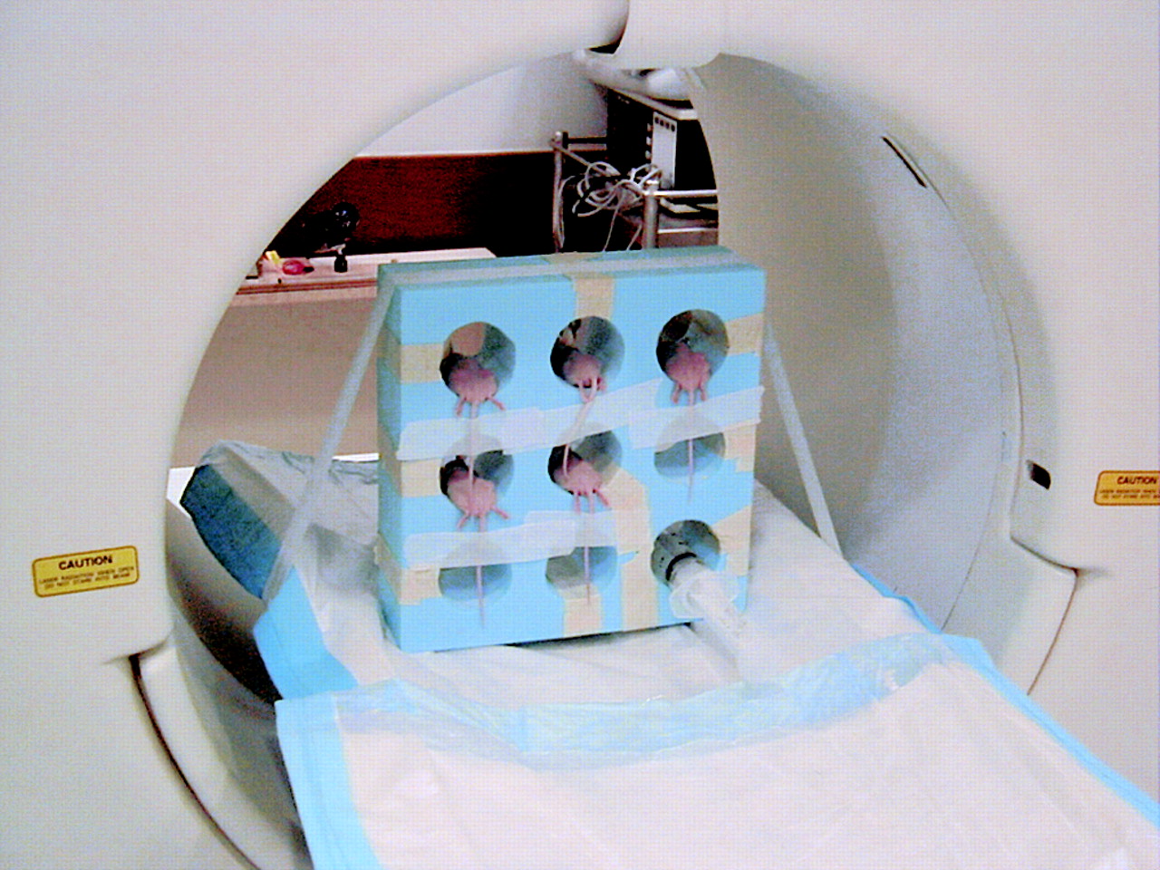
86Y images were obtained on a whole-body Advance PET scanner (General Electric, Milwaukee, WI). The mice were injected with 3.7 MBq 86Y-hu3S193 (20 μg) in 200 μL PBS + 0.5% HSA. The mice were anesthetized at 1 h, 24–26 h, and 48–50 h after injection and positioned on a circular Styrofoam (Dow Chemical) motel (Fig. 1) taped to the scanner couch. Acquisitions were performed for 20 min within a 300 to 650-keV energy window in 2-dimensional (2D) (septa-in) mode. Normalization, randoms, and scatter corrections were applied and the images were reconstructed by standard filtered backprojection using a Hanning filter with an 8-mm cutoff.

Animal set-up in Styrofoam motel on GE Advance PET scanner.
Tissue Processing
Mice were killed 1 or 2 d after injection and blood and tumors were harvested. Blood samples were centrifuged and the radioactive components in serum were analyzed by fractionation on an FPLC column. Selected tumor samples were mounted in cryomolds (Tissue Tek, Elkhart, IN) and embedded in optimal cutting tissue and frozen on dry ice. Sections were performed on a cryostat (Bright Instruments, Chelmsford, UK) and applied to glass slides for digital autoradiographic and immunohistochemical analysis. The glass slides with 8-μm tissue sections were placed on a phosphor storage plate (Bio-Rad) for a 24-h exposure and then read on a scanning laser to reveal the radiolabel distribution within the tumor tissue sections. These sections were further analyzed by immunohistochemistry as follows: Detection of the injected hu3S193 was conducted with a goat-antihuman mAb (1:100; Jackson Labs, West Grove, PA) followed by a biotinylated horse-antigoat mAb (1:200; Jackson Labs). Labeling of the secondary mAb was done with an avidin-biotin complex system (ABC-Elite; Vector Laboratories, Burlingame, CA). Diaminobenzidine tetrahydrochloride (DAB; BioGenex, San Ramon, CA) was used as a chromogen. Negative control slides omitting the goat-antihuman mAb were included. To analyze the presence of the Ley on the tumor cells independently from injected mAb, a separate set of slides was immunohistochemically stained by applying hu3S193 as a primary reagent, followed by a biotinylated goat-antihuman mAb and the ABC-kit and chromogen as described previously.
RESULTS
Radiolabeling Yields
The radiochemical yields for 86Y- and 111In-labeling and purification were 42% ± 2% (n = 2) and 76% ± 3% (n = 6), respectively. For 86Y-hu3S193, the specific activity was 74 ± 37 MBq/mg (n = 2), the radiochemical purity was 96% ± 4% (n = 2), and the immunoreactive fraction was 44% (n = 1). For 111In-hu3S193, the specific activity was 148 ± 74 MBq (n = 6), the radiochemical purity was 98% ± 3% (n = 6), and the immunoreactive fraction was 40% ± 14% (n = 5). In the FPLC radiochromatograms, only intact radiolabeled hu3S193 could be distinguished for both 86Y- and 111In-hu3S193. The lower labeling yield for 86Y versus 111In was assumed to be attributable to iron contamination. No assessment of the absolute chemical purity of 86Y was performed. 125I-labeling of hu3S193 resulted in 47% radiochemical yield and a specific activity of 74 MBq/mg (n = 1).
Cell Binding
After 111In- and 125I-hu3S193 binding to HCT-15 cells, 80% of the initially cell-bound radioactivity was recovered in the medium as high-molecular-weight species after an 8-h incubation. It was expected that a much higher yield of 125I low-molecular-weight activity would be observed because of the digestion of mAb on internalization and release of the radioiodine. However, the results showed only a small difference between 111In- and 125I-hu3S193, suggesting that <20% internalization had occurred within the 8-h incubation (Fig. 2). The advantage of the radiometals, 111In, and the isotopes of Y(III) is the nonspecific retention by cells upon intracellular disassociation of the metal from the chelate, leading to longer tumor retention relative to radiohalogenated immunoconjugates.
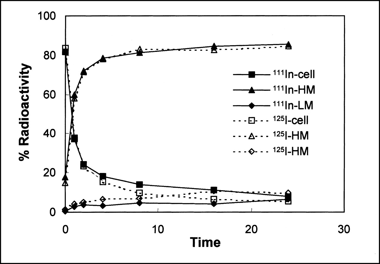
Cellular retention of 111In-hu3S193 (solid lines) and 125I-hu3S193 (dotted lines). Cell = cell-bound radioactivity; HM and LM = high- and low-molecular-weight radioactivity in supernatant. Error bars (n = 3) were omitted for clarity.
Biodistribution Kinetics
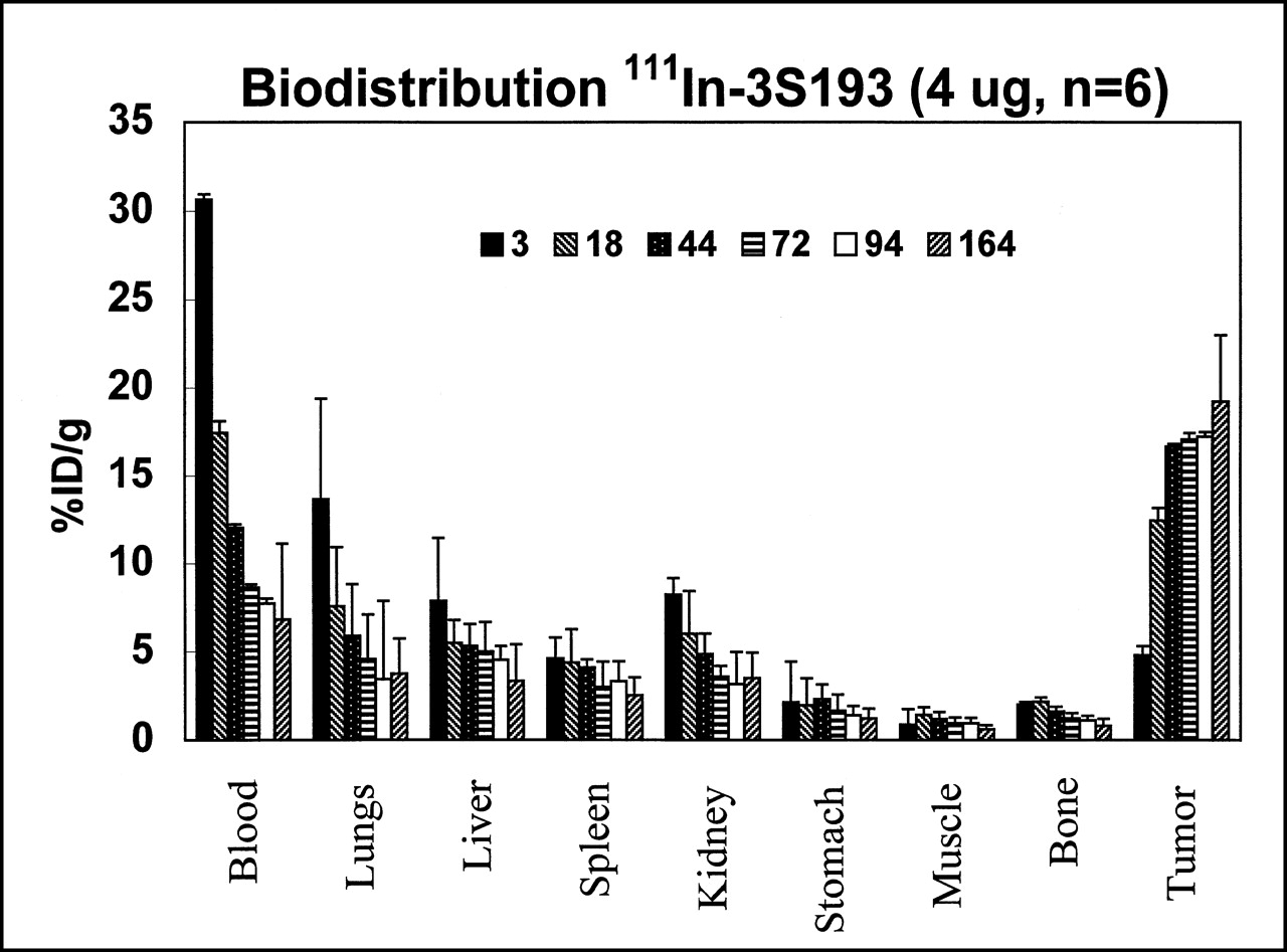
HCT-15 tumor uptake of 111In-hu3S193 reached maximum values 2 d after injection (Fig. 3). Tumor uptake reached 30 percentage injected dose per gram (%ID/g) in agreement with similar studies, with the same mAb targeting MCF-7 xenografts in BALB/c mice (18). Apart from blood, the highest normal tissue concentration was found in the lung, presumably caused by the high blood content in the lung. However, the activity cleared rapidly, falling from 14 %ID/g at 3 h to 3 %ID/g at 94 h. The lowest 111In concentration was found in muscle. Bone 111In concentration was low at all time points of analysis, indicating a stable binding of 111In to the CHX-A′′-DTPA chelate. The general biodistribution of the 111In-hu3S193 was in close agreement with the recent data of Finn et al. (17) and Clarke et al. (18,19).

Biodistribution kinetics of 111In-hu3S193 in nude mice carrying HCT-15 xenografts (n = 6) at: 3, 18, 44, 72, 94, and 164 h.
Comparison Between 86Y-hu3S193 and 111In-hu3S193
Coinjected 86Y-hu3S193 and 111In-hu3S193 showed generally similar distribution patterns at 2 and 4 d after injection (Table 1), with tumor uptake of both radiolabels reaching about 30 %ID/g after 2 d. However, 86Y concentration was significantly higher than that of 111In in all tissues other than tumor, spleen, muscle, and bone at 2 d, and lung and muscle at 4 d after injection. Results are presented as mean ± SD. A paired Student t test was used to analyze the 86Y-111In-hu3S193 double-tracer data (α < 0.05).
Comparative Biodistribution of Coinjected 86Y-3S193 and 111In-3S193
Four days after injection, the highest differences (about 30%) were found in liver, kidney, and spleen. At that time point, 86Y uptake in tumor and bone was 20% higher than corresponding values for 111In. The general trend of this data (Table 1) shows that most tissues, except muscle, exhibit a progressively higher %ID/g for 86Y with time after injection, relative to 111In. This may be attributable to the marginally lesser stability of the CHX-A′′-DTPA chelate to retain the 86Y, which results in a higher release of the 86Y radiometal, and, therefore, a prolonged whole-body clearance rate relative to 111In-labeled immunoconjugate. However, the expected increase in bone uptake of 86Y caused by complex instability that had been noted in previous reports was not observed in this experiment. Thus, it is not entirely clear that the source of the higher %ID/g recorded in the majority of tissues originated solely from complex instability.
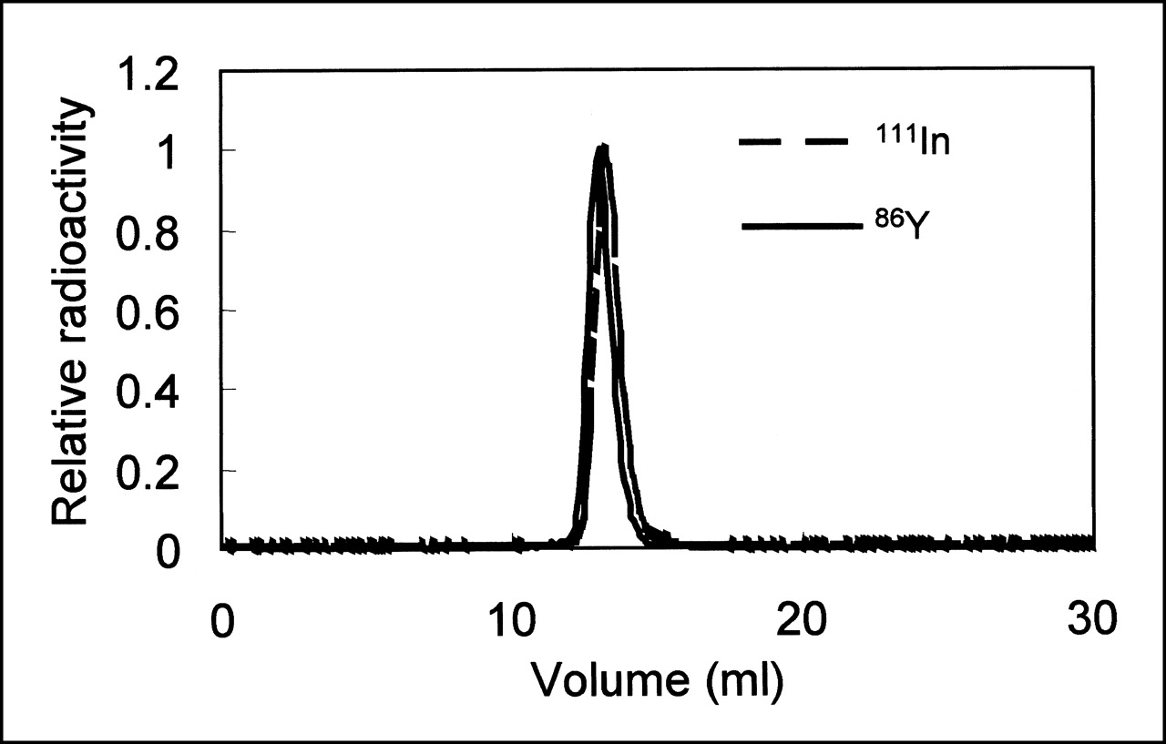
Analysis of the serum stability of each radioimmunoconjugate, 86Y-hu3S193, or 111In-hu3S193 at 2 d after injection showed that the only radioactive component was intact mAb, as evaluated by FPLC chromatography (Fig. 4). However, it should be noted that “free” radiometals, unless associated with blood proteins, would not be detected in this assay.

Serum analysis of animals 2 d after injection with 86Y-hu3S193 or 111In-hu3S193.
Radionuclide Imaging
Reconstructed coronal slices (4.3 mm thick) of 3 serial PET scans showing the biodistribution of 86Y-hu3S193 in 2 mice are shown in Figure 5. At 1 h after injection, only blood pools in the heart and liver were visualized. One day later, tumors were clearly distinguishable, and tumor 86Y accumulation was the dominant feature at 2 d after injection.

Serial coronal PET images of 2 mice injected with 86Y-hu3S193.
Serial planar images of the mice injected with 111In-hu3S193 are shown in Figure 6. These images display the biodistribution of the 111In-radioimmunoconjugate relative to the 86Y-hu3S193 PET images. Tumor sites are apparent within 24 h of injection, and tumor-to-liver contrast improves even further at 48 h. Because of the difficulties of partial volume effects for small tumors in rodent model systems with 86Y, direct tissue counting was performed but quantitative analysis of the images was not. This will be subject to further study on a dedicated small animal microPET imaging system (Concorde Microsystems, Knoxville, TN), which offers much greater resolution and, consequently, higher recovery coefficients and improved activity quantitation. One issue to be resolved with 3-dimensional PET imaging equipment (such as the Concorde microPET) is subtraction of coincidences, which result from a 511-keV annihilation photon with a prompt γ-photon emitted by 86Y (20). These are true coincidences (not randoms), which give rise to false lines of response because they occur between 2 γ-photons with no angular correlation. Such false coincidences are minimized when scanning in 2D (septa-in mode), as was the case in this study. Potential solutions for these effects, which reduce the quantitative accuracy and degrade PET images for 86Y, are under investigation (20).
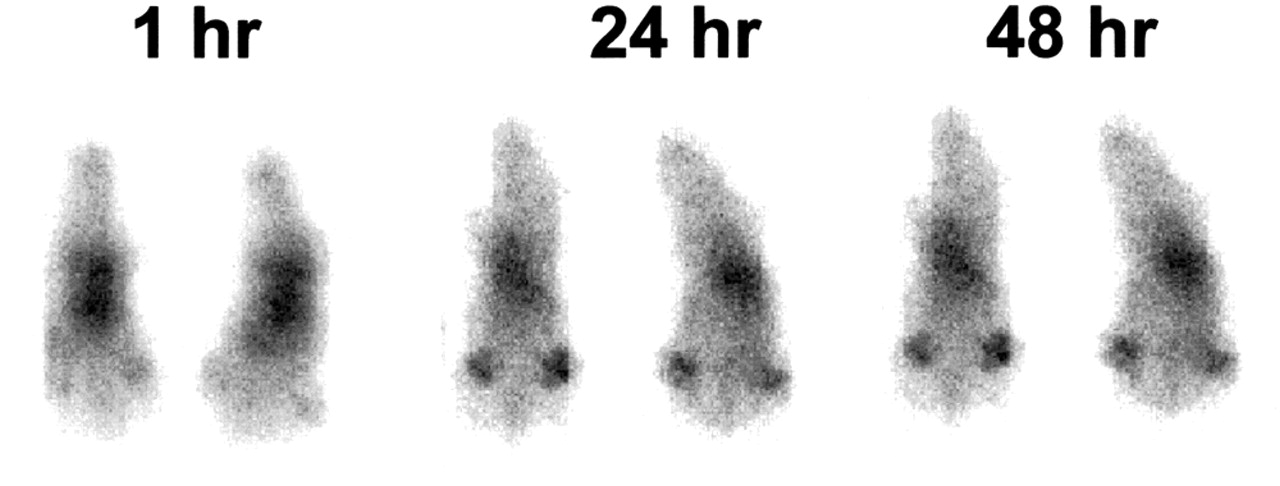
Serial planar images of 2 mice injected with 111In-hu3S193.
Autoradiography and Immunohistochemistry
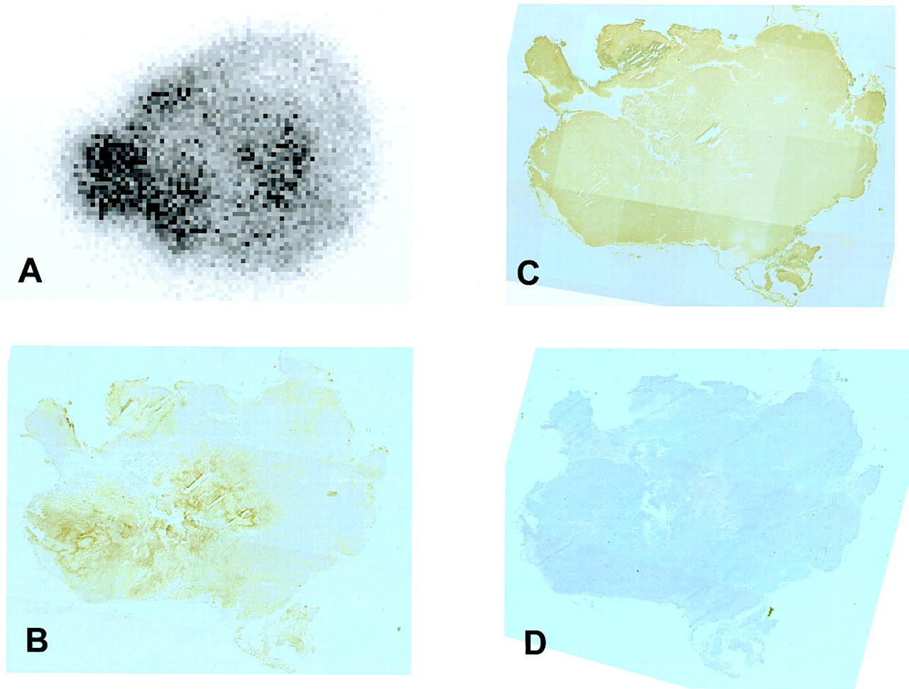
Tumor tissue section autoradiography was performed using the GS350 Molecular Imager System (Bio-Rad), a digital autoradiography device. An example autoradiograph for 1 of the tumors after 86Y-hu3S193 administration is shown in Figure 7A. The nonuniform distribution of activity reflected the limited diffusion of the radiolabeled mAb within the tumor at 24 h. Figure 7B shows immunohistochemical staining of the Ley mAb in an adjacent section taken from the same tumor. The similar radioactivity distribution and staining pattern provided evidence that 86Y remained associated with the mAb at that time. The distribution of Ley expression in the HCT-15 tumors was relatively uniform, as shown by the immunohistochemical staining for the Ley antigen in Figure 7C. Figure 7D shows the tumor histology by classical hematoxylin–eosin staining.

Four serial sections from HCT-15 tumor at 24 h after injection with 86Y-hu3S193. (A) Phosphor plate autoradiograph (100 × 100 μm2 resolution) showing spatial distribution of 86Y. (B) Horseradish peroxidase stain for injected hu3S193 mAb on adjacent section. (C) Horseradish peroxidase stain for Ley antigen on adjacent section. (D) Hematoxylin–eosin stain of adjacent section. Sections (B–D) were digitized frame-by-frame on microscope at ×40 magnification using motorized scanning stage.
DISCUSSION
90Y has many desirable properties for radionuclide therapies, but emits no γ-rays suitable for gamma camera imaging. Attempts to image the Bremsstrahlung photons emitted by the slowing down of the high-energy β-rays within the patient have been of inadequate quality for diagnosis and dosimetry. These difficulties have resulted in the use of 111In as a surrogate isotope for 90Y. Although the biodistribution of mAbs labeled with these 2 radionuclides may be similar, small differences may have radiotoxic implications when levels of activity are administered in therapeutic applications approaching dose-limiting toxicity. This study was designed to examine the potential of 86Y as a chemically equivalent surrogate for 90Y.
The results of this study show that the biodistributions of 111In-hu3S193 with 86Y-hu3S193 anti-Ley mAb are comparable within the 48-h time frame (>3.5 half-lives), indicating that PET imaging with 86Y is feasible. Despite 7 half-lives, counts per minute in the tumor, which were obtained with the well scintillation counter, were still highly significant (>24,000) and >10 times background in all tissues (except muscle) at 96 h.
The advantage of using 86Y is that the annihilation photon emissions detected by PET permit a more accurate determination of 86Y than single-photon imaging devices. Furthermore, the use of this γ-emitting yttrium isotope permits the use of solid scintillation counting methods of tissue samples, thereby avoiding the quantification problems encountered with liquid scintillation counting of a pure β−-emitter such as 90Y.
This study confirmed that 111In is a good analog for the isotopes of yttrium, especially at early time points (<48 h) after injection. However, the lower stability of the CHX-A′′-DTPA chelate for 86Y relative to 111In results in a progressively higher ratio of the 86Y radiometal to the parent compound. The marginally slower clearance kinetics of the yttrium radiometal resulted in a progressively higher %ID/g during a 4-d time period relative to 111In. The implications of this departure between the pharmacokinetics of the 2 radiometals would lead to dosimetry estimates based on 111In images that would underestimate the doses received by 90Y. For this reason, 86Y would be a more accurate surrogate for 90Y. Furthermore, the ability to perform quantitative PET imaging with 86Y radiopharmaceuticals offers a significant advantage over 111In. Although the 14.7-h half-life of 86Y is much lower than that of 111In (2.7 d), the far higher sensitivity of PET cameras should permit clinical imaging to at least 4 half-lives.
CONCLUSION
This study has shown the feasibility of radiolabeling the hu3S193 anti-Ley mAb with 86Y to obtain tumor uptake (30 %ID/g) equivalent to 111In and to obtain high-quality PET images at 48 h after injection with excellent tumor localization. Future studies will explore the utility of 86Y-labeled mAbs to determine the biodistribution and dosimetry of 90Y-labeled mAbs in patients.
Acknowledgments
The authors thank Chaitanya Divgi for contributing greatly to the experimental design. The authors also thank Andrew Scott from the Ludwig Institute for Cancer Research for many quality revisions of the manuscript. This study was funded by the Swedish Medical Research Council, the Swedish Institute, Department of Energy grant DE-FG02-95ER62039, and National Cancer Institute grants R01 CA78642 and 1R24CA83084.
Footnotes
Received Nov. 10, 2000; revision accepted Apr. 9, 2001.
For correspondence or reprints contact: John L. Humm, PhD, Department of Medical Physics, Memorial Sloan-Kettering Cancer Center, 1275 York Ave., New York, NY 10021.
REFERENCES
In this issue


Jump to section
Related Articles
Cited By...
- Site-Specific Radiometal Labeling and Improved Biodistribution Using ABY-027, A Novel HER2-Targeting Affibody Molecule-Albumin-Binding Domain Fusion Protein
- Advances in Immuno-Positron Emission Tomography: Antibodies for Molecular Imaging in Oncology
- Immuno-PET Quantitation of de2-7 Epidermal Growth Factor Receptor Expression in Glioma Using 124I-IMP-R4-Labeled Antibody ch806
- Immuno-PET: A Navigator in Monoclonal Antibody Development and Applications
- PET Imaging of Colorectal Cancer in Xenograft-Bearing Mice by Use of an 18F-Labeled T84.66 Anti-Carcinoembryonic Antigen Diabody
- Radioiodinated versus Radiometal-Labeled Anti-Carcinoembryonic Antigen Single-Chain Fv-Fc Antibody Fragments: Optimal Pharmacokinetics for Therapy
- Enhanced Efficacy of 90Y-Radiolabeled Anti-Lewis Y Humanized Monoclonal Antibody hu3S193 and Paclitaxel Combined-Modality Radioimmunotherapy in a Breast Cancer Model
- The Progress and Promise of Molecular Imaging Probes in Oncologic Drug Development
- 89Zr as a PET Surrogate Radioisotope for Scouting Biodistribution of the Therapeutic Radiometals 90Y and 177Lu in Tumor-Bearing Nude Mice After Coupling to the Internalizing Antibody Cetuximab
- Patient-Specific Dosimetry of Pretargeted Radioimmunotherapy Using CC49 Fusion Protein in Patients with Gastrointestinal Malignancies
- Pharmacokinetics and Biodistribution of 111In- and 177Lu-Labeled J591 Antibody Specific for Prostate-Specific Membrane Antigen: Prediction of 90Y-J591 Radiation Dosimetry Based on 111In or 177Lu?
- Current Status of Therapy of Solid Tumors
- The Promise of Immuno-PET in Radioimmunotherapy
- Quantitation of Small-Animal 124I Activity Distributions Using a Clinical PET/CT Scanner
- Quantitative 89Zr Immuno-PET for In Vivo Scouting of 90Y-Labeled Monoclonal Antibodies in Xenograft-Bearing Nude Mice
- Pharmacokinetics and Biodistribution of 86Y-Trastuzumab for 90Y Dosimetry in an Ovarian Carcinoma Model: Correlative MicroPET and MRI
- Molecular imaging in living subjects: seeing fundamental biological processes in a new light
- 90Y-DOTA-hLL2: An Agent for Radioimmunotherapy of Non-Hodgkin's Lymphoma
- Improved Prediction of Myelotoxicity Using a Patient-Specific Imaging Dose Estimate for Non-Marrow-Targeting 90Y-Antibody Therapy