


Abstract
Because of their better penetration, smaller targeting proteins may be superior to antibodies for radioimmunotherapy of solid tumors. Therefore, Affibody molecules (6.5 kDa) have a potential for being suitable as targeted moiety for radiolabeled therapeutic proteins. Previous studies have demonstrated that a fusion of an Affibody molecule with an albumin-binding domain (ABD) provides a strong noncovalent binding to albumin in vivo. This strong noncovalent binding can be used for reduction of the renal uptake of the Affibody molecule while maintaining a size smaller than that of an antibody, which is important when using residualizing radionuclide labels conjugated to Affibody molecules. The goal of this study was to design and evaluate a new targeting Affibody–ABD fusion protein with improved biodistribution properties for radionuclide therapy. Methods: A novel Affibody-based construct, ZHER2:2891-ABD035-DOTA (ABY-027), was created by fusion of the reengineered HER2-binding Affibody molecule ZHER2:2891 to the N terminus of the high-affinity ABD035, and a maleimido-derivative of DOTA was conjugated at the C terminus of the construct. Binding and processing of 177Lu-ABY-027 by HER2-expressing cells were evaluated in vitro. Targeting of HER2-expressing SKOV-3 xenografts was evaluated in BALB/C nu/nu mice and compared with targeting of previously reported ABD-(ZHER2:342)2. Results: The binding affinity (dissociation constant) of ABY-027 to HER2 (74 pM) was the same as for the parental ZHER2:2891 (76 pM). ABY-027 was stably labeled with 177Lu and 111In with preserved specific binding to HER2-expressing cells in vitro. In vivo receptor saturation experiments demonstrated that targeting of SKOV-3 xenografts in BALB/C nu/nu mice was HER2-specific. 177Lu-ABY-027 demonstrated substantially (2- to 3-fold) lower renal and hepatic uptake than previously assessed HER2-specific Affibody-based albumin-binding agents. Tumor uptake of radiolabeled ABY-027 at 48 h after injection was 2-fold higher than that for previously reported ABD-(ZHER2:342)2. Conclusion: An optimized molecular design of an ABD fusion protein resulted in an Affibody molecule construct with better properties for therapy. Fully preserved in vivo targeting of the fusion protein was shown in xenografted mice. Site-specific coupling of DOTA provides a uniform conjugate and creates the potential for labeling with a broad range of therapeutic radionuclides. The biodistribution of 177Lu-ABY-027 in a murine model suggests it is more suitable for therapy than alternative approaches.
During the last decades, impressive progress in the treatment of disseminated cancer has been achieved. Conventional chemotherapy has been supplemented with new therapeutic modalities that exploit the recognition of unique molecular features of cancer cells or the microenvironment in tumor tissue, resulting in increased selectivity of several therapies and reduced toxicity to healthy tissues. One cancer-associated molecular target for therapy is the human epidermal growth factor receptor type 2 (HER2, Neu, ErbB2). HER2 is a member of the epidermal growth factor tyrosine kinase receptor family involved in the regulation of cell proliferation, motility, and apoptosis suppression. HER2 is overexpressed in many types of carcinomas and is considered to be part of the malignant phenotype (1). A recent review reports a HER2 overexpression frequency of 25% for breast cancer, 20%–70% for hormone-refractory prostate cancers, and 35%–55% for colorectal cancer (1). In contrast, HER2 expression in healthy adult tissues is low or nondetectable. Treatment of HER2-expressing breast cancer with trastuzumab, a HER2-specific antibody, significantly prolongs survival of breast cancer patients (2). Unfortunately, many HER2-expressing breast cancers have primary resistance, and others develop treatment-induced resistance to the HER2-binding monoclonal antibody trastuzumab within 1 y (3). Resistance often develops despite sustained HER2 expression (3).
The persistence of HER2 expression may be used for targeting a payload to the tumor cells, including cytotoxic agents and radionuclides (4). Radionuclide therapy offers the advantage of a cross-fire effect occurring when nuclides delivered to cancer cells irradiate neighboring malignant cells, which can overcome issues associated with heterogeneous expression of a particular malignant target or nonuniform penetration of targeting agents into the tumor tissue (5). Radioimmunotherapy of non-Hodgkin lymphoma using the radiolabeled antibodies 131I-tositumomab and 90Y-ibritumomab tiuxetan has demonstrated high response rates and constitutes an encouraging example of targeted radionuclide therapy (6). However, targeted radionuclide therapy of solid tumors using immunoglobulins remains a challenge (7). The major reasons are the long residence time of the large (150 kDa) immunoglobulins in circulation, resulting in irradiation of healthy tissues, such as bone marrow, and slow extravasation and penetration into the tumor, limiting the therapeutic effect (8). The slow diffusion and poor penetration of antibodies into solid tumors has raised interest in smaller targeting proteins as carriers for radionuclides and other payloads. Proteins with a mass below the cutoff for renal clearance (~60 kDa) are rapidly lost from the bloodstream (9). Despite the short circulatory half-life, high-affinity proteins may yield high levels of solid tumor targeting, probably because of greatly superior tumor penetration, as exemplified with a 6.5-kDa HER2-binding Affibody (Affibody AB) molecule (10,11).
Our approach is to make targeted therapeutic agents based on Affibody molecules. Affibody molecules constitute a new class of affinity proteins based on a 58-amino-acid residue protein domain derived from staphylococcal protein A (12,13). Affibody molecules combine small size (∼6.5 kDa) with high affinity and specificity for the target. The most studied is the ZHER2:342 Affibody molecule, binding to HER2 with high affinity (dissociation constant [KD], 22 pM) (11). The clinical utility for imaging of HER2 expression in patients with metastasized breast cancer was demonstrated using a chemically manufactured variant of ZHER2:342 labeled with 111In or 68Ga (14). The main obstacle for using Affibody molecules with residualizing radiometal labels for radionuclide therapy is their predominant renal excretion associated with nearly quantitative tubular reabsorption (15–17). The renal concentration of radioactivity in a murine model at 4 h after injection was as high as 250 percentage injected dose per gram (%ID/g) for Affibody variants labeled with 111In or 177Lu using a DOTA chelator (15,16).
One of the approaches for the generation of the new targeting agents is to assemble the targeting molecule from smaller parts, providing the desired biologic and pharmacologic properties. In our previous studies (17,18), we demonstrated that fusion of the anti-HER2 Affibody molecule dimer (ZHER2:342)2 to a 5-kDa, non–cysteine-containing, 3-helix-bundle albumin-binding domain (ABD) modified the pharmacokinetics. The residence time in blood was appreciably extended, and the renal uptake was reduced 20- to 30-fold in comparison with non–ABD-fused Affibody molecules. The 177Lu-CHX-A″-DTPA-ABD-(ZHER2:342)2 conjugate demonstrated a curative effect in murine microxenografts with high HER2 expression (17). In a parallel study, Dennis et al. showed that a fusion protein of a Fab fragment and an albumin-binding peptide was superior to the Fab fragment in terms of tumor penetration (19). Serum albumin has previously been shown to preferentially locate in tumors, compared with normal tissue (20), and the favorable tumor penetration of an albumin-associated fusion protein might be a result of combining favorable localization with high-affinity binding to a protein on the tumor cell surface.
In the present study, the improved HER2-targeted Affibody molecule ZHER2:2891 has been combined with an engineered ABD fusion protein ABD035, which has a high affinity (KD, 50–500 fM) for human serum albumin (21). ABD035 also displays higher affinity for murine serum albumin than the parent ABD, found in the HER2-binding fusion protein used in our previous study. The Affibody molecule ZHER2:2891 is an engineered derivative of ZHER2:342 used in that study. The target-binding surface has been retained, but the nonbinding surface has been optimized and is distinctly different from the parental molecule (22). ZHER2:2891 was recently validated in a clinical trial and was shown to target HER2-expressing metastatic lesions in breast cancer patients (23). In the fusion protein, denoted ABY-027, the Affibody moiety was inserted N-terminally of the ABD, to separate the HER2 and the albumin-binding surfaces as far as possible. In the Affibody molecule, the HER2-binding surface is located on helices 1 and 2, whereas in the ABD the albumin-binding surface is located on helices 2 and 3 of the triple helical protein domain. Last, the chelating group used for labeling the fusion protein was site-specifically introduced, using a unique cysteine residue specifically introduced for this purpose.
The goal of this study was to evaluate the biodistribution and targeting properties of the new potential anti-HER2 therapeutic agent ZHER2:2891-ABD035-C-DOTA (further denoted ABY-027).
MATERIALS AND METHODS
General
177Lu-lutetium and 111In-chloride were from Covidien. Buffers used for conjugation and labeling were purified from metal contaminations using a Chelex 100 resin (Bio-Rad Laboratories). The NAP-5 size-exclusion columns were from GE Healthcare. The radioactivity was measured using an automated γ-counter with a 7.62-cm (3-in) NaI(Tl) detector (1480 WIZARD; Wallac Oy). The radioactivity distribution on the instant thin-layer chromatography strips was measured by a Cyclone Storage Phosphor System and analyzed using the OptiQuant image analysis software (PerkinElmer). CHX-A″-DTPA-ABD-(ZHER2:342)2 was produced and labeled with 111In as described earlier (17,18).
Production of ZHER2:2891-ABD035-DOTA (ABY-027)
A DNA fragment encoding ZHER2:2891 (22) was subcloned into a pET (Novagen)-derived expression vector containing a gene encoding ABD035 (21) with a C-terminal cysteine, resulting in the expression vector pAY02107. Soluble protein was produced in Escherichia coli and purified using human serum albumin (HSA)-Sepharose (CNBr-activated Sepharose 4FF; Amersham Biosciences AB) as described earlier (17). The ZHER2:2891-ABD035 was conjugated to maleimide-DOTA (Macrocyclics) as described by Ahlgren et al. (24). The conjugate was analyzed by high-performance liquid chromatography and online mass spectrometry (HPLC-MS) using an Agilent 1100 HPLC/MSD equipped with Zorbax 300SB-C18 (4.6 × 150, 3.5 μm) column. The binding kinetics and affinity of ABY-027 for HER2-Fc chimera and for HSA were measured using a Biacore 2000 instrument (GE Healthcare) according to methods described earlier (21,22).
Labeling Chemistry
For labeling, an aliquot of 50 μg of ABY-027 in 0.5 M sodium acetate containing ascorbic acid (10 mg/mL), pH 5.5, was mixed with a predetermined amount (65–76 MBq) of 177Lu or 111In and incubated at 60°C or 80°C for 30 min. A metal-to-ligand ratio of 1:5 was used for labeling. For routine quality control of the labeling, silica gel–impregnated glass fiber sheets for instant thin-layer chromatography (Gelman Sciences Inc.), eluted with 0.2 M citric acid, were used.
For a stability test, 2 aliquots of 177Lu-ABY-027 were mixed with a 500-fold molar excess of ethylenediaminetetraacetic acid, incubated for 60 min at ambient temperature and analyzed as described above. An amount of non–protein-bound 177Lu was compared with a standard not exposed to ethylenediaminetetraacetic acid.
In Vitro Studies
The HER2-expressing ovarian carcinoma cell line SKOV-3 (American Type Culture Collection, LGC Standards AB), displaying approximately 1.6 × 10⁶ HER2 receptors per cell (25), was used in this study. All experiments were performed in triplicate.
The binding specificity for HER2-expressing cells of the 177Lu-ABY-027 conjugates labeled at 60°C or 80°C was tested using methods described earlier (17). For HER2 blocking, a 1,000-fold excess of nonlabeled Affibody molecule was used. Cells were incubated with labeled conjugate (0.5 nM) for 1 h at 37°C, and the percentage of cell-bound radioactivity was determined in blocked and nonblocked samples.
The antigen-binding capacity of 177Lu-ABY-027 labeled at 60°C or 80°C was determined using methods described earlier (17). A solution of 177Lu-ABY-027 in cell culture medium (1 mL, 0.06 pmol) was added to cell pellets to provide an approximately 100-fold molar excess of receptor over conjugate. The cells were incubated for 5 h at 4°C, and the percentage of cell-bound radioactivity was determined.
The cellular retention and processing of 177Lu-ABY-027 by HER2-expressing cells were studied according to the method described earlier (26). Cells were incubated with radiolabeled Affibody conjugate (5 nM) for 1 h at 4°C. The medium was then exchanged, and incubation was continued at 37°C. The membrane-associated radioactivity was determined at preselected time points. The percentage of small (<5 kDa) radiocatabolites in the incubation medium was determined by analysis using NAP-5 size-exclusion columns.
In Vivo Studies
The animal studies were approved by the Local Ethics Committee for Animal Research. The tumor was grafted by subcutaneous injection of approximately 107 SKOV-3 cells in the hind legs of female BALB/c nu/nu mice. Tumors were allowed to grow for 4–5 wk before experiments. In all biodistribution studies, groups of 4–6 animals were used. At predetermined time points, the animals were euthanized by heart puncture under anesthesia (ketamine [Ketalar, 10 mg/mL; Pfizer] and xylazin [Rompun, 1 mg/mL; Bayer], 20 μL of solution per gram of body weight). Blood and organ samples were excised and weighed, and their radioactivity was measured. The tissue uptake values were calculated as %ID/g.
For comparison, 2 groups of mice were injected with 50 kBq of 111In-ABY-027 (10 μg) and 111In-CHX-A″-DTPA-ABD-(ZHER2:342)2 (16 μg) in 100 μL of phosphate-buffered saline (PBS), and biodistribution was measured at 48 h after injection. The average tumor weight was 0.2 ± 0.1 g.
To evaluate the influence of the injected ABY-027 protein dose on tumor uptake and biodistribution of 177Lu-ABY-027, 3 groups of mice were injected with 20 kBq in 100 μL of PBS per mouse. The protein dose was adjusted to 1, 10, or 50 μg per mouse by mixing with nonlabeled ABY-027. The biodistribution was compared at 48 h after injection. The average tumor weight was 0.14 ± 0.07 g.
To evaluate the biodistribution and tumor-targeting capacity of the labeled compound, and to obtain input data for dosimetry calculations, 24 mice were injected with 120 kBq of 177Lu-ABY-027 (protein dose, 10 μg in 100 μL of PBS) each. The average tumor weight was 0.10 ± 0.05 g. At predetermined time points (4, 24, 48, 72, 168, and 336 h after injection), a group of 4 mice was euthanized, and the concentration of the radioactivity in tumors and tissues was determined. To confirm HER2-specific tumor targeting of 177Lu-ABY-027, 4 mice were injected with 1 mg each of nonlabeled ABY-027. One hour later, each mouse was injected with 120 kBq of 177Lu-ABY-027 (protein dose, 10 μg in 100 μL of PBS). The animals were euthanized at 48 h after injection, and the biodistribution of radioactivity was determined.
Dosimetry Calculations
The dosimetry calculations were performed according to methods described earlier (17). Briefly, the organ uptake values from the biodistribution study, noncorrected for physical half-life, were time-integrated by the trapezoid method to obtain the residence time per gram of tissue. The extrapolated area was less than a few percent in all organs. In the absorbed dose calculations, S values for 177Lu were obtained from RADAR (RAdiation Dose Assessment Resource) phantoms (unit density spheres) (http://www.doseinfo-radar.com/). The S value for a 1-g sphere (0.0233 mGy/MBq⋅s) was used generally to calculate all organ doses. This simplified dosimetry calculation is motivated by the fact that the low-energy β-particles in the 177Lu decay are locally absorbed, and photons and other penetrating radiations are contributing to a low extent, meaning that the cross-talk between different organs in the mouse is negligible.
Statistics
Statistical analyses were performed by unpaired, 2-tailed t tests using Prism (version 4.00; GraphPad Software) for Windows (Microsoft). A P value below 0.05 was considered significant.
RESULTS
Characterization of ZHER2:2891-ABD035-DOTA (ABY-027)
The identity and purity of ZHER2:2891-ABD035-cys and ZHER2:2891-ABD035-DOTA were characterized by HPLC-MS and sodium dodecyl sulfate polyacrylamide gel electrophoresis. Both analyses confirmed the expected mass of the constructs (12,122 and 12,646 Da, respectively). The purity of ZHER2:2891-ABD035-DOTA was above 98%, as determined by HPLC-MS. No variants other than the 1:1 chelator-to-protein ratio were detected. The binding kinetics of ABY-027 to the extracellular domain of HER2 were measured using surface plasmon resonance. The association rate constant (ka) was (8.78 ± 0.06) × 106 M−1s−1, the dissociation rate constant (kd) was (6.45 ± 0.03) × 10−4 s−1, and the dissociation constant (KD = kd/ka) was 74 pM. A retained high-albumin-binding affinity was confirmed and shown to be similar to that of the single-domain ABD035 used as control. Because of high affinity, it was not possible to determine the kd using surface plasmon resonance
Labeling Chemistry
The labeling of ABY-027 was rapid and efficient under the selected conditions. Labeling with 177Lu at 60°C provided yields of 94% ± 7% after 15 min and 98% ± 2% after 30 min (n = 6). At 80°C, the yields were 96% ± 3% and 97% ± 1% after 15 and 30 min, respectively. The specific radioactivity was 1.3–1.5 MBq/μg (16.4–19.3 GBq/μmol), depending on the specific radioactivity of 177Lu-lutetium chloride. Challenge with a 500-fold molar excess of ethylenediaminetetraacetic acid during 1 h did not reveal any measurable release of radioactivity. Labeling yields were 98.8% for 111In-ABY-027 and 99.1% for 111In-CHX-A″-DTPA-ABD-(ZHER2:342)2.
Cellular Binding and Retention
The binding of 177Lu-ABY-027, labeled at 60°C or 80°C, was significantly decreased by preincubation with an excess of nonlabeled compound that demonstrated receptor-mediated binding (Fig. 1).The binding-competent fraction of 177Lu-ABY-027 labeled at 60°C during 30 min was 88% ± 1%. Increasing the labeling temperature up to 80°C significantly decreased the binding-competent fraction to 81% ± 2% (P = 0.02). Because labeling at 80°C did not provide higher yield or more rapid labeling, further biologic experiments were performed with conjugates labeled at 60°C.
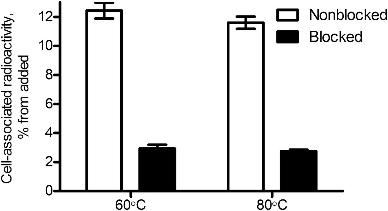
In vitro specificity test for 177Lu-ABY-027 labeled at 60°C and 80°C on SKOV-3 cells (average of 3 ± SD).
Cellular retention and processing experiments demonstrated good retention of 177Lu radioactivity after interrupted incubation of SKOV-3 cells with 177Lu-ABY-027 (Fig. 2A): half of the cell-associated radioactivity was retained at 24 h after interrupted incubation. Most of the radioactivity release occurred during the first 4 h. Only half of the cell-associated radioactivity had been internalized after 24 h. Size-exclusion chromatography of incubation medium showed that after 24 h of incubation approximately 40% of the radioactivity in the medium was associated with low-molecular-weight (<5 kDa) radiocatabolites, indicating that some of the conjugate was processed intracellularly and radiocatabolites were excreted (Fig. 2B).

(A) Cellular processing of 177Lu-ABY-027 in SKOV-3 cells after interrupted incubation (average of 3 samples ± SD). In each sample, radioactivity was normalized by cell-associated radioactivity at time of interruption. (B) Size-exclusion analysis of incubation medium after interrupted incubation of 177Lu-ABY-027 with SKOV-3 cells (average of 2 ± SD).
Tumor Uptake and Biodistribution
The direct comparison of the biodistribution pattern of 111In-ABY-027 and 111In-CHX-A″-DTPA-ABD-(ZHER2:342)2 in tumor-bearing mice (Table 1) demonstrated a 2-fold-higher tumor uptake and 3-fold-lower radioactivity retention in kidneys for 111In-ABY-027. The radioactivity concentration in other studied tissues was slightly higher for 111In-ABY-027. Data for 111In-CHX-A″-DTPA-ABD-(ZHER2:342)2 were in good agreement with published results (17,18).
Comparison of Biodistribution of Radiolabeled Potential Affibody-Based Therapeutics in Tumor-Bearing Mice
Data on the influence of ABY-027 protein dose on tumor uptake and biodistribution of 177Lu-ABY-027 in mice bearing SKOV-3 xenografts are presented in Figure 3. No dose-dependent significant difference in organ accumulation of radioactivity at 48 h after injection of 1, 10, or 50 μg of ABY-027 can be seen, except from a slightly (but significantly) higher retention of radioactivity in the carcass in mice injected with 1 μg of conjugate. The apparent lower tumor concentration of 177Lu in mice injected with 50 μg of conjugate was not significantly separate from the value obtained with 10 μg (P > 0.05). In further experiments, a dose of 10 μg per animal was used.
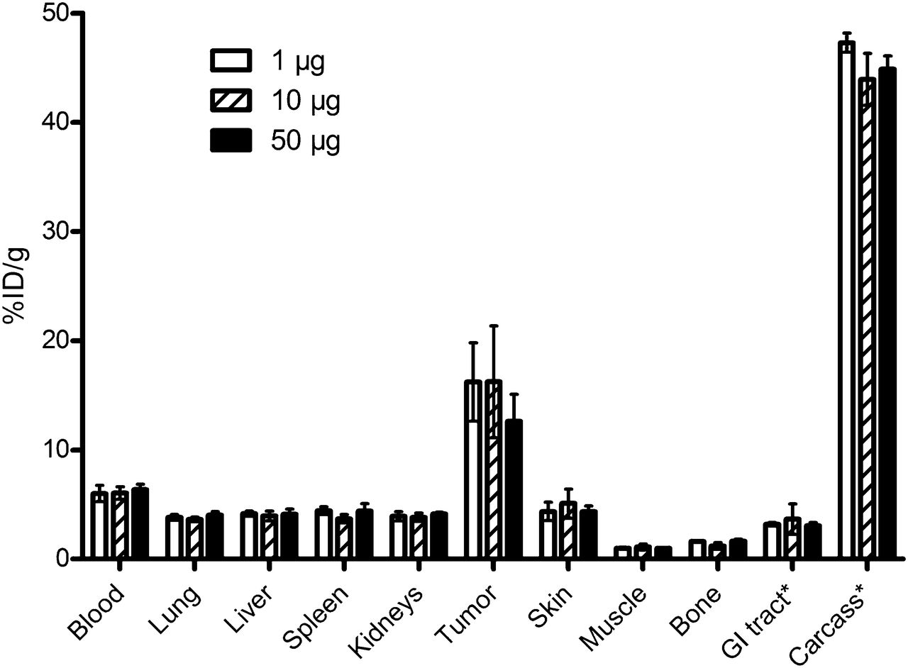
Uptake of radioactivity at 48 h after injection of 1, 10, or 50 μg of 177Lu-ABY-027 in SKOV-3–xenografted mice (average of 4–6, %ID/g ± SD). *For gastrointestinal (GI) tract (with content) and carcass, data are presented as %ID per whole sample.
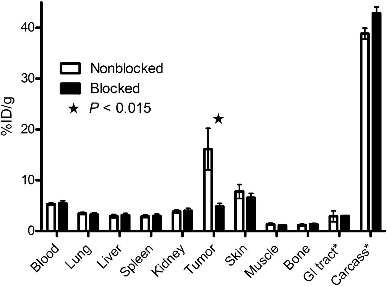
Data on the biodistribution of 177Lu-ABY-027 in BALB/C nu/nu mice bearing SKOV-3 xenografts at different time points after injection are presented in Figure 4 and Table 2. The mean tumor weight was 98 ± 42 mg in these experiments. Figure 4 presents the results of the targeting specificity test. The tumor radioactivity concentration at 48 h after injection was decreased from 17 ± 7 to 5 ± 1 %ID/g (P < 0.015) by preinjection of 1 mg of nonlabeled ABY-027. There was no significant difference in the radioactivity concentrations in all other organs and tissue samples. The decrease of 177Lu-ABY-027 uptake in the tumor by presaturation of HER2 confirms that the tumor-targeting is receptor-mediated.

In vivo binding specificity of 177Lu-ABY-027, 48 h after injection (average of 4, %ID/g ± SD). Blocked group was preinjected with 1 mg of nonlabeled ABY-027. *For gastrointestinal (GI) tract (with content) and carcass, data are presented as %ID per whole sample.
Biodistribution of 177Lu-ABY-027 (Injected Dose, 10 μg) in BALB/C nu/nu Mice Bearing HER2-Expressing SKOV-3 Xenografts
177Lu-ABY-027 demonstrated efficient targeting of HER2-expressing SKOV-3 xenografts in nude mice (Table 2). Already at 24 h after injection, the tumor radioactivity concentration was equal to the radioactivity concentration in blood and higher than in all other studied organs and tissue samples. The concentration of 177Lu in tumors peaked at 48 h after injection, followed by slow washout. Up to 14 d after injection, the concentration of the radioactivity in the tumor exceeded the concentrations in any other organs. The blood clearance was slow, with an elimination half-life of 41 h. The concentration of radioactivity in excretory organs (kidneys and liver) was lower than that in tumors and blood, peaking between 24 and 48 h after injection.
Dosimetry Calculations
The fraction of decays per gram of the administered activity (the residence time) was calculated by integrating the kinetic data (area under the curve) obtained in BALB/c nu/nu mice bearing SKOV-3 tumor xenografts and is presented in Table 3. The contribution of extrapolated data was small, less than 10%. The residence time was multiplied with a conversion factor to obtain the absorbed dose given in Table 3. The data on a previous ABD-fused Affibody molecule variant, 177Lu-CHX-A″-DTPA-ABD-(ZHER2:342)2 (17), are provided for comparison. The data were obtained in the same xenograft model and calculated using the same method. The comparison suggests that the ratio of doses to tumor and bone is equal for both conjugates. At the same time, tumor-to-liver (4.9 for 177Lu-ABY-027 vs. 2.8 for 177Lu-CHX-A″-DTPA-ABD-(ZHER2:342)2) and tumor-to-kidney (3.9 for 177Lu-ABY-027 vs. 1.4 for 177Lu-CHX-A″-DTPA-ABD-(ZHER2:342)2) dose ratios were appreciably higher for 177Lu-ABY-027.
Dosimetry of 177Lu-ABY-027 in Mice in Comparison with 177Lu-CHX-A″-DTPA-ABD-(ZHER2:342)2 (17)
DISCUSSION
Targeted delivery of cytotoxic radionuclides is one potential option for treatment of HER2-expressing malignant tumors that are resistant to receptor-blocking therapies. Numerous attempts have been made, with smaller proteins having better extravasation and penetration in tumors than antibodies. However, several studies have demonstrated that the use of small HER2-targeting proteins such as Fab′2 (27), diabodies (28), DARPins (29), nanobodies (30), and Affibody molecules (16,24) labeled with radiometals all resulted in a high concentration of radioactivity in the kidneys. In each case, the renal concentration of radioactivity exceeded the tumor concentration severalfold, suggesting a high risk of severe nephrotoxicity as a side effect of targeted radionuclide therapy.
We have previously shown that fusion of a dimeric anti-HER2 Affibody molecule with an ABD provided a protein capable of strong binding to albumin in vivo (17,18). This binding prevented rapid glomerular filtration and tubular reabsorption and endowed a conjugate with favorable targeting properties for radiotherapy. Dosimetry calculations demonstrated that the dose to the tumor exceeded the dose to the kidney 1.4-fold, and curative treatment in a microxenograft model was possible (17). However, a further reduction of renal uptake would be desirable, and the present study was based on the hypothesis that a stronger binding to albumin might reduce the renal uptake of the radiolabeled conjugate. Furthermore, a strong binding to albumin is also expected to minimize losses due to lysosomal catabolism after pinocytosis of albumin carrying the conjugate, by enabling maximal use of neonatal Fc receptor–mediated salvage of albumin (31). Thus, ABD035 with femtomolar affinity for albumin may offer a pharmacokinetic advantage as a fusion partner to new targeting agents.
The surface plasmon resonance experiment showed that ABY-027 binds to HER2 with an affinity (KD, 74 pM) identical to the affinity of parental ZHER2:2891 (KD, 76 pM). This finding supported our assumption that the HER2-targeting moiety should preferably be situated at the N terminus of the fusion protein to preserve the affinity by minimizing sterical interference from ABD. The construct was efficiently and stably labeled with 177Lu at 60°C with preserved HER2-binding capacity (Fig. 1). The cellular processing experiment suggested a moderate internalization rate (Fig. 2), although more rapid than that for the parent anti-HER2 Affibody molecule (24).
As expected, a clear effect on the half-life of ABD binding to the serum albumin was demonstrated. The elimination half-life of the parental ZHER2:2891 Affibody molecule from blood was increased 80-fold, from 0.5 h (24) to 41 h. Furthermore, in SKOV-3–xenografted mice biodistribution experiments with ABY-027 labeled with 177Lu showed more than a 3-fold reduced renal accumulation of radioactivity, compared with 177Lu-CHX-A″-DTPA-ABD-(ZHER2:342)2 (17), with retained tumor uptake. Targeting of HER2-expressing xenografts was specific, because saturation of HER2 by preinjection of an excess of nonlabeled ABY-027 caused a significant decrease of tumor uptake (Fig. 4). At the same time, targeting was equally efficient in a broad range of injected protein doses (Fig. 3). Thus, 111In-ABY-027 provides an efficient expansion of the plasma half-life and a reduction of renal uptake of Affibody molecules as well as specific and improved targeting of HER2-expressing xenografts in comparison with previously reported ABD-(ZHER2:342)2 (Table 1). Importantly, the circulatory half-life of 177Lu-ABY-027 in mice was 2- to 2.5-fold shorter than the half-life of radiolabeled anti-HER2 antibodies (25,32), and the molecular weight of 177Lu-ABY-027-albumin adduct (~80 kDa) is approximately half the weight of an antibody. These together should reduce problems seen when using antibodies for radioimmunotherapy, such as an excessively long residence time in blood and poor tumor penetration. It has been previously shown that the penetration of a Fab–albumin-binding peptide–albumin complex (size, ~120 kDa) was much better than what would be assumed from its size and at least equal to that of the noncomplexed Fab (19). We have previously shown a curative effect of targeted radionuclide therapy in mice carrying SKOV-3 xenografts using an anti-HER2 Affibody molecule fused with the first-generation ABD (17), in contrast to the mAb pertuzumab yielding only prolonged survival in the same tumor model (25).
As discussed above, the current Affibody ABD fusion protein is a development from the first-generation HER2-binding ABD fusion protein (17,18). Recently, Hoppman et al. (33) evaluated the alternative approach of direct chemical coupling of ZHER2:342 to HSA. We have made a comparison of the biodistribution of these different constructs, using the data from 48 h after injection because this was the only common time point (Table 1). The study of the construct 111In-DOTA-ABD-(ZHER2:342)2 was performed in mice xenografted with the low-HER2-expressing cell line LS174T, compared with the other 3 constructs studied, possibly explaining the lower tumor values in xenografts of the high-HER2-expressing cell line SKOV-3. Furthermore, in addition xenografts based on the same cell line may show batch-to-batch variability and variation in biodistribution pattern of the same conjugate labeled with different radionuclides (34). Therefore, comparisons are preferably done head-to-head in the same batch of xenografts, as with 111In-ABY-027 and 111In-CHX-A″-DTPA-ABD-(ZHER2:342)2 in the present study. However, data from normal tissues should be directly comparable. Despite multiple HER2 bindings, the HSA conjugate did not provide the highest tumor accumulation, compared with the fusion proteins analyzed in the high-HER2-expressing xenograft model. Furthermore, the high levels in the liver and spleen, as well as the faster clearance, suggest negative effects of multiple random conjugations on the properties of albumin. On the contrary, the novel fusion protein based on affinity-matured ABD provided the lowest kidney values and also the lowest values in the liver and spleen. A comparison of dosimetry of 177Lu-ABY-027 and 177Lu-CHX-A″-DTPA-ABD-(ZHER2:342)2 (17) are presented in Table 3. Both calculations were based on the same methodology, and the biodistribution input data were obtained in the same animal model, BALB/C nu/nu mice bearing SKOV-3 xenografts. A curative effect was obtained after injection of 21.6 MBq of 177Lu-CHX-A″-DTPA-ABD-(ZHER2:342)2 (17). With 177Lu-ABY-027, the same dose to tumor would be achieved with 33.1 MBq. With the specific activity used in the present study, this administration of such activity would require injection of 22–25 μg of 177Lu-ABY-027 per mouse. As shown in the present study, injection of such a protein dose is in a range where small differences in the injected protein dose insignificantly affect tumor uptake. A comparison at equal dose to tumor shows that the dose to blood and bone would be similar for the 2 constructs and that 177Lu-ABY-027 would give a 2.8- and 1.8-fold-lower dose to kidney and liver, respectively.
CONCLUSION
ZHER2:2891-ABD035-DOTA (ABY-027) is a fusion protein having a biodistribution suitable for therapy and showing retained HER2-specific binding. The capacity of specific targeting of HER2-expressing xenografts was shown in vivo. In a murine model, 177Lu-ABY-027 provided an appreciable reduction of renal and hepatic uptake of radioactivity in comparison with alternative approaches. Site-specific conjugation of DOTA provides a uniform conjugate and creates the potential for labeling with a broad range of therapeutic radionuclides. The unique cysteine can also be used for site specific coupling of other effector functions.
DISCLOSURE
The costs of publication of this article were defrayed in part by the payment of page charges. Therefore, and solely to indicate this fact, this article is hereby marked “advertisement” in accordance with 18 USC section 1734. This work was supported by grants from Swedish Research Council (Vetenskapsrådet) and Swedish Cancer Society (Cancerfonden). Affibody AB holds intellectual property rights and trademarks for Affibody molecules, which might be considered as a potential conflict of interest. However, we believe that we have presented data and considerations in a scientifically strict and unbiased way and refer to results published in established international peer-review journals. No other potential conflict of interest relevant to this article was reported.
Footnotes
Published online Mar. 25, 2013.
- © 2013 by the Society of Nuclear Medicine and Molecular Imaging, Inc.
REFERENCES
- Received for publication July 1, 2012.
- Accepted for publication November 27, 2012.


Jump to section
Related Articles
Cited By...
- Structure-activity analysis of truncated albumin-binding domains suggests new lead constructs for potential therapeutic delivery
- Human Epidermal Growth Factor Receptor 3-Specific Tumor Uptake and Biodistribution of 89Zr-MSB0010853 Visualized by Real-Time and Noninvasive PET Imaging
- Diabody Pretargeting with Click Chemistry In Vivo
- An engineered affibody molecule with pH-dependent binding to FcRn mediates extended circulatory half-life of a fusion protein
- Positron Emission Tomography Imaging with 18F-Labeled ZHER2:2891 Affibody for Detection of HER2 Expression and Pharmacodynamic Response to HER2-Modulating Therapies