


Abstract
Radioimmunotherapy (RIT) of solid tumor is often limited in efficacy because of restrictions in achieved tumor dose. In an effort to overcome this, the combination of RIT with other therapeutic modalities was investigated in an animal model of breast carcinoma. The rationale for this combined-modality RIT (CMRIT) was to increase the therapeutic efficacy of RIT through the use of paclitaxel to arrest cells in the radiosensitive G2/M phase of the cell cycle. Methods: In this study, the biodistribution and therapeutic efficacy of 90Y-radiolabeled humanized anti-Lewis Y hu3S193 monoclonal antibody (90Y-hu3S193) RIT in combination with paclitaxel chemotherapy was explored in a Lewis Y–expressing MCF-7 tumor xenografted BALB/c nude mouse model of breast cancer. Results: Biodistribution studies demonstrated excellent tumor targeting and limited normal tissue uptake by 90Y-hu3S193. A therapeutic study with established tumors assessed 90Y-hu3S193 as a single agent and demonstrated significant antitumor effects in all animals receiving a single intravenous 1.85 or 3.70 MBq dose of this treatment compared with phosphate-buffered saline placebo controls (P = 0.0008 vs. P < 0.0001). Complete responses were observed in all animals in the 3.70 MBq study arm for the duration of the study. Single-dose 90Y-hu3S193 plus paclitaxel (600 μg) CMRIT displayed improved efficacy over single-modality therapies, with a significant difference (P < 0.0001) between the mean percentage change in tumor volume in mice receiving 0.46 MBq 90Y-hu3S193 alone and when combined with 600 μg paclitaxel. Conclusion: The significant efficacy of 90Y-hu3S193 and paclitaxel CMRIT at low radiation doses in this model of breast carcinoma indicates its therapeutic potential and warrants further investigation into this promising therapeutic approach.
Despite advances in the treatment of breast cancer, it remains a predominant cause of death from cancer in women, with the 5-y survival reduced from 97% to 23% when distant metastases occur (1). Although current therapies such as chemotherapy can effectively treat >60% of patients, cure of advanced and metastatic disease is uncommon, and novel methods of treatment are needed (2–4). Monoclonal antibody (mAb) therapy of cancer is one such treatment that may prove beneficial in the management of metastatic breast cancer (3,5–7). A number of target antigens for mAb therapy have been identified in breast cancer, including HER-2, MUC-1, carcinoembryonic antigen, and Lewis Y (Ley); therefore, this disease is an ideal candidate for mAb therapy (8).
The Ley antigen belongs to the family of type 2 blood group–related difucosylated oligosaccharide antigens and has the specific chemical structure Fucα1 → 2Galβ1 → 4(Fucα1 → 3) GlcNAcβ1 → 3Gal (9). The expression of Ley antigen is associated with 60%−90% of carcinomas of epithelial cell origin, including breast, colon, lung, gastric, prostate, and ovarian cancer (5,10,11). The high frequency and relatively homogeneous nature of Ley expression in both primary and metastatic tumors, along with its high density and altered expression on tumor cells, have led to its targeting in cancer immunotherapy (5,9,12). Murine 3S193 was originally developed as described by Kitamura (9). To allow clinical applications of this antibody, it was humanized by Complementarity Determining Region (CDR) grafting to reduce immunogenicity. The humanized form of 3S193 (hu3S193) retains the murine antibody's specificity, demonstrates improved immune effector functions of antibody-dependent cell-mediated cytotoxicity and complement-dependent cytotoxicity, and also significantly slows the growth of newly established MCF-7 xenografts (13). Preclinical biodistribution studies with radiolabeled hu3S193 have demonstrated superior intracellular uptake and tumor retention of radiometal isotopes over radiohalides, demonstrating the suitability of hu3S193 for radioimmunotherapy (RIT) with radiometal isotopes (5).
Although RIT has demonstrated some efficacy in the treatment of aggressive metastatic tumors such as breast cancer in animal models, clinical studies have shown that significant tumor regression rates have not been observed through the use of this single therapy (14). Novel therapies are required in the treatment of breast cancer, and the relatively high therapeutic index of RIT makes this therapy an ideal candidate for combination with other therapies in synergistic treatment aimed at increasing therapeutic efficacy while minimizing toxicity (4,14,15).
Tumor cells are most sensitive to radiation during the G2/M phase of the cell cycle; therefore, therapies that target this stage of the cell cycle may increase the efficacy of RIT (16). One such agent is the anticancer drug paclitaxel (Taxol; Bristol Myers-Squibb), a member of plant-derived antineoplastic compounds known as taxanes (15). Paclitaxel is a microtubule stabilizer, which causes cells to be arrested in the radiosensitive G2/M phase and also causes the induction of apoptosis through other mechanisms, making it ideal for use in combined-modality RIT (CMRIT) (4). In a therapeutic study assessing the efficacy of 131I-labeled hu3S193 as a single agent, and in combination with a subtherapeutic dose of paclitaxel, the combination of RIT with chemotherapy resulted in a significant antitumor effect in 80% of treated mice, whereas no response was observed in placebo or control groups (17).
Currently, the radiometal 90Y is considered to be a suitable alternative to 131I and has characteristics close to ideal for use in RIT, such as a half-life of 1–3 d, major energy emission ranging between 0.5 and 30 cell diameters, and decay to a ground state (18,19). 90Y is a pure high-energy β-emitter, having a maximum energy of 2.27 MeV, compared with the maximum emission of 131I of 0.61 MeV, and a half-life of 64 h (2.67 d), compared with approximately 8 d for 131I (20). The greatest advantage that 90Y possesses over 131I is a longer radiation track length of 12 mm compared with just 2 mm for 131I, which allows for irradiation of large tumors and those with heterogeneous expression of the target antigen. Furthermore, 90Y is also retained intracellularly after antibody internalization and is therefore able to deliver a higher and extended dose of radiation compared with the rapidly degraded 131I (18).
As RIT has demonstrated promise in the treatment of malignancy, this study aimed to assess the efficacy of 90Y RIT in an experimental xenograft model of breast cancer. In an effort to maximize the effectiveness of RIT while reducing associated toxicities, the synergistic effects of low-dose RIT with the chemotherapeutic agent paclitaxel was also explored in CMRIT studies.
MATERIALS AND METHODS
mAbs
Humanized 3S193 (hu3S193), a CDR grafted IgG1 antibody specific for the Ley antigen (13), and isotype control huA33 (21) were produced by the Biologic Production Facility, Ludwig Institute for Cancer Research (Melbourne).
Cell Culture
MCF-7, a Ley-positive breast adenocarcinoma cell line obtained from the American Type Culture Collection, and the colon carcinoma cell line SW1222 (Ludwig Institute for Cancer Research, New York, NY) were grown in RPMI 1640 medium supplemented with 10% fetal calf serum (CSL Ltd.), 5% penicillin/streptomycin (penicillin G 5,000 units/mL and streptomycin sulfate 5,000 μg/mL; CSL Ltd.), and 5% l-glutamine (200 mmol/L stock; JRH Biosciences) in 175-cm2 flasks (Nunc; Nunclon), and incubated at 37°C in 5% CO2 incubators (Forma Scientific). Cell viability, as determined by trypan blue exclusion, exceeded 90% in all experiments.
Antibody Labeling
The radiolabeling of hu3S193 and isotype control huA33 with 90Y radioisotope was achieved through the bifunctional metal ion-chelating agent C-functionalized trans-cyclohexyl-diethylenetriaminepentaacetic acid (CHX-A″-DTPA) (22). The procedure is a modification of the previously published method by Nikula et al. (23). Five microliters of 90Y (89.2 MBq; MDS Nordion) in 0.04 mol/L HCl were mixed with 10 μL 2 mol/L sodium acetate. To this solution, 4.4 μL of 1 mol/L HCl were added, with 10 μL of this mixture added to 26 μL of CHX-A″-DTPA–chelated antibody. The reaction mixture was incubated for 30 min at room temperature, and the 90Y-CHX-A″-DTPA–hu3S193 (90Y-hu3S193) was purified using a Sephadex G-50 column (Pharmacia) equilibrated with saline.
Radioconjugate Characterization
The immunoreactivity of the 90Y-hu3S193 was determined by linear extrapolation to binding at infinite antigen excess according to the cell-binding assay of Lindmo et al. (24) and by single-point immunoreactivity assays performed as previously described (5). Instant thin-layer chromatography (ITLC) was performed to determine the amount of free radionuclide (90Y) versus that bound to the hu3S193 mAb after the radiolabeling procedure (23), as described (5). Single-point immunoreactivity assays were also performed to assess the in vivo stability of the 90Y-hu3S193 antibody incubated in serum over a 7 d period, with the assay performed at 0 (no incubation), 72, and 168 h. Briefly, 10 μg of 90Y-hu3S193 were added to 200 μL of healthy donor human serum and incubated at 37°C over a 7 d period. At each time point, 21.8 μL (∼1 μg) of the incubated antibody in sera were diluted 1:50 in saline. Twenty microliters of the diluted 90Y-hu3S193 were then added to 30 × 106 MCF-7 cells for the single-point immunoreactivity assay.
Animal Model
Tumor xenografts were established in female BALB/c nude (nu/nu) mice, 5 to 6 wk old, obtained from the Animal Resource Centre, Western Australia. The in vivo growth of MCF-7 xenografts is estrogen dependent, necessitating estrogen implants on the day of cell inoculation into the mice to sustain the xenografts as previously described (5). MCF-7 cells (25 × 106 in 150 μL RPMI medium) were injected subcutaneously into the left mammary line of the BALB/c nude mice to form a cell pellet from which the tumor xenograft was established. In the biodistribution studies, Ley-negative SW1222 colon carcinoma control tumors were established approximately 2 wk before the studies because of their more rapid growth rates. To establish the SW1222 tumors, 10 × 106 cells in 150 μL RPMI medium were injected subcutaneously in the right mammary line of the mice, on the flank opposite to the primary MCF-7 tumor. Tumor growth was regularly measured using digital callipers and the formula: tumor volume (TV) = (length × width2)/2 of the tumor, where length is the longest axis and width is the measurement at right angles to length (5). TV is expressed in cubic millimeters, allowing construction of tumor growth curves over time. All animal studies were approved by the Animal Ethics Committee of the Austin Hospital, (Heidelberg Victoria, Australia).
Biodistribution Study
The biodistribution study was performed to assess the in vivo distribution of the 90Y-hu3S193 antibody in xenografted BALB/c nude mice. The first component of this study comprised 15 BALB/c mice bearing MCF-7 and control SW1222 xenografts with a mean MCF-7 TV ± SD of 480 ± 143 mm3. Mice were injected with 90Y-hu3S193 (5 μg protein, 0.28 MBq activity) in 100 μL saline by tail vein injection. Groups of 5 mice were culled at 4, 24, or 72 h after 90Y-hu3S193 injection. In a second biodistribution study, 20 BALB/c MCF-7 and SW1222 xenografted mice with a mean MCF-7 TV ± SD of 398 ± 187 mm3 were injected with 0.28 MBq 90Y-hu3S193 as for the initial biodistribution study. Five mice were assigned to each time point, with this second study assessing biodistribution at 48, 168, 240, and 288 h after injection of the radiolabeled antibody. Immediately after euthanasia, the mice were bled via cardiac puncture, and the blood was collected in γ-counter tubes. Both the MCF-7 and SW1222 control tumors were collected, along with liver, spleen, kidney, muscle, skin, bone (femur), lung, heart, stomach, brain, and the small bowel. Once removed, all tissues were counted on a Cobra II Auto Gamma Counter (Packard Instruments), and the percentage injected dose per gram (%ID/g) was calculated. The ratio of %ID/g in the MCF-7 tumor relative to the blood was also calculated to allow a comparison of 90Y-hu3S193–specific uptake into the tumor from the blood.
The physical distribution of 90Y-hu3S193 in tumors at 4, 24, and 72 h after injection in the initial biodistribution study was also assessed using autoradiography as previously described (5). To enable comparison between 90Y-hu3S193 biodistribution and Ley expression, immunohistochemistry and histology analysis was performed on the same series of tumor sections prepared for autoradiography as previously described (5).
Therapy Studies
The combined therapy of 90Y-hu3S193 with paclitaxel was investigated in BALB/c nude mice bearing established MCF-7 tumors. In the first component of the study, groups received radiolabeled antibody alone: mice were injected with 0.46, 0.92, 1.85, or 3.70 MBq 90Y-hu3S193 (75 μg protein) or equivalent doses of the isotype control antibody, 90Y-huA33, by tail vein injection. Five mice were used for each dose level of 90Y-hu3S193, whereas 4 mice were included in each 90Y-huA33 control grouping. A separate group of mice received vehicle (phosphate-buffered saline [PBS]) at injection volumes equivalent to the radiolabeled antibodies.
In the second component of this study examining the efficacy of combinational therapy between RIT and paclitaxel chemotherapy, only the lower 3 subtherapeutic doses of radiolabeled antibody were used to assess any synergistic effects of the combined therapies. The lower doses were chosen because 3.70 MBq of 90Y-hu3S193 administered as a single agent has previously been shown to be effective in reducing TV in MCF-7 xenografts (Fook T. Lee, unpublished data). Paclitaxel was administered intraperitoneally at a 600 μg dose (6 mg/mL) in an aqueous solution containing 100 μL paclitaxel diluted in 400 μL saline for injection (0.9% saline; Pharmacia & Upjohn). This dose has been shown to have no significant efficacy when administered alone and did not demonstrate toxicity in BALB/c nude mice (4,17).
In parallel to the groups of mice receiving 0.46, 0.92, or 1.85 MBq of radiolabeled antibody (75 μg protein hu3S193) with 600 μg paclitaxel, a control arm received only 600 μg paclitaxel as a secondary control in this study. All 16 groups of mice in this study received treatment 16 d after establishment of the MCF-7 xenografts, when the mean TV ± SD was 141 ± 33 mm3 (range, 67–211 mm3). The day of RIT treatment was designated day 0 of the study, with those mice in the combined-modality study receiving the 600 μg dose of paclitaxel on day 1 of the study. The TV and health of animals were monitored daily. Individual mice, or whole treatment groups, were culled when the TV exceeded 1 g (1,000 mm3) or when toxicity became apparent. Because of the estrogen pellets' duration of 90 d release, the study was concluded on day 70, 86 d after implantation of the pellets.
A control study comprising 3 groups of 5 mice was performed to examine the efficacy of unlabeled hu3S193 on MCF-7 tumor growth. Mice received PBS, 75 μg hu3S193 alone, or 75 μg of the mAb in combination with 600 μg paclitaxel. Mice in this study received treatment 15 d after xenograft establishment when the mean TV ± SD was 164 ± 36 mm3 (range, 124–233 mm3). Combination therapy groups received paclitaxel 24 h after antibody administration as for the initial study.
Tumor Response and Toxicity Definitions
TVs were expressed as a percentage change relative to the initial TV of a mouse on day 0 of the study. This was calculated using the formula (25):
In mice where an at least a 50% decrease in TV was observed for at least 7 d, a partial response (PR) was recorded. In mice where a complete regression was recorded for a 7 d period (i.e., TV reduction = 100%), a complete response (CR) was recorded. If any animal demonstrated a CR lasting until the end of the 70-d study, these mice were recorded as cured (25). Toxicity was defined as a >15% body weight loss in any animal, with all animals euthanized for reasons other than excessive tumor burden (>1,000 mm3) undergoing postmortem examination.
Statistical Analysis
Statistical analysis of the therapy study results was performed using an unpaired 2-tailed t test on mean TVs ± SD and percentage change of TVs from day 0 to day 45 of the study, corresponding to the termination of the PBS control group. At this time point, tests were performed between the PBS control group and all other treatment groups, between equivalent doses of radiolabeled hu3S193 and the huA33 control antibody and also between doses of 90Y-hu3S193 alone and in combination with paclitaxel. Significance was set at the 95% level, with results declared statistically significant if P < 0.05.
RESULTS
Antibody Labeling
The immunoreactivity of the 90Y-hu3S193 immunoconjugate with the Ley antigen was determined to be 53.7%. The association constant (Ka) of 90Y-hu3S193 was 1.1 × 10⁷ mol/L−1, and the number of antibody-binding sites per MCF-7 cell was calculated to be 4.3 × 10⁶, consistent with earlier results (17). Radiochemical purity (ITLC) analysis of 90Y-hu3S193 indicated a >99% radiochemical purity for the 90Y-hu3S193 immunoconjugate. During incubation at 37°C in serum, a slight decrease to 97.1% was recorded by day 3, which further declined to 91.2% radiochemical purity by day 7. The single-point immunoreactivity of 90Y-hu3S193 on day 0 was determined to be approximately 45%. Upon incubation at 37°C in serum, a decrease in immunoreactivity to approximately 18% was observed after 3 d, with a further decrease to approximately 10% after incubation for 7 d.
Biodistribution Studies
Results from the biodistribution study are shown in Figures 1 and 2. Peak tumor uptake of 90Y-hu3S193 antibody was 58.04 ± 15.18 %ID/g at 72 h after injection, rising steadily from 17.52 ± 3.89 %ID/g recorded at 4 h after injection. At 72 h after injection, uptake in the control SW1222 tumor was only 7.71 ± 2.05 %ID/g, indicating the specific uptake of the hu3S193 antibody into the Ley-positive MCF-7 tumors. At the final time point of the biodistribution study (t = 288 h), the %ID/g for the MCF-7 tumor was 38.5%, indicating prolonged tumor retention of 90Y-hu3S193 (Fig. 2). Blood clearance for 90Y-hu3S193 occurred progressively, falling from 48.68 ± 5.10 %ID/g at 10 min after injection to only 3.13 ± 0.68 %ID/g at 288 h after injection (Fig. 2). As shown in Figure 1, normal tissues displayed low levels of uptake consistent with blood-pool activity.
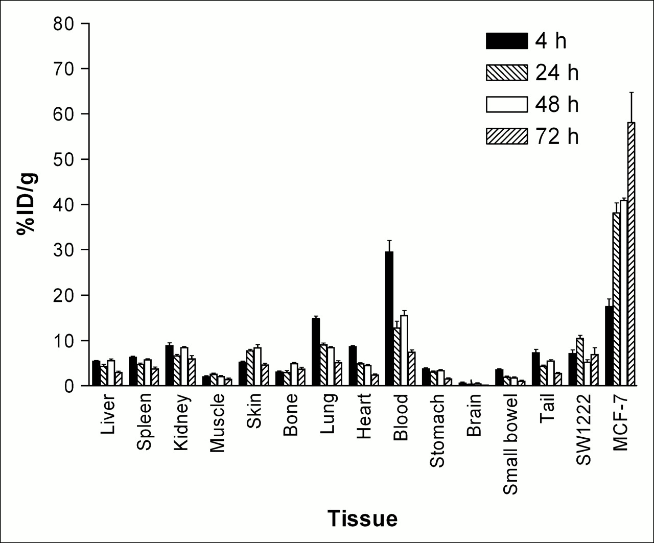
Biodistribution of 90Y-hu3S193 in normal murine tissues, blood, MCF-7 tumors, and control SW1222 tumors at selected time points after injection into BALB/c nude mice. Results (%ID/g) are expressed as mean ± SD (n = 4–5).

Biodistribution of 90Y-hu3S193 in blood (▪), MCF-7 tumors (▴), and control SW1222 tumors (▾), with %ID/g expressed as mean ± SD (n = 4–5).
Autoradiography, Immunohistochemistry, and Histology
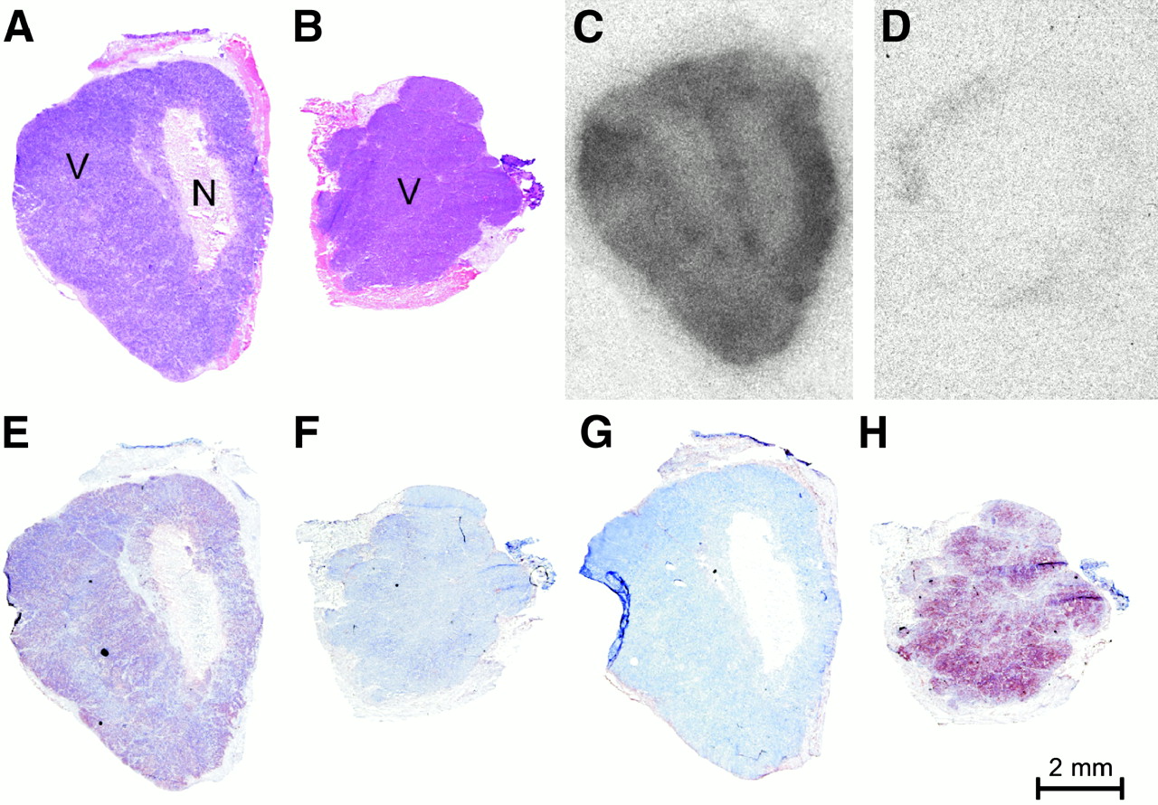
The distribution of 90Y radioactivity, antigen expression, and viability of tumor xenografts is shown in Figure 3. At 72 h after injection, corresponding to maximal tumor uptake, hematoxylin staining indicated large areas of viable MCF-7 tumor, which was disrupted by a large region of clearly necrotic tissue (Fig. 3A). Ley expression was evident in viable tumor, with no positive staining evident in the necrotic region of the tumor, or peripheral murine tissue (Fig. 3E). In all viable areas of the tumor, distribution of radioactivity by autoradiography was relatively uniform, although some more peripheral areas demonstrated intense localization (Fig. 3C). This distribution of radioactivity was also evident at earlier time points examined, with an increase in radioactive intensity over time apparent (data not shown). A marked difference was apparent between a control SW1222 colon tumor xenograft with no Ley and high A33 antigen expression (Fig. 3H) and little radioactivity observed.

Uptake of 90Y-hu3S193 in tumor xenografts. Analysis of MCF-7 xenograft at 72 h after injection by hematoxylin–eosin staining (A), autoradiography (C), Ley immunohistochemistry (E), and huA33 immunohistochemistry (G). SW1222 control tumor xenograft at 72 h after injection by hematoxylin–eosin staining (B), autoradiography (D), Ley immunohistochemistry (F), and huA33 immunohistochemistry (H). V = viable tumor; N = necrotic.
Therapy Studies
All treatments were tolerated well, and the maximum tolerated dose (MTD) of 90Y-hu3S193 RIT was not reached in this study. Six mice died, or were culled during the study, with 2 mice euthanized on day 24 (0.92 MBq 90Y-huA33 and 1.85 MBq 90Y-hu3S193), with metastases identified on postmortem examination. A further 3 mice receiving 1.85 MBq 90Y-hu3S193, 0.92 MBq 90Y-huA33 and paclitaxel, and 0.46 MBq 90Y-huA33 and paclitaxel were also euthanized or died on days 32, 33, and 52, respectively, due to weight loss possibly attributable to radiotoxicity or micrometastatic disease. On day 70, 1 mouse treated with 1.85 MBq 90Y-hu3S193 and paclitaxel was euthanized because of weight loss, with metastatic disease apparent on postmortem examination. No other animals had significant weight loss.
RIT Dose Escalation
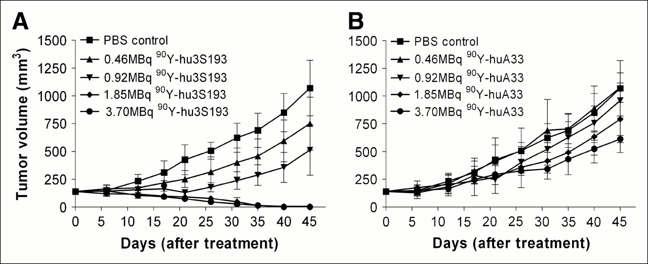
A dose–response effect was apparent in the RIT study, with those mice receiving the highest dose of 90Y-hu3S193 demonstrating a more marked suppression of tumor growth (Table 1; Fig. 4). At all RIT doses, a significant difference was evident between 90Y-hu3S193 and 90Y-huA33, with mice that received 90Y-hu3S193 achieving more significant suppression of tumor growth, especially at higher doses (Table 2; Fig. 4). The greatest efficacy in the single agent component of this study was observed with the highest dose of 90Y-hu3S193, 3.70 MBq (Fig. 4A). The corresponding group of 90Y-huA33 also achieved improved survival, not reaching TVs exceeding 1,000 mm3 until day 68 of the study (Fig. 4B). By day 38 of the study, all mice receiving 3.70 MBq 90Y-hu3S193 had complete resolution of tumor xenografts (100% CR), with this therapy demonstrating remarkable efficacy. This clearly significant decrease in TV was sustained for the remainder of the study, with no tumor regrowth observed; accordingly, these mice were defined as cured. Despite receiving the highest dose of radiation administered in this study, no toxicity was observed, with increases in body weight recorded as evidence of improved health. Mice receiving 1.85 MBq 90Y-hu3S193 also demonstrated defined responses, with 2 mice achieving a CR and 1 achieving a PR on day 45 of the study.

MCF-7 xenograft growth (mm3) in response to single-dose treatment on day 0 with 90Y-hu3S193 (A) or isotype control 90Y-huA33 (B). Results are expressed as mean ± SD (n = 4–5).
TV and Percentage Change in TV on Day 45 of Study
Comparison of Percentage Change in TV Between 90Y-hu3S193 and 90Y-huA33 Antibodies on Day 45
CMRIT
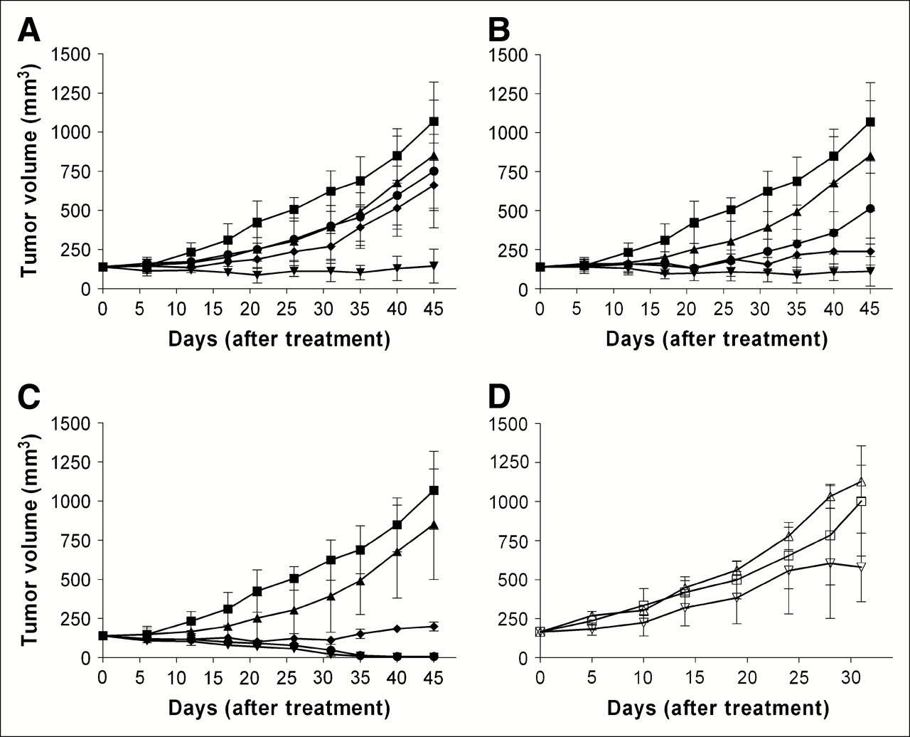
The results of combining RIT and paclitaxel chemotherapy are presented in Figure 5. Paclitaxel as a single agent produced minimal and insignificant tumor suppression that was attributable to the limited subtherapeutic effect of the 600 μg paclitaxel dose. The lowest RIT dose of 0.46 MBq 90Y-hu3S193 combined with paclitaxel was observed to cause significant inhibition of tumor growth on day 45 compared with placebo (P = 0.004), and all mice in this group survived to completion of the study (Fig. 5A). A significant difference from PBS control using 0.46 MBq 90Y-hu3S193 was observed only on the combination with paclitaxel, not as a single agent (Table 1). Furthermore, the combination of paclitaxel with 0.46 MBq 90Y-hu3S193 RIT resulted in a significant increase in antitumor efficacy compared with RIT alone on day 45 (P < 0.0001). As observed in the dose-escalation study, little efficacy was observed for 0.46 MBq 90Y-huA33, with tumor growth similar to that of mice treated with paclitaxel alone (Fig. 5A).

MCF-7 xenograft curves after CMRIT in mice receiving PBS control (▪), 600 μg paclitaxel (▴), 90Y-hu3S193 and 600 μg paclitaxel (▾), 90Y-huA33 and 600 μg paclitaxel (⧫), and 90Y-hu3S193 (•) at doses of 0.46 MBq (A), 0.92 MBq (B), and 1.85 MBq (C) of 90Y-hu3S193 or 90Y-huA33. (D) MCF-7 xenograft growth after single treatments on day 0 of PBS control (□), 75 μg hu3S193 (▵), and 75 μg hu3S193 and 600 μg paclitaxel (▿). Results are expressed as mean ± SD (n = 4–5).
Aside from 1 animal dying early from weight loss/metastatic disease, all animals receiving 0.92 MBq 90Y-hu3S193 with paclitaxel survived until completion of the study. This combination produced significant antitumor effects compared with the control vehicle (Fig. 5B), which were more pronounced than isotype control results (P = 0.003 vs. P = 0.032). As observed for 0.46 MBq, 0.92 MBq 90Y-hu3S193 and paclitaxel CMRIT mediated a significant improvement in antitumor efficacy relevant to RIT alone (P = 0.0109). Two PRs were recorded in the 5 animals receiving 90Y-hu3S193 and paclitaxel CMRIT on day 45 after treatment, whereas none was recorded for mice receiving RIT as a single agent.
All mice receiving 1.85 MBq of RIT with paclitaxel survived until day 70 of the study, although 1 animal was culled at this time because of metastatic disease burden. Treatment with 1.85 MBq 90Y-hu3S193 and paclitaxel produced significant reductions in the TV on day 45 (Fig. 5C), compared with placebo controls (P = 0.002), with all animals (5/5) recorded as achieving CRs on day 40 of the study. Some regrowth of tumors was recorded after day 45 of the study, although a mean reduction of 85% from the initial TV was still present at completion of the study (day 70). Treatment at 1.85 MBq 90Y-huA33 with paclitaxel demonstrated a significant difference from PBS controls, although no overall reduction in the TV from day 0 was recorded.
The control study assessing the efficacy of a single dose of unlabeled hu3S193 (75 μg) on tumor growth, both as a single agent and in combination with paclitaxel, did not produce any significant inhibition of tumor growth (Fig. 5D).
DISCUSSION
CMRIT is a promising regimen in the treatment of cancer where single agent therapies often do not have significant efficacy (4,26,27). This approach aims to increase the therapeutic efficacy while not increasing associated toxicities of either single agent. In this study, the antitumor efficacy of 90Y-labeled anti-Ley hu3S193 in combination with paclitaxel has clearly been demonstrated to be superior to either therapy alone and produced marked and durable tumor responses.
At the highest dose of 1.85 MBq 90Y-hu3S193 with 600 μg paclitaxel, 2 cures and 3 PRs resulted from the treatment, whereas only PRs were observed in mice receiving 1.85 MBq 90Y-hu3S193 as a single agent. This result compares favorably with a previous study assessing the combined efficacy of 131I-hu3S193 and 600 μg paclitaxel, where only PRs were observed, even though a 3.70-MBq dose of radiation was administered (17). The greater antitumor efficacy observed in this study, at radiation doses half of that used for 131I-hu3S193, and with minimal toxicity, indicates the potent effects of the high-energy β-emissions from 90Y RIT, and also results from the longer retention of radiometals at the tumor site compared with radiohalides. In an earlier study using 90Y-labeled chimeric L6 and paclitaxel CMRIT, up to 50% of mice were cured of tumor xenografts using a radiation dose of 9.62 MBq, demonstrating the potency of this therapy in the treatment of aggressive breast cancer xenografts (4).
After radiolabeling with 90Y via the metal ion chelate CHX-A″-DTPA, the immunoreactivity of 53.7% of the 90Y-hu3S193 antibody for the Ley antigen as determined by the Lindmo assay is an improvement over previous studies, which determined immunoreactivity as low as 20%, indicating an improved radioconjugate for use in the Ley antigen–antibody system (5). The Ka of 90Y-hu3S193 of 1.1 × 10⁷ mol/L−1, as determined by Scatchard analysis, agrees closely with previously reported values using 111In-hu3S193, and is within the range expected for antibodies against carbohydrate antigens (5,28,29).
The biodistribution study of 90Y-hu3S193 in BALB/c nude mice was performed to assess the in vivo properties of this radioconjugate. Though unable to accurately reflect the likely biodistribution in humans, because of a lack of endogenous antigen expression analogous to that in humans, valuable insights into the quantitative uptake and retention of antibody into tumors can be gained. High and specific levels of 90Y-hu3S193 uptake were observed throughout biodistribution studies of this radioconjugate, with peak MCF-7 tumor uptake of 58.0 ± 15.2 %ID/g recorded at 72 h after injection This was significantly higher than uptake in the Ley-negative SW1222 control tumors (7.7 ± 2.0 %ID/g), indicating the specificity of MCF-7 tumor uptake. The high tumor uptakes obtained in this study are an improvement over a previous assessment of 90Y-hu3S193 biodistribution, where peak uptake was limited to 22.47 %ID/g recorded at 48 h after injection (5), and may reflect the superior immunoreactivity of the radioconjugate used in the current study. In another Ley-expressing tumor model using pretargeted anti-Ley B3–streptavidin and 111In-labeled biotin, peak uptake of 21.8 ± 2.1 %ID/g was observed at 24 h after injection, indicating the uptake observed using 90Y-hu3S193 to be superior and more prolonged than that of other anti-Ley mAbs (30). Results from other biodistribution studies, assessing 111In-labeled anti-HER2 4D5 and anti-MUC1 PAM4 antibodies, have shown maximal uptakes of approximately 30 and 40 %ID/g, respectively, at 48 h after injection, demonstrating that high levels of tumor localization are possible in mouse xenograft models (18,31). In an animal model biodistribution study of the 111In-labeled anti-GD3 mAb KM871 using the same CHX-A″-DTPA chelate as used in this study, peak tumor uptake of 41.9 ± 7.0 %ID/g was observed at 72 h after injection, indicating that this chelate system is stable in vivo and is able to effect significant and prolonged targeting of tumor xenografts (32).
The prolonged tumor retention observed in this study agrees with other assessments of radiometal-labeled antibodies,and is thought to be related to the internalization and subsequent retention of radioactive metabolites in tumor lysosomes due to enzymatic cleavage (33,34). Recent investigations have shown that hu3S193 mAb does undergo internalization (Andrew M. Scott and Kiki Tahtis, unpublished data), and 90Y-hu3S193 undergoes intracellular trafficking to tumor lysosomes. Antibodies radiolabeled with 90Y have demonstrated similar retention when compared with 111In, which is often used as a surrogate in biodistribution studies because of its γ-emissions, allowing imaging of tumors (35).
The rationale of RIT using β-emitting radioisotopes such as 90Y is the delivery of specific, targeted radiation to both antigen-positive and adjacent antigen-negative tumor cells, while limiting the systemic exposure associated with external beam radiotherapy (16). As reported here, a clear dose-dependent relationship in the efficacy of a single dose of 90Y-hu3S193 was apparent in established tumors, and a MTD of this treatment was not reached. In the single-agent study, maximal efficacy was observed with a single 3.70-MBq dose of 90Y-hu3S193, which resulted in the complete resolution of established tumor xenografts (n = 5) on day 38, which was maintained until completion of the study on day 70. This efficacy observed using RIT as a single modality, within its MTD, indicates its comparative superiority over paclitaxel as respective single modalities. Significantly, even at maximal radiation dose, no evidence of radiotoxicity was observed, and these mice were observed to gain weight. In previous studies of 90Y-labeled antibodies using a breast cancer model, myelotoxicity was assessed by blood count measurements. Moderate decreases were observed after doses of 9.62 MBq (260 μCi), with cell counts recovering after 21 d (4,26). The transient and moderate nature of the myelotoxicity observed in these studies, at doses twice as high as the maximal dose used in this study, suggests that significant myelotoxicity is unlikely to occur at the doses investigated in this study; therefore, direct measurements of radiotoxicity were not performed in this study. It should be noted, though, that a different antibody and radioisotope chelate were used in these studies and, therefore, direct correlation is difficult.
Paclitaxel is known for its radiosensitizing properties, which are observed far below levels required for cytotoxic effects. This is most beneficial in CMRIT, as side effects from the systemic administration of the drug must be minimal for effective therapy (15). The radiosensitizing properties of paclitaxel arise from 2 distinct abilities of the drug. The foremost is the arrest of tumor cells at the G2/M interface, a recognized radiosensitive stage of the cell cycle (27,36). The arrest at G2/M mediated by paclitaxel results in mitotic block, a situation that itself results in the induction of apoptosis (37). The second property is the ability of paclitaxel to hyperphosphorylate and subsequently inhibit Bcl-2, a protein significantly involved in the regulation of apoptosis (38). The radiation delivered by RIT is a continuous low-dose irradiation, which itself is associated with the induction of apoptosis (16). Although peak tumor uptake of RIT occurred at 72 h after injection, paclitaxel was administered 24 h after RIT, allowing comparisons with previous studies by DeNardo (4) and Clarke (17), in which this dosing regime had demonstrated clear efficacy. Therefore, the combination of the apoptotic inducing irradiation of cells by 90Y-hu3S193 with the dual radiosensitizing and proapoptotic mechanisms of paclitaxel have been exploited in an effort to achieve maximal antitumor efficacy at the lowest possible dose.
The efficacy of 90Y-hu3S193 with paclitaxel CMRIT at low-radiation doses is encouraging, as this minimizes the potential for associated toxicities. Although excellent antitumor responses have been observed with higher 90Y-hu3S193 doses in this study, direct correlation to clinical efficacy in humans within acceptable levels of toxicity is not possible. Examination of antitumor agents in nude mice models generally overestimates the likely clinical efficacy of the treatment. Additionally, the MTDs of these therapies are also overestimated, as mice are more resistant to radiation than humans at equivalent doses (25). In clinical trials of 90Y-labeled mAbs, MTDs have ranged between 555 and 1,480 MBq/m2 (15−40 Ci/m2), whereas murine models commonly use equivalent doses far in excess of this (39). The relatively low dose of 1.85 MBq and below, as used in the 90Y-hu3S193 CMRIT study, approaches the equivalent MTDs observed in patients, although these doses are far lower than those in other reported animal model studies in which administered doses were >7.4 MBq (200 Ci) (26,27,30). To date, only a single phase I clinical RIT trial using an anti-Lewis–Y mAb, B3, has been reported, where myelosuppression resulted in a MTD of 20 mCi. In a phase I clinical trial, hu3S193 labeled with 111In demonstrated excellent tumor imaging, without any evident normal tissue uptake. The specific localization of hu3S193 to tumor sites in patients suggests that normal tissue toxicity may be limited in future trials of hu3S193 RIT, although some hematologic toxicity is likely with escalation to high-dose RIT (19,40).
CONCLUSION
The use of a single dose of 90Y-hu3S193 with subtherapeutic paclitaxel in CMRIT has displayed significant efficacy, and further examination of the therapeutic potential of this therapy in the treatment of Ley-expressing tumor metastases is warranted.
Acknowledgments
We are grateful for the technical assistance of Cathrine Hall during these investigations. This research was supported in part by a Melbourne Research Scholarship, University of Melbourne, Melbourne, Australia, and the Intramural Research Program of the National Cancer Institute, National Institutes of Health, Center for Cancer Research, Bethesda, Maryland.
References
- Received for publication October 7, 2005.
- Accepted for publication November 18, 2005.


Jump to section
Related Articles
Cited By...
- Targeted Chemoradiation in Metastatic Colorectal Cancer: A Phase I Trial of 131I-huA33 with Concurrent Capecitabine
- Radioimmunotherapy with {alpha}-Particle-Emitting 213Bi-C-Functionalized trans-Cyclohexyl-Diethylenetriaminepentaacetic Acid-Humanized 3S193 Is Enhanced by Combination with Paclitaxel Chemotherapy
- A Phase I Biodistribution and Pharmacokinetic Trial of Humanized Monoclonal Antibody Hu3s193 in Patients with Advanced Epithelial Cancers that Express the Lewis-Y Antigen
- Adjuvant and Combined Radioimmunotherapy: Problems and Prospects on the Road to Minerva