
Main menu

  • Home
  • Content
    • Current
    • Ahead of print
    • Past Issues
    • JNM Supplement
    • SNMMI Annual Meeting Abstracts
    • Continuing Education
    • JNM Podcasts
  • Subscriptions
    • Subscribers
    • Institutional and Non-member
    • Rates
    • Journal Claims
    • Corporate & Special Sales
  • Authors
    • Submit to JNM
    • Information for Authors
    • Assignment of Copyright
    • AQARA requirements
  • Info
    • Reviewers
    • Permissions
    • Advertisers
  • About
    • About Us
    • Editorial Board
    • Contact Information
  • More
    • Alerts
    • Feedback
    • Help
    • SNMMI Journals
  • SNMMI
    • JNM
    • JNMT
    • SNMMI Journals
    • SNMMI

User menu

  • Subscribe
  • My alerts
  • Log in
  • Log out
  • My Cart

Search

  • Advanced search
Journal of Nuclear Medicine
  • SNMMI
    • JNM
    • JNMT
    • SNMMI Journals
    • SNMMI
  • Subscribe
  • My alerts
  • Log in
  • Log out
  • My Cart
Journal of Nuclear Medicine

Advanced Search

  • Home
  • Content
    • Current
    • Ahead of print
    • Past Issues
    • JNM Supplement
    • SNMMI Annual Meeting Abstracts
    • Continuing Education
    • JNM Podcasts
  • Subscriptions
    • Subscribers
    • Institutional and Non-member
    • Rates
    • Journal Claims
    • Corporate & Special Sales
  • Authors
    • Submit to JNM
    • Information for Authors
    • Assignment of Copyright
    • AQARA requirements
  • Info
    • Reviewers
    • Permissions
    • Advertisers
  • About
    • About Us
    • Editorial Board
    • Contact Information
  • More
    • Alerts
    • Feedback
    • Help
    • SNMMI Journals
  • View or Listen to JNM Podcast
  • Visit JNM on Facebook
  • Join JNM on LinkedIn
  • Follow JNM on Twitter
  • Subscribe to our RSS feeds
Meeting ReportInstrumentation & Data Analysis Track

Clinical image quality comparison of a 3-ring vs 4-ring configuration of a commercial SiPM array PET/CT

John Sunderland, Lisa Dunnwald, John Richmond, Michael Graham, Yusuf Menda and Janet Pollard
Journal of Nuclear Medicine May 2018, 59 (supplement 1) 1762;
John Sunderland
1University of Iowa Iowa City IA United States
2University of Iowa Iowa City IA United States
  • Find this author on Google Scholar
  • Find this author on PubMed
  • Search for this author on this site
Lisa Dunnwald
1University of Iowa Iowa City IA United States
2University of Iowa Iowa City IA United States
  • Find this author on Google Scholar
  • Find this author on PubMed
  • Search for this author on this site
John Richmond
1University of Iowa Iowa City IA United States
2University of Iowa Iowa City IA United States
  • Find this author on Google Scholar
  • Find this author on PubMed
  • Search for this author on this site
Michael Graham
1University of Iowa Iowa City IA United States
2University of Iowa Iowa City IA United States
  • Find this author on Google Scholar
  • Find this author on PubMed
  • Search for this author on this site
Yusuf Menda
1University of Iowa Iowa City IA United States
2University of Iowa Iowa City IA United States
  • Find this author on Google Scholar
  • Find this author on PubMed
  • Search for this author on this site
Janet Pollard
1University of Iowa Iowa City IA United States
2University of Iowa Iowa City IA United States
  • Find this author on Google Scholar
  • Find this author on PubMed
  • Search for this author on this site
  • Article
  • Figures & Data
  • Info & Metrics
Loading

Abstract

1762

Objectives: Enhanced time-of-flight (TOF) timing resolution with silicon photomultiplier array (SiPM) technology coupled with point response function (PRF) and regularized reconstructions (RR) are creating a new generation of PET scanners with unprecedented signal-to-noise characteristics. Commercial options for some of these scanners allow for 3, 4, or 5 detector ring configurations with substantially different sensitivities. Although sensitivities associated with 4 or 5-ring systems (20-25 cm axial field of view (FOV)) allow for unprecedented throughput opportunities, 3-ring systems with advanced reconstructions may provide an affordable entry into the high-performance world of SiPM PET/CT. The objective of this work is to compare clinical image quality (noise and SUVmax measurements) of 15 clinical oncology patients acquired on both a clinical 3-ring and 4-ring SiPM array PET/CT system reconstructed with standard and advanced reconstructions.

Methods: 15 oncology subjects were injected with FDG and imaged on a 4-ring PET/CT system for 4 min. per bed position using list mode acquisition. Subsequently, a detector ring was removed, the scanner was fully recalibrated, and 14 subjects returned for a follow-up 4 min. per bed position list mode acquisition on the 3-ring system. Paired injected doses were within ± 5% and uptake times within ± 2 min. of original scan. For both the 3 and 4-ring systems, the list mode data was replayed to simulate 1, 2, 3, and 4 minutes per bed position acquisitions. Images were reconstructed using a 3D OSEM reconstruction with TOF, a PRF, and a RR, resulting in 12 image sets per subject per scanner configuration. To characterize noise properties, 7 30mm spherical VOIs were placed in uniform regions of the liver. SUVmean and standard deviation (SD) of voxels were calculated. The 7 coefficient of variations (SD/SUVmean, COV) were averaged to generate a single metric representative of image noise. COVs were calculated as a function of acquisition time and reconstruction approach to quantitatively determine at what min/bed position a 3-ring system matched the noise characteristics of a 4-ring system, and to what extent advanced reconstructions could compensate. Those subjects with positive clinical FDG scans (n=7) each had up to 5 positive index lesions assessed for SUVmax (n=18 lesions total) for each reconstruction as a metric for image signal.

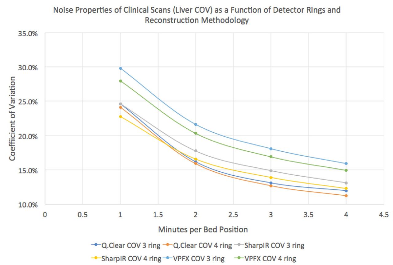
Results: Under no conditions were 1 min scans of acceptable image quality. All images on both systems collected for more than 3 minutes were, subjectively, of excellent quality. COVs in the liver for the 3 and 4-ring scanner systems several reconstructions are plotted as a function of min. per bed position in the figure below. Adding approximately 30 seconds per bed position to an OSEM or PRF reconstruction on a 3-ring system results in comparable image noise characteristics to a 4-ring system. However, the limited axial FOV of the 3-ring system requires approximately 1.5x more bed positions for similar coverage. Total scan time, therefore, is substantially higher to achieve identical image quality. RR, however, has virtually identical COV properties on the 3 and 4-ring systems, suggesting successful noise suppression; however, the 1.5X time penalty remains. PRF reconstructions appear to buy 1 minute per bed position for similar COV characteristics. A 3-ring system equipped with RR at 2:40 seconds per bed position results in a COV superior to a 4-ring system operating with OSEM and TOF at 4 minutes per bed position (identical total scan times). PRF reconstructions also boosted SUVmax values 14±7%, and RR an additional 12±9%, adding additional lesion conspicuity.

Conclusions: Expected noise penalties associated with a 3-ring SiPM array PET/CT scanner vs. a 4-ring system have been characterized on 14 FDG oncology studies. Advanced reconstructions substantially improve noise properties and lesion conspicuity that result in excellent image quality on both 3 and 4-ring SiPM systems.

Figure
  • Download figure
  • Open in new tab
  • Download powerpoint
Previous
Back to top

In this issue

Journal of Nuclear Medicine
Vol. 59, Issue supplement 1
May 1, 2018
  • Table of Contents
  • Index by author
Article Alerts
Sign In to Email Alerts with your Email Address
Email Article

Thank you for your interest in spreading the word on Journal of Nuclear Medicine.

NOTE: We only request your email address so that the person you are recommending the page to knows that you wanted them to see it, and that it is not junk mail. We do not capture any email address.

Enter multiple addresses on separate lines or separate them with commas.
Clinical image quality comparison of a 3-ring vs 4-ring configuration of a commercial SiPM array PET/CT
(Your Name) has sent you a message from Journal of Nuclear Medicine
(Your Name) thought you would like to see the Journal of Nuclear Medicine web site.
Citation Tools
Clinical image quality comparison of a 3-ring vs 4-ring configuration of a commercial SiPM array PET/CT
John Sunderland, Lisa Dunnwald, John Richmond, Michael Graham, Yusuf Menda, Janet Pollard
Journal of Nuclear Medicine May 2018, 59 (supplement 1) 1762;

Citation Manager Formats

  • BibTeX
  • Bookends
  • EasyBib
  • EndNote (tagged)
  • EndNote 8 (xml)
  • Medlars
  • Mendeley
  • Papers
  • RefWorks Tagged
  • Ref Manager
  • RIS
  • Zotero
Share
Clinical image quality comparison of a 3-ring vs 4-ring configuration of a commercial SiPM array PET/CT
John Sunderland, Lisa Dunnwald, John Richmond, Michael Graham, Yusuf Menda, Janet Pollard
Journal of Nuclear Medicine May 2018, 59 (supplement 1) 1762;
Twitter logo Facebook logo LinkedIn logo Mendeley logo
  • Tweet Widget
  • Facebook Like
  • Google Plus One
Bookmark this article

Jump to section

  • Article
  • Figures & Data
  • Info & Metrics

Related Articles

  • No related articles found.
  • Google Scholar

Cited By...

  • No citing articles found.
  • Google Scholar

More in this TOC Section

Instrumentation & Data Analysis Track

  • Deep Learning Based Kidney Segmentation for Glomerular Filtration Rate Measurement Using Quantitative SPECT/CT
  • The Benefit of Time-of-Flight in Digital Photon Counting PET Imaging: Physics and Clinical Evaluation
  • Preclinical validation of a single-scan rest/stress imaging technique for 13NH3 cardiac perfusion studies
Show more Instrumentation & Data Analysis Track

Instrumentation posters

  • Feasibility of Low-Dose18F-FDG PET in Pediatric Patients using SiPM-based Detector PET: Quantitative and Qualitative Image Evaluation
  • Image quality of brain SPECT recorded with the whole-body Veriton CZT camera and a focal brain configuration of detectors, as compared with conventional SPECT and PET systems
  • Initial clinical experience with rapid high definition Na18F digital photon counting PET/CT imaging for whole-body osteoblastic disease assessment
Show more Instrumentation posters

Similar Articles

SNMMI

© 2025 SNMMI

Powered by HighWire