
Main menu

  • Home
  • Content
    • Current
    • Ahead of print
    • Past Issues
    • JNM Supplement
    • SNMMI Annual Meeting Abstracts
  • Subscriptions
    • Subscribers
    • Institutional and Non-member
    • Rates
    • Corporate & Special Sales
    • Journal Claims
  • Authors
    • Submit to JNM
    • Information for Authors
    • Assignment of Copyright
    • AQARA requirements
  • Info
    • Permissions
    • Advertisers
    • Continuing Education
  • About
    • About Us
    • Editorial Board
    • Contact Information
  • More
    • Alerts
    • Feedback
    • Help
    • SNMMI Journals
  • SNMMI
    • JNM
    • JNMT
    • SNMMI Journals
    • SNMMI

User menu

  • Subscribe
  • My alerts
  • Log in
  • My Cart

Search

  • Advanced search
Journal of Nuclear Medicine
  • SNMMI
    • JNM
    • JNMT
    • SNMMI Journals
    • SNMMI
  • Subscribe
  • My alerts
  • Log in
  • My Cart
Journal of Nuclear Medicine

Advanced Search

  • Home
  • Content
    • Current
    • Ahead of print
    • Past Issues
    • JNM Supplement
    • SNMMI Annual Meeting Abstracts
  • Subscriptions
    • Subscribers
    • Institutional and Non-member
    • Rates
    • Corporate & Special Sales
    • Journal Claims
  • Authors
    • Submit to JNM
    • Information for Authors
    • Assignment of Copyright
    • AQARA requirements
  • Info
    • Permissions
    • Advertisers
    • Continuing Education
  • About
    • About Us
    • Editorial Board
    • Contact Information
  • More
    • Alerts
    • Feedback
    • Help
    • SNMMI Journals
  • Follow JNM on Twitter
  • Visit JNM on Facebook
  • Join JNM on LinkedIn
  • Subscribe to our RSS feeds
Meeting ReportPhysics, Instrumentation & Data Sciences

Feasibility of Low-Dose18F-FDG PET in Pediatric Patients using SiPM-based Detector PET: Quantitative and Qualitative Image Evaluation

Boxue Shu, Tongxiao Xue, Weiru Zhou, Chenwei Li, Runze Wu, Zhen Li and Pengbo Xie
Journal of Nuclear Medicine May 2019, 60 (supplement 1) 1391;
Boxue Shu
2Xi‘An Gao Xing Hospital Xi’An China
  • Find this author on Google Scholar
  • Find this author on PubMed
  • Search for this author on this site
Tongxiao Xue
2Xi‘An Gao Xing Hospital Xi’An China
  • Find this author on Google Scholar
  • Find this author on PubMed
  • Search for this author on this site
Weiru Zhou
2Xi‘An Gao Xing Hospital Xi’An China
  • Find this author on Google Scholar
  • Find this author on PubMed
  • Search for this author on this site
Chenwei Li
1Shanghai United Imaging Healthcare Co., Ltd. Shanghai China
  • Find this author on Google Scholar
  • Find this author on PubMed
  • Search for this author on this site
Runze Wu
1Shanghai United Imaging Healthcare Co., Ltd. Shanghai China
  • Find this author on Google Scholar
  • Find this author on PubMed
  • Search for this author on this site
Zhen Li
1Shanghai United Imaging Healthcare Co., Ltd. Shanghai China
  • Find this author on Google Scholar
  • Find this author on PubMed
  • Search for this author on this site
Pengbo Xie
1Shanghai United Imaging Healthcare Co., Ltd. Shanghai China
  • Find this author on Google Scholar
  • Find this author on PubMed
  • Search for this author on this site
  • Article
  • Figures & Data
  • Info & Metrics
Loading

Abstract

1391

Objectives: Pediatric patients are more susceptible to the radiation due to longer life expectancy and vigorous cell reproduction. As newly introduced PET with higher sensitive detector, it is possible to maintain the image quality of FDG-PET on pediatric patients and reduce the injected tracer dose, which can lower the radiation dose to the patient. Therefore, this research aims to analyze the quantitative and qualitative image quality of FDG-PET on simulated low-dose imaging studies and find the optimal low-dose recommendation for pediatric patients.

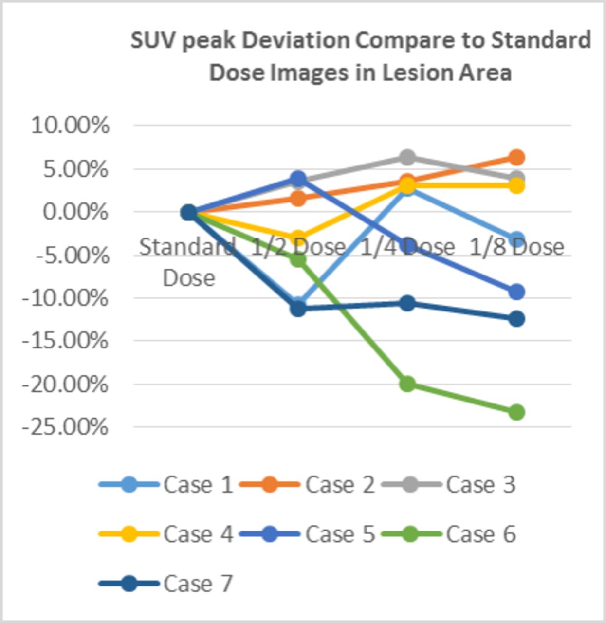
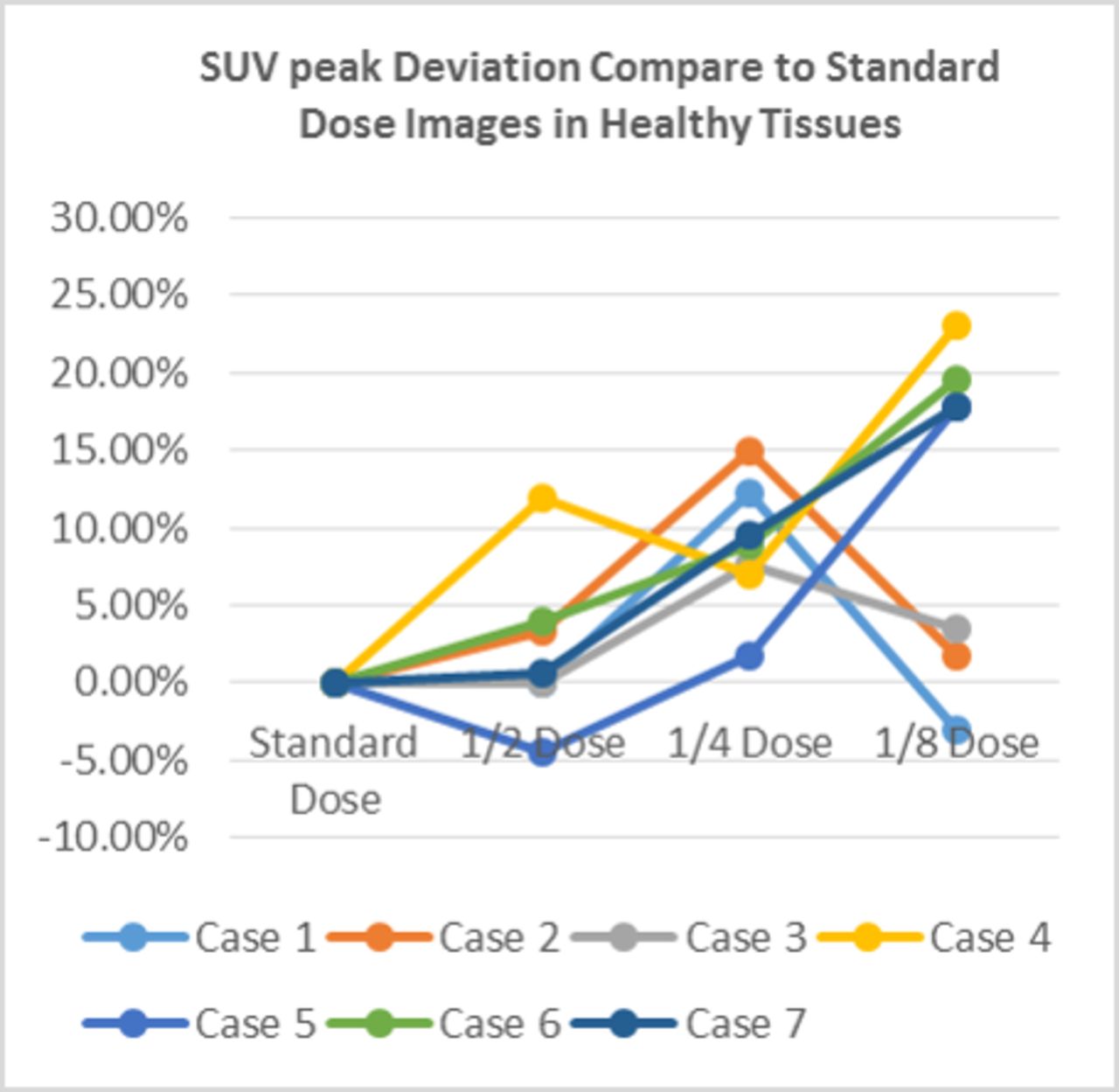
Methods: 7 cases (age 2 to 14 years old) were acquired on a SiPM-based detector PET/CT scanner (uMI780, United Imaging Healthcare, Shanghai, China). The acquisition settings were 3-minute PET acquisition per bed position and the injection dose was around 0.1mCi/kg. All cases were retrospectively reconstructed with different levels of simulated dose reduction and then were compared with full dose images in sequence. Simulated 1/2 dose, 1/4 dose, 1/8 dose images were produced via reconstruction using 90s (1/2 dose), 45s (1/4 dose) and 22.5s (1/8 dose) data from the acquired list data. A combination of two physicians’ evaluation of various low-dose images and quantitative measurement of SUV mean value, SUV peak and volume were used for image quality evaluation. Two physicians (3- and 7-year experience) assessed image quality using a Likert-scale to rank the quality of images and to see if the low-dose images were clinically acceptable (1, unacceptable image quality; 2, suboptimal image quality; 3, acceptable image quality; 4, good image quality; 5, excellent image quality). VOIs with radius of 20mm were placed at the same position of different levels of low-dose images on normal hepatic tissue to compare the variability and bias of SUV peak and SUV mean among different images. The VOIs were placed on the lesions, and the volume was as large as possible to cover the whole lesion.

Results: A total of 21 lesions in 7 cases were successfully identified and no lesion was omitted compared to standard-dose images. The image quality was all clinically acceptable according to the qualitative evaluation of two physicians. The Likert-scale grades of 1/2 dose, 1/4 dose and 1/8 dose images were 4.14±0.69, 3.85±1.21,3.28±0.75, respectively. The deviations of quantitative measurement between low-dose and standard dose images are all under 25%. For 1/2 dose images, the difference of SUV peak, SUV mean and lesion volume are lower than 15% compared with full-dose images. The deviation of 1/4 dose images ranges from 0% to 19.91%. 1/8 dose images have the most dramatic variation from 0 % to 26.97%. Conclusions: Our results demonstrates that dose reduction is feasible without compromising the diagnostic image quality or influence quantitative measurement. Considering the fluctuation of 1/2 dose images is minimum and combining with the results of two physicians, 1/2 dose reduction (0.05 mCi/kg) might be potentially feasible and worthy for further exploration. The values are calculated based on the following equations: [(A-B)/B][asterisk]100% A: SUVpeak, SUVmean, Volume of low dose B: SUVpeak, SUVmean, Volume of standard dose

Figure
  • Download figure
  • Open in new tab
  • Download powerpoint
Figure
  • Download figure
  • Open in new tab
  • Download powerpoint
Figure
  • Download figure
  • Open in new tab
  • Download powerpoint
Figure
  • Download figure
  • Open in new tab
  • Download powerpoint
Figure
  • Download figure
  • Open in new tab
  • Download powerpoint
Previous
Back to top

In this issue

Journal of Nuclear Medicine
Vol. 60, Issue supplement 1
May 1, 2019
  • Table of Contents
  • Index by author
Article Alerts
Sign In to Email Alerts with your Email Address
Email Article

Thank you for your interest in spreading the word on Journal of Nuclear Medicine.

NOTE: We only request your email address so that the person you are recommending the page to knows that you wanted them to see it, and that it is not junk mail. We do not capture any email address.

Enter multiple addresses on separate lines or separate them with commas.
Feasibility of Low-Dose18F-FDG PET in Pediatric Patients using SiPM-based Detector PET: Quantitative and Qualitative Image Evaluation
(Your Name) has sent you a message from Journal of Nuclear Medicine
(Your Name) thought you would like to see the Journal of Nuclear Medicine web site.
Citation Tools
Feasibility of Low-Dose18F-FDG PET in Pediatric Patients using SiPM-based Detector PET: Quantitative and Qualitative Image Evaluation
Boxue Shu, Tongxiao Xue, Weiru Zhou, Chenwei Li, Runze Wu, Zhen Li, Pengbo Xie
Journal of Nuclear Medicine May 2019, 60 (supplement 1) 1391;

Citation Manager Formats

  • BibTeX
  • Bookends
  • EasyBib
  • EndNote (tagged)
  • EndNote 8 (xml)
  • Medlars
  • Mendeley
  • Papers
  • RefWorks Tagged
  • Ref Manager
  • RIS
  • Zotero
Share
Feasibility of Low-Dose18F-FDG PET in Pediatric Patients using SiPM-based Detector PET: Quantitative and Qualitative Image Evaluation
Boxue Shu, Tongxiao Xue, Weiru Zhou, Chenwei Li, Runze Wu, Zhen Li, Pengbo Xie
Journal of Nuclear Medicine May 2019, 60 (supplement 1) 1391;
del.icio.us logo Digg logo Reddit logo Twitter logo Facebook logo Google logo Mendeley logo
  • Tweet Widget
  • Facebook Like
  • Google Plus One
Bookmark this article

Jump to section

  • Article
  • Figures & Data
  • Info & Metrics

Related Articles

  • No related articles found.
  • Google Scholar

Cited By...

  • No citing articles found.
  • Google Scholar

More in this TOC Section

Physics, Instrumentation & Data Sciences

  • Deep Learning Based 3D Dose Estimation from Prompt Gamma Distribution for Proton Therapy Monitoring
  • Data-driven respiratory signal estimation from finely sampled projection data in conventional cardiac perfusion SPECT imaging.
  • Clinical feasibility of quantitative holmium-166 SPECT in presence of technetium-99m colloid
Show more Physics, Instrumentation & Data Sciences

Instrumentation Posters

  • Image quality of brain SPECT recorded with the whole-body Veriton CZT camera and a focal brain configuration of detectors, as compared with conventional SPECT and PET systems
  • Initial clinical experience with rapid high definition Na18F digital photon counting PET/CT imaging for whole-body osteoblastic disease assessment
Show more Instrumentation Posters

Similar Articles

SNMMI

© 2022 Journal of Nuclear Medicine

Powered by HighWire